Abstract
Commitment of hemopoietic progenitors to the T-cell lineage is a crucial requirement for T-cell development, yet the timing and developmental cues regulating this process remain controversial. Here we have devised a technique to analyze the T-cell/B-cell lineage potential of precursors that have been recruited to the fetal mouse thymus but which have yet to contact the thymic epithelial microenvironment. We show that lymphoid progenitors arriving at the thymus are not bipotent T/B precursors, and provide evidence that intrathymic Notch signaling is not the mechanism determining T/B lineage choice in migrant precursors. Rather, we provide evidence that Notch signaling influences T/B lineage choice in lymphoid precursors through interactions with defined stromal components within the fetal liver. Collectively, our data redefine our understanding of the role and timing of Notch signaling in relation to lineage choices in lymphoid precursors.
Introduction
T cells develop in the thymus from migrant progenitors originating in hemopoietic tissues. In the mouse, the thymic primordium is first colonized by precursors at around day 11 to 12 of gestation, with some evidence suggesting that subsequent entry of precursors into the thymus occurs in distinct waves.1 Although several studies have isolated and characterized early T-cell precursor populations from extrathymic sites such as fetal blood and fetal liver,2-4 it is controversial whether precursors recruited to the developing thymus are multipotential hemopoietic stem cells (HSCs), common lymphoid progenitors (CLPs), or cells already restricted to the T-cell lineage. However, a recent study on adult mice has suggested that CLPs are not recruited to the thymus from the bone marrow, and that the only cells in the adult circulatory system that can give rise to T cells are of a Lin–Scal+ckithi phenotype, so-called “LSK” cells, which can also give rise to B cells.5 Direct analysis of the nature of thymus-colonizing cells is further complicated by extrapolation between fetal and adult situations, where the nature of the thymus-colonizing cells may be different (for a review, see Kincade et al6 ), and may also be confused by the presence of recirculating B-restricted progenitors in the thymic vasculature in the case of the adult.
Clonal assays of single cells isolated from within the developing embryonic thymus have shown that these cells are not bipotent T/B progenitors,7 which contrasts with recent studies indicating that early thymic progenitors in the adult thymus can give rise to both T cells and B cells.8 In the adult, cells with B-cell potential have been demonstrated within the CD44+25– (DN1) population representing the least mature thymocytes.9 Whilst this was initially suggestive of entry of CLPs or even HSCs with both T and B potential, the recent demonstration of phenotypic and developmental heterogeneity within DN1 cells10 makes it difficult to exclude entry of CD44+ committed B-cell progenitors along with cells of T-restricted potential as an explanation for these findings. The development of B cells rather than T cells in the thymus of mice colonized by Notch-1–deficient bone marrow precursors has also been interpreted as indicating that lymphoid progenitors entering the thymus have both T- and B-cell potential, but then adopt a B-cell rather than a T-cell fate in the absence of Notch signaling.11 Again, these observations do not exclude the possibility that both T-restricted and B-restricted progenitors separately enter the thymus. In this scenario, in the absence of Notch signaling, the former are unable to develop, whereas the latter are able to reveal their B-cell potential, which would otherwise be inhibited by interaction by Notch ligands present within the thymic microenvironment.12 This interpretation also raises the possibility that whereas Notch signaling within the thymus is essential to reveal T-lineage commitment, T/B lineage choice itself may occur at an earlier stage and thus is not necessarily dependent on intrathymic Notch activation.
In this study we have sought to clarify the status of the first wave of precursors recruited to the developing fetal thymus in terms of both phenotype and developmental potential. By isolating precursors from the prevascular 12-day thymic rudiment, we have excluded possible involvement of more mature cells in the circulation and focused on precursors that have been specifically recruited to the thymus. Separating these cells into those isolated from the perithymic mesenchyme and those in the intraepithelial regions of the thymus has also enabled us to compare the status of precursors immediately before and immediately after contact with the epithelial microenvironment where they interact with Notch ligands expressed by thymic epithelial cells.12 We show that CD45+ precursors isolated from the epithelial region of the thymus are able to give rise to both αβ and γδ T cells but not B cells, in marked contrast to fetal liver progenitors, which generate both T cells and B cells. Importantly, we show that this loss of B-cell potential in migrant progenitors occurs before interaction with Notch ligand–expressing thymic epithelial cells. Moreover, by correlating Notch ligand expression and Notch activation in precursors within the embryonic day 12 (E12) fetal liver, we provide evidence that Notch plays a role in T/B lineage choice at a prethymic level. Collectively, our findings extend our understanding of the timing of Notch signaling in relation to the developmental potential of early lymphoid precursors, and argue against current models suggesting a role for intrathymic Notch activation in mediating initial T/B lineage choice.
Materials and methods
Mice
BALB/c mice were bred and maintained under specific pathogen-free (SPF) conditions in the Biomedical Services Unit, Birmingham University. The day of vaginal plug detection was designated as day 0.
Antibodies and immunoconjugates
The following were used for flow cytometric analysis: anti–interleukin 7 receptor (IL-7R) α-biotin (A7R34), anti–c-kit–phycoerythrin (PE) Cy5 (2B8), anti-Flt3PE (A2F10), anti–CD4-PE (RM4-5) (all supplied by eBioscience, San Diego, CA) and anti–CD8–fluorescein isothiocyanate (FITC) (53-6.7), anti–CD45-FITC (30-F11), anti–T-cell receptor beta (TCRβ) chain (H57-597), anti-TCRγδ (GL3) (supplied by BD Pharmingen, San Diego, CA), anti–intercellular adhesion molecule 1 (ICAM-1)–PE (YN1/1.7.4; eBioscience), anti–vascular cell adhesion molecule 1 (VCAM-1) biotin (clone 429; eBioscience), anti–EpCAM1 (epithelial cell adhesion molecule) (clone G8.8; a kind gift of Professor A. Farr), anti–platelet-derived growth factor receptor alpha (PDGFRα) (APA5; eBioscience). For B-cell assays, anti–CD45-biotin (30-F11; BD Pharmingen) was used in conjunction with CD19-PE. Streptavidin-PE Cy7 (Chemicon, Temecula, CA) was used to visualize biotin-conjugated primary antibodies. The following were coated onto anti–rat immunoglobulin (Ig) magnetic beads (Dynal, Wirral, United Kingdom): anti-TER119 (BD Pharmingen) and anti-CD45 (M1/9; ATCC, Manassas, VA).
Immunohistochemistry
Frozen sections of freshly dissected embryonic day 12 thymuses, with perithymic mesenchyme attached, were prepared as described.2 Sections were fixed in ice-cold acetone, and stained using FITC-conjugated anti–pan cytokeratin (clones C-11, PCK-26, CY-90, Ks-1A3, M20, and A53-B/A2), rabbit antifibronectin (Sigma, St Louis, MO) to reveal mesenchyme, and rat anti-CD45 (M1/9; ATCC) to identify migrant precursors. Primary antibodies were detected using anti–rat Ig biotin (Amersham, Arlington Heights, IL) followed by extravidin TRITC (tetramethylrhodamine-5 [and -6] isothiocyanate; Sigma), and anti–rabbit Ig–AMCA (Dako, Carpinteria, CA). Sections were mounted in antifade glycerol solution (Citifluor, Canterbury, United Kingdom). Sections were viewed using a Zeiss Axioplan microscope (Welwyn Garden City, United Kingdom).
Isolation of progenitors from E12 fetal liver, thymus, and perithymic mesenchyme
Haematopoietic progenitors present in the E12 perithymic mesenchyme and fetal thymic epithelium were isolated on the basis of the leukocyte marker CD45. E12 thymic lobes surrounded by their mesenchymally derived covering were isolated from BALB/c embryos by microdissection. These were incubated at 37°C for 20 minutes in 2.5 mg/mL collagenase/dispase (R&D Systems, Minneapolis, MN) in RPMI plus 10% fetal calf serum (FCS), placed under a dissecting microscope and aspirated gently using a glass pipette to separate perithymic mesenchyme from the inner epithelial rudiment. Perithymic mesenchyme and intact epithelial rudiments were transferred into fresh solutions of collagenase/dispase and enzymatic treatment continued until cell suspensions were produced. CD45+ cells were then sorted from such suspensions using a MoFlo high-speed cell sorter to a purity of 99.5% or greater. To obtain hematopoietic precursors from fetal liver, organs from day-12 embryos were disaggregated mechanically, stained with a lineage marker kit (Pharmingen) together with CD45 and IL-7Rα. Lin–CD45+IL-7Rα+ cells were then sorted using a MoFlo to a purity of 99.5% or greater.
Isolation of perithymic mesenchyme, thymic epithelium and fetal liver stromal cells from E12 organ rudiments
For the isolation of stromal cells from E12 thymic rudiments, freshly isolated lobes with surrounding mesenchyme still attached were disaggregated by incubation in 0.25% trypsin, and then labeled with antibodies to EpCAM1 and PDGFRα. EpCAM1+PDGFRα– thymic epithelial cells and EpCAM1–PDGFRα+ mesenchymal cells were then sorted using a MoFlo cell sorter to a purity of 99.5% or greater. To isolate stromal cells from E12 fetal liver, freshly dissected livers were disaggregated for 10 minutes at 37°C using 1 mg/mL collagenase/dispase (R&D Systems) containing 0.1 mg/mL DNAse in calcium and magnesium-free phosphate-buffered saline (PBS). Cell suspensions were then stained with labeled antibodies for CD45, EpCAM1, VCAM-1, and ICAM-1, and appropriate stromal cell populations were then sorted from CD45– cells using a MoFlo high-speed cell sorter to a purity of 99.5% or greater.
Assessment of B-cell potential in OP9 coculture assays
OP9 bone marrow stromal cells (a kind gift of J.-C. Zuniga-Pflucker, University of Toronto, Canada) were seeded in wells of a 6-well plate at 2 × 10⁵ cells/well in α–minimum essential medium (α-MEM) supplemented with 20% FCS, 100 U/mL penicillin, and 100 mg/mL streptomycin. The following day, 2 × 10³ CD45+ cells isolated from fetal liver, perithymic mesenchyme, or E12 thymic epithelial rudiment were seeded onto confluent OP9 monolayers in fresh α-MEM also containing recombinant IL-7 and Flt-3 ligand (R&D Systems) both at 5 ng/mL. Medium was changed after 3 days and cocultures were harvested after 6 days by vigorous pipetting. All resultant populations were pretreated with antibody specific to CD16/32 (93; eBioscience) to prevent nonspecific Fc binding, prior to dual staining for CD45 and CD19.
Assessment of T-cell potential by fusion of dGuo lobes with E12 tissues
To assay T-cell precursor potential in cells isolated from E12 tissues, fragments of E12 perithymic mesenchyme, E12 thymic epithelial rudiments, and E12 fetal liver were placed on a 0.8-μm pore size polycarbonate filter in organ culture, alongside an alymphoid 2-deoxyguanosine (2-dGuo) fetal thymic lobe.13 Lobes were cultured for 12 days in organ culture conditions, during which time migration of precursors from E12 tissues to 2-dGuo–treated lobes occurs. Cultures were harvested by gentle teasing using fine cataract knives, and recovered cells were analyzed by flow cytometry on a Becton Dickinson LSR machine with forward and side scatter gates set so as to exclude nonviable cells.
Reverse transcriptase (RT)–PCR analysis
High-purity cDNA was obtained from purified mRNA using μMacs One-step cDNA kit, according to the manufacturer's instructions (Miltenyi Biotech, Auburn, CA). β-actin was used as the housekeeping gene for sample normalization, prior to amplifying the target genes of interest. Reactions were conducted in a Peltier Thermal Cycler PTC-200 (MJ Research, Genetic Research Instrumentation, Braintree, Essex, United Kingdom), as described.14 Polymerase chain reaction (PCR) products taken from the exponential phase of the reactions were analyzed by ethidium bromide gel electrophoresis and identified by fragment size. Densitometrical analysis was determined using Syngene Gel Documentation Gene Tools software, and graphs show standard error of ratios of mRNA for the genes of interest relative to β-actin. Primer sequences and amplicon sizes were as follows: β-actin: forward 5′-ATC TAC GAG GGC TAT GCT CTC C-3′, reverse 5′-CTT TGA TGT CAC GCA CGA TTT CC-3′ (148 bp); Notch-1: forward 5′-CCCAGCAGGTGCAGCCACAG-3′, reverse 5′-GGT GAT CTG GGA CGG CAT GG -3′ (461 bp); Delta-like-1: forward 5′-GTC ACA GAG CTC TGC AGG AG-3′, reverse 5′-TGT GGG CAG TGC GTG CTT CC-3′ (472 bp); Delta-like-4: forward 5′-CAG AGA CTT CGC CAG GAA AC-3′, reverse 5′-ATC CAT TCT TGC ACG GAG AG-3′ (501 bp); Jagged-1: forward 5′-GTC AGA GTT CAG AGG CGT CC-3′, reverse 5′-TGT CAT CCT CCT CCA CTT CC-3′ (319 bp); Jagged-2: forward 5′-GTC CTT CCC ACA TGG GAG TT-3′, reverse 5′-GTT TCC ACC TTG ACC TCG GT-3′ (591 bp); Deltex-1: forward 5′-CAC TGG CCC TGT CCA CCC AGC CTT GGC AGG-3′, reverse 5′-GAG GCATGT GCCAGG CTAGAG GCAAGG CAA-3′ (943 bp); Hes-1: forward 5′-GCC AGT GTC AAC ACG ACA CCG G-3′, reverse 5′-TCA CCT CGT TCA TGC ACT CG-3′ (310 bp); Presenilin-1: forward 5′-CCA TCC TCC TGG TGG TCC TG-3′, reverse 5′-TAC GAA GTG GGC CTT TGG GA-3′ (369 bp).
Results
Identification and isolation of intrathymic and perithymic precursors recruited to the fetal thymus
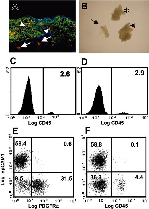
The murine fetal thymic rudiment undergoes initial colonization by migrant precursors at around day 12 of gestation before vascularization of the rudiment is established.3 At this stage, CD45+ precursors can be identified both within the cytokeratin+ network of the thymic epithelial rudiment (Figure 1A) and outside the thymic epithelium, within the cytokeratin– fibronectin+ perithymic mesenchyme (Figure 1A). Interestingly, at earlier stages in development (E11), T-cell precursors are found only within the surrounding perithymic mesenchyme (Itoh et al15 and data not shown), indicating that CD45+ cells first have to migrate through the surrounding perithymic mesenchyme before entering the epithelial compartment. Thus, during initial colonization of the prevascular fetal thymic rudiment, it is possible to define 2 major stromal compartments, epithelium and mesenchyme, and 2 populations of CD45+ precursors that have been attracted to the thymus, those that have already entered the epithelial environment and those still within the perithymic mesenchyme that have yet to contact thymic epithelial cells. The ability to characterize and separately isolate each of these lymphoid and stromal populations would enable direct analysis of lineage commitment within the lymphoid populations relative to precursors in the hemopoietic tissues and would facilitate investigation of the role of prethymic and thymic stromal microenvironments in directing this process.
To explore the possibility of separately isolating lymphoid precursors located in the perithymic and intrathymic compartments, we isolated thymus rudiments by microdissection from E12 mouse embryos with the perithymic mesenchyme still attached (Figure 1B, arrowhead). Following brief exposure to collagenase, lobes were carefully drawn up into a fine capillary pipette to strip the perithymic mesenchyme from the inner epithelial core. This procedure led to the separation of epithelium and mesenchyme along the basement membrane dividing these compartments to produce clean epithelial cores devoid of mesenchyme (Figure 1B, arrow) and mesenchymal fragments uncontaminated by epithelium (Figure 1B, asterisk). To determine whether lymphoid cells could be detected in these separated populations, epithelial cores and mesenchymal fragments were separately pooled and further disaggregated to generate single cell suspensions followed by staining for CD45. As shown in Figure 1C-D, distinct populations of CD45+ progenitors could be identified in both isolated mesenchymal (Figure 1C) and isolated epithelial (Figure 1D) preparations representing pre- and postentry populations of cells recruited to the thymus. MoFlo sorting yielded populations of more than 99.5% purity (not shown) from such preparations, providing material for functional, phenotypic, and RT-PCR analysis.
The initial wave of thymus colonization can be anatomically separated into 2 distinct stromal compartments. Frozen section from E12 BALB/c thymic rudiment (A; original magnification × 400) showing the cytokeratin+ epithelial core compartment (green) surrounded by fibronectin+ perithymic mesenchyme (blue). Note that CD45+ lymphoid progenitors (red) are present in both the epithelial compartment and the outer layers of mesenchyme. Combined enzymatic and mechanical separation of isolated intact lobes (B, arrowhead; original magnification × 50) produces smooth epithelial cores (arrow) stripped of mesenchyme and separate mesenchyme fragments (asterisk) devoid of epithelium. Following complete disaggregation of separated mesenchyme and epithelial components, small but distinct populations of CD45+ lymphoid precursors can be detected by flow cytometry in each preparation (mesenchyme, C; epithelium, D) allowing the identification and isolation of CD45+ precursor subsets before and after contact with thymic epithelium. Vertical bars show levels of background staining (panels C and D). Total disaggregation of whole freshly isolated E12 thymic lobes and surface labeling for EpCAM1 and PDGFRα (E) identifies nonoverlapping populations of epithelial cells and mesenchymal cells, respectively. Note that, apart from a small cohort of CD45+ cells representing the earliest thymic migrants (F), the thymus at this stage consists predominantly of EpCAM1+PDGFRα– epithelium and EpCAM1–PDGFRα+ mesenchyme. Numbers indicate percentages of positive cells. Data are typical of 5 separate experiments.
The initial wave of thymus colonization can be anatomically separated into 2 distinct stromal compartments. Frozen section from E12 BALB/c thymic rudiment (A; original magnification × 400) showing the cytokeratin+ epithelial core compartment (green) surrounded by fibronectin+ perithymic mesenchyme (blue). Note that CD45+ lymphoid progenitors (red) are present in both the epithelial compartment and the outer layers of mesenchyme. Combined enzymatic and mechanical separation of isolated intact lobes (B, arrowhead; original magnification × 50) produces smooth epithelial cores (arrow) stripped of mesenchyme and separate mesenchyme fragments (asterisk) devoid of epithelium. Following complete disaggregation of separated mesenchyme and epithelial components, small but distinct populations of CD45+ lymphoid precursors can be detected by flow cytometry in each preparation (mesenchyme, C; epithelium, D) allowing the identification and isolation of CD45+ precursor subsets before and after contact with thymic epithelium. Vertical bars show levels of background staining (panels C and D). Total disaggregation of whole freshly isolated E12 thymic lobes and surface labeling for EpCAM1 and PDGFRα (E) identifies nonoverlapping populations of epithelial cells and mesenchymal cells, respectively. Note that, apart from a small cohort of CD45+ cells representing the earliest thymic migrants (F), the thymus at this stage consists predominantly of EpCAM1+PDGFRα– epithelium and EpCAM1–PDGFRα+ mesenchyme. Numbers indicate percentages of positive cells. Data are typical of 5 separate experiments.
To assess the possibility of isolating the 2 major stromal compartments of the E12 rudiment, we next analyzed expression of the pan-epithelial marker EpCAM-1 and the fibroblast marker PDGFRα on disaggregated suspensions of whole isolated E12 lobes. As shown in Figure 1E, between them, these 2 markers encompassed approximately 90% of the cells in disaggregated E12 lobes, defining 2 nonoverlapping populations of epithelium and mesenchyme, separate from the minority of CD45+ lymphoid progenitors (Figure 1F), providing a basis for purification of the 2 major stromal populations forming distinct anatomical compartments (Figure 1A) to which colonizing progenitors are exposed. To determine the efficiency of separation of epithelial and mesenchyme compartments, we analyzed expression of the epithelialrestricted gene FoxN116 and the FGF7, which is expressed by mesenchyme but not epithelium,17 by RT-PCR to monitor the efficiency of separating the mesenchymal and epithelial compartments of the embryonic thymus. Importantly, FoxN1 was abundant in epithelial preparations but absent from mesenchymal preparations, whereas FGF7 was present in mesenchyme but not in epithelium (not shown), indicating epithelial and mesenchymal tissues had been successfully separated.
Progenitors recruited to the fetal thymus have a lineage-restricted rather than HSC phenotype, prior to contact with the thymic epithelium
Cells that may be recruited to the thymus include self-renewing hemopoietic stem cells (HSCs), and their presumed immediate descendents, non–self-renewing multipotent progenitors (MPPs), and common lymphoid progenitors (CLPs) (reviewed in Kincade et al6 ). Both HSCs and MPPs are characterized by high expression of ckit (ckithi) that is down-regulated following lineage commitment. These 2 populations can be distinguished from each other by Flt3 expression, which is absent from HSCs but expressed on MPPs.18 CLPs, which have been detected in the bone marrow in adults, are ckitloIL-7Rα+ and express high levels of Flt-3. These cells have the potential to give rise to both T cells and B cells in experimental assays but do not appear to colonize the adult thymus under normal physiologic conditions.5 To investigate the nature of the precursors representing the first wave of thymus-colonizing cells, we used flow cytometry to analyze CD45+ cells within the fetal liver, perithymic mesenchyme, and epithelial rudiment.
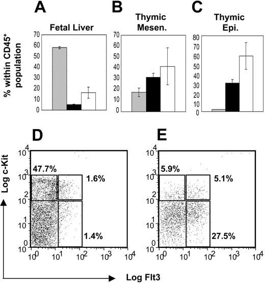
Analysis of CD45+ cells within E12 fetal liver preparations showed that approximately 60% were ckithi, consistent with HSC/MPP status (Figure 2A). A different pattern of ckit expression was seen when CD45+ cells in the epithelial compartment of the thymus were examined (Figure 2C), with the majority of cells being ckitlo, consistent with their having initiated lineage commitment. Unexpectedly, cells in the perithymic mesenchyme (Figure 2B) showed a pattern of ckit expression similar to that seen in the epithelial compartment rather than that in fetal liver, consistent with the idea that lineage commitment may be initiated by the time precursors are recruited to the thymus and before they contact thymic epithelial cells.
To further define the status of thymus-colonizing progenitors, we also analyzed the expression of IL-7Rα and Flt3. While only approximately 15% of CD45+ cells from E12 fetal liver were IL-7Rα+ (Figure 2A), approximately 40% and 60% of CD45+ cells isolated from perithymic (Figure 2B) and intrathymic (Figure 2C) regions, respectively, were positive for IL-7Rα. A similar pattern was seen for CD45+Flt3+ cells, which showed a 6-fold relative enrichment in both mesenchymal (Figure 2B) and epithelial regions (Figure 2C) of the thymus as compared with fetal liver (Figure 2A). We next analyzed a combination of CD45, Flt3, and c-kit expression in precursors within the fetal liver and thymus, to enable a more detailed comparison of precursor subsets in these sites. Interestingly, while CD45+ cells in the fetal liver were enriched for cells of a ckithiflt3– “HSC-like” phenotype with few ckitloflt3+ “CLP-like” cells (Figure 2D), a reciprocal pattern was observed in thymus-colonizing cells. Thus, while few CD45+ckit+Flt3– cells were found, we detected an enrichment of CD45+ckitloFlt3+ cells (Figure 2E) at the thymus. Compared with E12 fetal liver, this relative enrichment of IL-7Rα+ cells and ckit+Flt3+ cells in the CD45 populations of the thymus and particularly in the perithymic region provides further evidence for the preferential recruitment of a subset of precursors from the FL that have already lost HSC status and initiated lineage commitment prior to contact with the thymic epithelium.
Migrant thymic precursors show evidence of lineage restriction prior to contact with thymic epithelium. Cell suspensions prepared from E12 fetal liver (A), isolated perithymic mesenchyme (B) and isolated thymic epithelium (C) were stained for CD45 in combination with either Flt3 or IL-7Rα. Data are expressed as an average of 3 separate experiments ± SEM. ▦ shows the proportion of CD45+ expressing high levels of c-kit; ▪, the proportion expressing Flt3; and □, the proportion expressing IL-7Rα. Precursors in E12 fetal liver (D) and E12 fetal thymus (E) were also analyzed by 3-color flow cytometry for expression of CD45, Flt3, and ckit. Panels D and E show c-Kit/Flt3 expression in CD45+ cells. Note that in contrast to fetal liver, CD45+ cells in the fetal thymus are enriched for ckitloFlt3+ cells. Numbers in graphs indicate percentage of positive cells. Data shown are a result of 3 separate experiments.
Migrant thymic precursors show evidence of lineage restriction prior to contact with thymic epithelium. Cell suspensions prepared from E12 fetal liver (A), isolated perithymic mesenchyme (B) and isolated thymic epithelium (C) were stained for CD45 in combination with either Flt3 or IL-7Rα. Data are expressed as an average of 3 separate experiments ± SEM. ▦ shows the proportion of CD45+ expressing high levels of c-kit; ▪, the proportion expressing Flt3; and □, the proportion expressing IL-7Rα. Precursors in E12 fetal liver (D) and E12 fetal thymus (E) were also analyzed by 3-color flow cytometry for expression of CD45, Flt3, and ckit. Panels D and E show c-Kit/Flt3 expression in CD45+ cells. Note that in contrast to fetal liver, CD45+ cells in the fetal thymus are enriched for ckitloFlt3+ cells. Numbers in graphs indicate percentage of positive cells. Data shown are a result of 3 separate experiments.
T/B lineage choice occurs prior to contact with the thymic epithelium
While the above findings suggest that precursors undergoing recruitment to the thymus may have initiated lineage commitment prior to contact with the thymic epithelial microenvironment, functional assays are required to provide definitive evidence on T-cell and B-cell potential within these populations. To determine T-cell potential, fragments of E12 fetal liver, isolated perithymic mesenchyme, or isolated thymic epithelial rudiments with their contained CD45+ progenitors were fused with alymphoid 2dGuo-treated lobes in fetal thymus organ cultures. After the indicated culture period, colonized lobes were harvested and analyzed for T-cell differentiation. As shown in Figure 3A, this experimental approach of fusing tissue fragments with alymphoid thymus lobes was found to efficiently support T-cell development from precursors within all E12 tissues analyzed, with cells of CD4+8+, CD4+8– and CD4–8+ phenotypes as well as cells expressing αβTCR (Figure 3B) or γδTCR (Figure 3C) being detected.
To compare B-cell potential within the same precursor pools, CD45+ cells were purified from E12 fetal liver and from the mesenchymal and epithelial compartments of the thymus separated as described under “Isolation of progenitors from E12 fetal liver, thymus, and perithymic mesenchyme.” These cells were cocultured with OP9 bone marrow stromal cells in the presence of IL-7, conditions that support B-cell development from lymphoid progenitors.19 As expected, purified CD45+ precursors obtained from E12 fetal liver were found to expand in OP9 cultures and gave rise to CD19+ B cells (Figure 3D). In marked contrast, very few viable cells were recovered from age-matched OP9 cultures initiated with similar numbers of CD45+ precursors obtained from within the E12 thymic epithelium (Figure 3E). Typically, from an input of 2 × 10³ CD45+ E12 cells, approximately 6 × 10⁵ cells were recovered from fetal liver cultures, while less than 1 × 10⁴ cells were recovered from cultures initiated with thymus-derived cells (Figure 3E) with a 1000-fold difference in the generation of CD19+ B cells being observed between fetal liver precursors and those cells resident within or surrounding the thymus. Importantly, CD45+ cells isolated from the perithymic mesenchyme also failed to give rise to CD19+ cells in OP9 cultures (Figure 3E). As precursors initially colonizing the thymus migrate through the perithymic mesechyme prior to thymus entry, this finding suggests that these cells have already lost B-cell potential by the time they are recruited to the thymus and before they have contacted the thymic epithelium.
T/B lineage choice in migrant thymic progenitors occurs prior to thymus entry and contact with Notch ligands on thymic epithelium. CD45+ cells from E12 fetal liver, perithymic mesenchyme, and thymic epithelium introduced into alymphoid 2-dGuo–treated thymus lobes have the ability to give rise to differentiated T-cell progeny defined by CD4 and CD8 (A) including cells of both αβ and γδ T lineages (B-C). The B-cell potential of E12 precursors from these sources was also compared by culturing them on monolayers of OP9 bone marrow stromal cells in multiwell plates. Results are presented as the CD19+ cells within the CD45+ fraction of cells recovered from each well (D). Vertical lines in the histograms represent background staining controls. Average total numbers of CD45+CD19+ cells recovered per well for each precursor input population are shown in panel E. ▦ indicates E12 fetal liver; □, E12 perithymic mesenchyme; and ▪, thymic epithelium. Note the lack of B-cell development from thymus-derived precursors. Each experiment was performed 4 times with similar results. Data are expressed as an average ± SEM.
T/B lineage choice in migrant thymic progenitors occurs prior to thymus entry and contact with Notch ligands on thymic epithelium. CD45+ cells from E12 fetal liver, perithymic mesenchyme, and thymic epithelium introduced into alymphoid 2-dGuo–treated thymus lobes have the ability to give rise to differentiated T-cell progeny defined by CD4 and CD8 (A) including cells of both αβ and γδ T lineages (B-C). The B-cell potential of E12 precursors from these sources was also compared by culturing them on monolayers of OP9 bone marrow stromal cells in multiwell plates. Results are presented as the CD19+ cells within the CD45+ fraction of cells recovered from each well (D). Vertical lines in the histograms represent background staining controls. Average total numbers of CD45+CD19+ cells recovered per well for each precursor input population are shown in panel E. ▦ indicates E12 fetal liver; □, E12 perithymic mesenchyme; and ▪, thymic epithelium. Note the lack of B-cell development from thymus-derived precursors. Each experiment was performed 4 times with similar results. Data are expressed as an average ± SEM.
Correlation of Notch signaling with T/B lineage choice in prethymic and intrathymic microenviroments
Recent studies have emphasized the role of Notch signaling in T-cell/B-cell lineage choice, suggesting that in the absence of Notch signaling, lymphoid progenitors presumed to have both T- and B-cell potential adopt a B-cell fate, whereas active Notch signaling inhibits B-cell development and supports T-cell development.11,20,21 Since activation of Notch signaling depends on interaction of Notch with members of the Jagged and Delta-like family members of Notch ligands,21 we sought to compare the staging of T-cell/B-cell lineage choice with the availability of Notch ligands to migrant lymphoid precursors.
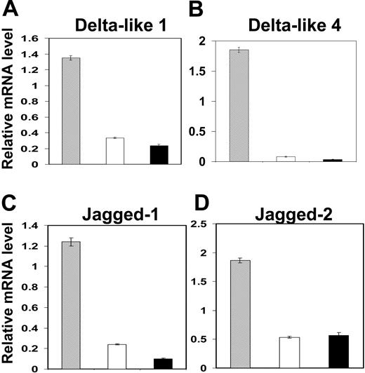
Migrant thymic precursors do not express Notch ligands. Freshly sorted CD45+ precursors obtained from the perithymic and intrathymic regions of the thymus were analyzed for expression of Notch ligands DL1 (A), DL4 (B), Jagged-1 (C), and Jagged-2 (D) by RT-PCR. Note that in comparison to thymic stromal cells that express readily detectable levels of Notch ligands (▦), migrant thymic precursors from the mesenchyme (□) or epithelium (▪) of the E12 thymus were found to express little if any of the Jagged and Delta families of Notch ligands. Data shown are representative of 3 separate experiments and are expressed as mean ± SEM.
Migrant thymic precursors do not express Notch ligands. Freshly sorted CD45+ precursors obtained from the perithymic and intrathymic regions of the thymus were analyzed for expression of Notch ligands DL1 (A), DL4 (B), Jagged-1 (C), and Jagged-2 (D) by RT-PCR. Note that in comparison to thymic stromal cells that express readily detectable levels of Notch ligands (▦), migrant thymic precursors from the mesenchyme (□) or epithelium (▪) of the E12 thymus were found to express little if any of the Jagged and Delta families of Notch ligands. Data shown are representative of 3 separate experiments and are expressed as mean ± SEM.
One possibility is that lymphoid progenitors themselves express Notch ligands and that recruitment to the thymus brings these cells into juxtaposition where mutual Notch signaling can occur. However, as shown in Figure 4, we were unable to detect Notch ligand expression in CD45+ cells in either the epithelial or mesenchymal compartments of the thymus. This suggests that Notch activation in migrant precursors does not occur via lateral T-cell precursor/T-cell precursor interactions, indicating the need for inductive signaling through interaction with stromal cells.
In view of our finding that T/B lineage choice has already taken place before progenitors contact the thymic epithelium, it was important to determine whether Notch ligands are expressed in the perithymic mesenchyme as well as in the thymic epithelium. To this end, we compared Notch ligand expression in the 2 major stromal compartments of the E12 rudiment as defined in the first section.
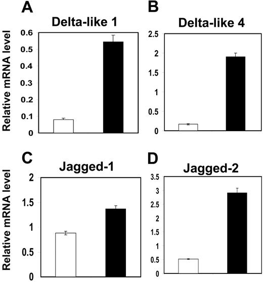
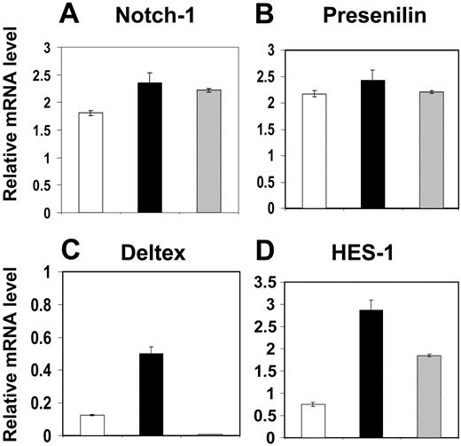
As shown in Figure 5, the Notch ligands DLL-1, DLL-4, and Jagged 1 and Jagged 2 were all detected in E12 thymic epithelial cells. In contrast, with the exception of Jagged 1, Notch ligand expression in the perithymic mesenchyme was low. This pattern of ligand distribution correlated well with the levels of Hes1 and Deltex expression, indicative of active Notch signaling,12 in CD45+ progenitors isolated from the different stromal compartments. Thus, Hes1 and Deltex expression were readily detectable in precursors located within the Notch ligand–expressing thymic epithelium, whereas CD45+ cells located in the perithymic mesenchyme showed a lower level of Hes1 and Deltex expression (Figure 6). Taken together with the observations in the previous section indicating that T/B lineage choice has already occurred by the time progenitors are recruited to the thymus, our data argue strongly against a role for intrathymic Notch signaling in determining T/B lineage choice, at least during initial thymus colonization in the fetus.
Compartmentalization of Notch ligand expression in the E12 thymus. PDGFRα+ mesenchyme (□) and EpCAM1+ epithelial cells (▪) were obtained from E12 thymus and analyzed for expression of Notch ligands by RT-PCR (A-D). Note that in comparison to thymic epithelial cells, with the possible exception of Jagged-1 (C), thymic mesenchymal cells do not express appreciable levels of the Notch ligands analyzed. Data shown are representative of 3 separate experiments and are expressed as mean ± SEM.
Compartmentalization of Notch ligand expression in the E12 thymus. PDGFRα+ mesenchyme (□) and EpCAM1+ epithelial cells (▪) were obtained from E12 thymus and analyzed for expression of Notch ligands by RT-PCR (A-D). Note that in comparison to thymic epithelial cells, with the possible exception of Jagged-1 (C), thymic mesenchymal cells do not express appreciable levels of the Notch ligands analyzed. Data shown are representative of 3 separate experiments and are expressed as mean ± SEM.
Notch activation in fetal liver lymphoid progenitors correlates with Notch ligand expression by fetal liver stroma
Although it has been assumed that Notch signaling for T/B lineage choice occurs intrathymically,11 this is not supported by our present observation that T/B lineage choice is determined before intrathymic Notch signaling is activated. This leaves open the possibility that T/B lineage choice does not depend on Notch signaling, even though subsequent Notch signaling within the thymus is needed to reveal it by supporting T-cell maturation.12,22 Alternatively, it is possible that transient Notch signaling may occur in the prethymic hemopoietic microenvironment with the loss of B-cell potential and retention of T-cell potential being determined at this stage.
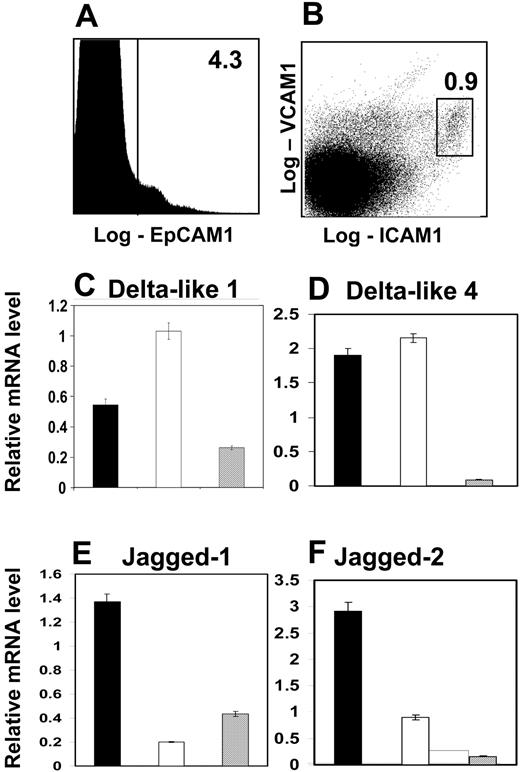
To investigate the potential for prethymic Notch signaling, we analyzed the availability of Notch ligands on stromal elements in the fetal liver, which represents the major hemopoietic compartment in the E12 embryo. Stromal components in the E12 fetal liver include epithelial cells derived from the endodermal liver buds and mesenchymal cells of mesoderm origin. These components were defined using a panel of antibodies to stromal cell populations, with candidate populations being isolated on the basis of EpCAM1 expression for epithelium, and a combination of VCAM-1 and ICAM-1 expression for mesenchymal cells (Figure 7A-B). While RT-PCR analysis of these purified populations (Figure 7C-F) revealed only low levels of Jagged expression in both the fetal liver stromal populations analyzed, VCAM+ICAM+ fetal liver stromal cells were found to express levels of Delta family members, including DLL-1, which can regulate Notch signaling for T-cell development,22,23 at levels similar to that seen in thymic epithelium (Figure 7C-F). This indicates the availability of Notch ligands in the prethymic fetal liver environment, and thus the possibility of Notch-driven lineage choice in lymphoid precursors at this stage. Consistent with this, we also found evidence of Notch activation, as indicated by Hes-1 expression, in Lin–IL-7Rα+ precursors isolated from E12 fetal liver (Figure 6D). Thus our findings provide direct evidence that Notch ligands are available on defined stromal elements within prethymic microenvironments, and that prior to thymus colonization and intrathymic Notch signaling, a phase of Notch signaling in lymphoid precursors can occur within the fetal liver.
Two phases of Notch activation occur in lymphoid precursors prior to and after thymus colonization. CD45+ precursors obtained from the perithymic (□) and intrathymic (▪) regions were compared with CD45+Lin–IL-7Rα+ fetal liver precursors (▦) for evidence of Notch signaling. Note that although all cell preparations were found to express comparable levels of Notch-1 (A) and presenilin (B), precursors from the fetal liver and from the thymic epithelial rudiment, but not the perithymic mesenchyme, show evidence of Notch signaling as indicated by expression of Hes-1 mRNA (D). Data shown are typical of 3 separate experiments and are expressed as mean ± SEM.
Two phases of Notch activation occur in lymphoid precursors prior to and after thymus colonization. CD45+ precursors obtained from the perithymic (□) and intrathymic (▪) regions were compared with CD45+Lin–IL-7Rα+ fetal liver precursors (▦) for evidence of Notch signaling. Note that although all cell preparations were found to express comparable levels of Notch-1 (A) and presenilin (B), precursors from the fetal liver and from the thymic epithelial rudiment, but not the perithymic mesenchyme, show evidence of Notch signaling as indicated by expression of Hes-1 mRNA (D). Data shown are typical of 3 separate experiments and are expressed as mean ± SEM.
VCAM-1+ICAM-1+stromal cells provide Notch ligands within the fetal liver. CD45– stromal cells from E12 fetal liver were subdivided into subsets on the basis of expression of EpCAM1, ICAM-1, and VCAM-1. (A) EpCAM1 expression in CD45–ICAM-1–VCAM-1– cells; (B) ICAM-1 and VCAM-1 expression in CD45–EpCAM1– cells. Percentages shown are of the CD45– population. Such populations were sorted by MoFlo and analyzed for expression of Notch ligands by PCR (C-F). Note that expression of DL1 and DL4 by ICAM-1+VCAM-1+ fetal liver stromal cells (□) is similar to that of thymic epithelial cells (▪). ▦ indicates CD45–VCAM1–ICAM1–EpCAM1+ fetal liver cells. Similar data were produced from 3 separate experiments, and are expressed as mean ± SEM.
VCAM-1+ICAM-1+stromal cells provide Notch ligands within the fetal liver. CD45– stromal cells from E12 fetal liver were subdivided into subsets on the basis of expression of EpCAM1, ICAM-1, and VCAM-1. (A) EpCAM1 expression in CD45–ICAM-1–VCAM-1– cells; (B) ICAM-1 and VCAM-1 expression in CD45–EpCAM1– cells. Percentages shown are of the CD45– population. Such populations were sorted by MoFlo and analyzed for expression of Notch ligands by PCR (C-F). Note that expression of DL1 and DL4 by ICAM-1+VCAM-1+ fetal liver stromal cells (□) is similar to that of thymic epithelial cells (▪). ▦ indicates CD45–VCAM1–ICAM1–EpCAM1+ fetal liver cells. Similar data were produced from 3 separate experiments, and are expressed as mean ± SEM.
Discussion
The thymus is known to provide a specialized microenvironment capable of supporting the efficient generation of a functional T-cell pool from migrant precursors. In recent years, many studies have investigated the nature of the precursors initially colonizing the thymus, in an attempt to understand the timing and mechanisms regulating commitment to the T-cell lineage (reviewed in Kincade et al6 ). In this study, we have devised a novel technique that allows the spatial separation of cells representing the first wave of migrant precursors to reach the thymus.
Using this approach, we provide direct evidence that progenitors colonizing the fetal thymus have already made a T/B lineage choice before making contact with the thymic epithelium. Thus, our results do not support the suggestion arising from adult studies that T/B lineage choice is determined by Notch signals resulting from Notch/Notch ligand interactions within the thymus. As mentioned earlier, evidence in favor of this model includes studies demonstrating the presence of B cells in the thymuses of mice repopulated with Notch-1–deficient bone marrow.11 However, an alternative explanation for these observations that has yet to be ruled out is that in the absence of Notch-1–dependent T-cell development in the thymus, small numbers of B-committed cells are able to expand intrathymically in the absence of inhibitory signals from Notch or from competition from developing thymocytes. Moreover, our finding that the choice between T- and B-cell lineages is made at a prethymic level suggests that precursors with T-but not B-cell potential reside within the fetal liver. Although we have not addressed this directly in this study, this notion is supported by previous studies using clonal assays to study lymphoid potential in defined precursor populations, where fetal liver precursors capable of giving rise to T cells but not B cells were identified.4-7 The results in our current study support these findings and extend them by investigating the role of Notch activation in relation to T/B lineage choice in extrathymic sites and within the thymus.
Rather than Notch activation regulating T/B lineage choice in the thymus, our data support a revised model in which Notch plays a role in influencing the developmental fate of lymphoid precursors within the prethymic hemopoietic microenvironment of the fetal liver. How Notch signaling resulting in loss of B-cell potential can occur within microenvironments capable of also supporting B-cell maturation24,25 is not clear. A testable hypothesis is that stromal microenvironments are functionally compartmentalized within the fetal liver, such that the ICAM-1+VCAM-1+ Notch ligand–bearing stromal cells identified here are excluded from areas of the fetal liver that support B-cell development. We are currently exploring the organization of stromal microenvironments within the fetal liver to test this possibility. Finally, our finding that Notch signaling is detectable in precursors resident in the fetal liver, yet is low in progenitors recently recruited to the perithymic mesenchyme, is consistent with the notion that Notch signaling occurring in fetal liver precursors has subsided by the time they have migrated to the perithymic region, as a result of loss of contact with Notch ligands. Notch signaling is then reactivated once progenitors make contact with Notch ligand–expressing thymic epithelial cells. Thus, rather than operating at a single development stage, these data support the notion that Notch signaling is regulated in a spatial and temporal fashion to allow the regulation of commitment and development of T-cell precursors at multiple stages.
Prepublished online as Blood First Edition Paper, April 21, 2005; DOI 10.1182/blood-2004-12-4881.
Supported by an MRC Programme grant (E.J.J. and G.A.) and by the European Union FP6 Eurothymaide Project.
An Inside Blood analysis of this article appears in the front of this issue.
The publication costs of this article were defrayed in part by page charge payment. Therefore, and solely to indicate this fact, this article is hereby marked “advertisement” in accordance with 18 U.S.C. section 1734.
We thank Roger Bird for expert cell sorting.


This feature is available to Subscribers Only
Sign In or Create an Account Close Modal