Abstract
Erythropoietin (Epo) is a glycoprotein hormone that promotes the production of red blood cells. Recombinant human Epo (rhEpo) is illicitly used to improve performance in endurance sports. Doping in sports is discouraged by the screening of athletes for rhEPO in urine. The adopted test is based on a combination of isoelectric focusing and double immunoblotting, and distinguishes between endogenous and recombinant human Epo. We show here that this widely used test can occasionally lead to the false-positive detection of rhEpo (epoetin-β) in postexercise, protein-rich urine, probably because the adopted monoclonal anti-Epo antibodies are not monospecific.
Introduction
Erythropoietin (Epo) is a glycoprotein hormone that is mainly produced by the kidney.1-3 It boosts the production of red blood cells by promoting the proliferation, differentiation, and survival of progenitor cells of the erythroid lineage. Recombinant human Epo (rhEpo) is widely used for the treatment of various forms of anemia. Since rhEpo increases the body's maximum oxygen consumption capacity and endurance by increasing red cell mass, it has also been embraced as an aid in endurance sports.4 However, this use of Epo was prohibited by the International Olympic Committee, which has led to the screening of athletes for rhEpo abuse.
Endogenous and recombinant human Epo isoforms have a somewhat different glycosylation pattern,5,6 and the resulting charge differences have been exploited to distinguish endogenous and recombinant isoforms by isoelectric focusing.7-10 Subsequently, the Epo isoforms can be visualized by a double-immunoblotting technique.11 This 2-step procedure forms the basis for a test that has been adopted by the World Anti-Doping Agency (WADA) to screen for rhEpo in urine samples.
As a result of a disputed case of alleged rhEpo abuse by an endurance athlete with postexercise proteinuria, we wondered whether the test for rhEpo can in such cases lead to false-positive results, perhaps as a result of cross-reactivity of the Epo-antibodies with unrelated proteins. A similar problem has recently been reported for the Epo receptor.12 We report here that anti-Epo antibodies are not monospecific and that their use can result in the false-positive detection of epoetin-β, a recombinant form of human Epo.5
Study design
NeoRecormon/epoetin-β, aranesp/darbepoetin-α, and mouse monoclonal anti–human Epo antibodies (clone AE7A5) were obtained from Roche (Vilvoorde, Belgium), Amgen (Brussels, Belgium), and R&D Systems (Abingdon, United Kingdom), respectively. An endurance athlete participated in this study voluntarily. In accordance with the Declaration of Helsinki, informed consent was obtained from the athlete who participated in this study. Urine samples were collected after a 4-km jog, followed by 4 running periods of 1000 m, separated by a short resting period. The samples were immediately supplemented with a protease inhibitor cocktail (Complete; Roche) and stored at –20°C. Details of sample treatment, isoelectric focusing (CleanGel IEF polyacrylamide gels; Amersham Biosciences, Diegem, Belgium), and double immunoblotting were as described by the group of Lasne.9,11 These papers also form the basis for the currently adopted WADA Epo-test. Deglycosylations were performed for 16 hours at 37°C with N-glycosidase F from Roche (2 units in 40 μL). The reaction was stopped by boiling in NuPAGE-SDS sample buffer and the proteins were separated on NuPAGE gels (10% Bis-Tris) with NuPage MOPS running buffer (Invitrogen, Merelbeke, Belgium). Standard tests were used to quantify total13 and specific14 urinary proteins, and to perform flow cytometric analysis.15
Results and discussion
The endurance athlete showed a normal creatinine clearance (2.12 mL/s [127 mL/min]) and no proteinuria at rest. After an overnight period without fluid intake, urinary osmolality reached 619 mOsm/kg (reference > 800 mOsm/kg). This mildly reduced urinary concentration capacity suggests a pre-existing tubulopathy. Following strenuous physical exercise, proteinuria varied between 0.3 g/L and 1.2 g/L. Flow cytometry revealed a marked hyaline cast count after exercise (9 casts/μL versus a reference value of < 0.3/μL).
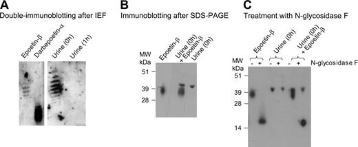
Urine samples from this athlete, obtained immediately after a strenuous interval training session (0 hours) and 1 hour later (1 hour), were analyzed for the presence of Epo. Neither of the samples were positive for endogenous Epo or Darbepoetin-α, but the 0-hours sample clearly contained bands that migrated like epoetin-β isoforms during isoelectric focusing (Figure 1A). In contrast, the 1-hour sample did not show any signal at these positions. Since recombinant human Epo has a half-life of more than 8 hours,1 this was a first indication that the bands detected at 0 hours could not be explained by the presence of epoetin-β.
False-positive detection of epoetin-β in urine. (A) Urine samples collected from the athlete immediately after a 4 × 1000 m sprint (0 hours) and 1 hour later (1 hour) contained 1.2 g and 0.3 g proteins/L, respectively. The samples were concentrated 200-fold and 800-fold, respectively, and processed for the detection of Epo by double immunoblotting after isoelectric focusing, as detailed in “Study design.” Also shown are the reference samples darbepoetin-α (1 ng) and epoetin-β (0.6 ng) (B) Immunoblotting of a postexercise urine sample (0 hours, 10-fold concentrated) after SDS-PAGE with anti-Epo antibodies. Also illustrated are epoetin-β (0.9 ng) and a mixture of the urine sample and 0.9 ng epoetin-β. (C) Immunoblotting of the same samples before and after a treatment with N-glycosidase F, as indicated.
False-positive detection of epoetin-β in urine. (A) Urine samples collected from the athlete immediately after a 4 × 1000 m sprint (0 hours) and 1 hour later (1 hour) contained 1.2 g and 0.3 g proteins/L, respectively. The samples were concentrated 200-fold and 800-fold, respectively, and processed for the detection of Epo by double immunoblotting after isoelectric focusing, as detailed in “Study design.” Also shown are the reference samples darbepoetin-α (1 ng) and epoetin-β (0.6 ng) (B) Immunoblotting of a postexercise urine sample (0 hours, 10-fold concentrated) after SDS-PAGE with anti-Epo antibodies. Also illustrated are epoetin-β (0.9 ng) and a mixture of the urine sample and 0.9 ng epoetin-β. (C) Immunoblotting of the same samples before and after a treatment with N-glycosidase F, as indicated.
To obtain additional information on the nature of the detected signals in the 0-hours sample, we performed an immunoblotting after sodium dodecyl sulfate–polyacrylamide gel electrophoresis (SDS-PAGE; Figure 1B), using the same anti-Epo antibodies. This immunoblotting visualized a major band of 42 kDa which had, however, a higher mass than that detected for the epoetin-β isoforms (32 kDa-39 kDa). The distinct migration was confirmed in a mixing experiment whereby epoetin-β was added to the 0-hours sample, resulting in the visualization of 2 distinct bands. Furthermore, the removal of N-linked carbohydrates by a preincubation with N-glycosidase F decreased the apparent mass of epoetin-β isoforms, from between 32 kDa and 39 kDa to 18 kDa, as detected by immunoblotting, but such a treatment did not cause a shift in the mass of the detected band in a postexercise urine sample (Figure 1C). The lack of an effect of N-glycosidase F in the latter case cannot be due to an inhibition of N-glycosidase F by urine components since an 18-kDa band was generated when epoetin-β was added to this urine sample before the glycosidase treatment. Thus, immunoblotting before and after a glycosidase treatment confirmed that the major urinary protein that was visualized with the anti-Epo antibodies was not Epo.
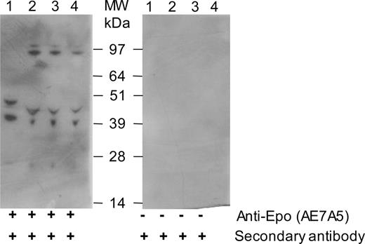
Lack of specificity of the AE7A5 anti-Epo antibodies. Four urine samples (lanes 1-4), obtained from the same athlete on 4 different days immediately after a 4 × 1000 m sprint, were concentrated 20-fold by ultrafiltration and subjected to immunoblotting after SDS-PAGE with monoclonal (clone AE7A5) anti-Epo antibodies (left blot). The right blot was treated identically except for the absence of anti-Epo antibodies. The protein concentration in the nonconcentrated samples amounted to 1.2 (lane 1), 1.1 (lane 2), 0.47 (lane 3), and 0.9 (lane 4) g/L.
Lack of specificity of the AE7A5 anti-Epo antibodies. Four urine samples (lanes 1-4), obtained from the same athlete on 4 different days immediately after a 4 × 1000 m sprint, were concentrated 20-fold by ultrafiltration and subjected to immunoblotting after SDS-PAGE with monoclonal (clone AE7A5) anti-Epo antibodies (left blot). The right blot was treated identically except for the absence of anti-Epo antibodies. The protein concentration in the nonconcentrated samples amounted to 1.2 (lane 1), 1.1 (lane 2), 0.47 (lane 3), and 0.9 (lane 4) g/L.
Immunoblotting on more concentrated urine samples yielded, in addition to a band of 42 kDa, bands of 48 kDa, 93 kDa, and 125 kDa, although the latter 2 were only detected in 3 of 4 tested postexercise urine samples from the athlete (Figure 2). Importantly, none of these bands were detected in the absence of the primary antibodies, showing that they did not result from the interaction of urine proteins with the secondary antibodies. It should also be pointed out that the immunoblots shown in Figure 1B-C and Figure 2 were obtained with urine samples that were less concentrated than those that are routinely used for Epo-tests which leads, if anything, to an underestimation of the problem of nonspecificity of the anti-Epo antibodies. In any case, our data clearly show that the monoclonal anti-Epo antibodies (clone AE7A5) visualize multiple polypeptides during immunoblotting of protein-rich urine samples. Some of these proteins may have a similar isoelectric point as the epoetin-β isoforms, which possibly accounts for the false-positive detection of epoetin-β. Recently, Kahn et al also reported the nonspecific binding of these anti-Epo antibodies to several proteins in the urine of a nonathletic volunteer.16
The athlete that we tested was only false-positive for epoetin-β in 2 of 7 postexercise urine samples (not illustrated). We also want to point out that the false-positive detection of epoetin-β may be restricted to (very) few athletes, as it may be linked to the extent and type of proteinuria. The extent of proteinuria correlates more with the intensity than the duration of exercise and has a half-time decay of about 1 hour. The athlete we tested showed a mixed glomerular-tubular proteinuria, which is characterized by a broad spectrum of urinary proteins. Some of these proteins show some structural homology with epoetin-β,16 which possibly accounts for their cross-reactivity with the anti-Epo antibodies. In a WADA report, the possible existence of such analytic interferences was already predicted.17 The false-positive detection of epoetin-β may be prevented by sampling before or at least 1 hour after exercise, which is particularly important for athletes who present with pronounced exercise-induced proteinuria. Additional tests can be performed to identify false-positive test results, such as 2-dimensional electrophoresis,16 deglycosylation assays (as described in this study), or indirect assays.18
Prepublished online as Blood First Edition Paper, February 21, 2006; DOI 10.1182/blood-2006-01-0028.
M. Beullens designed research and performed Epo-tests and immunoblot analyses; J.R.D. designed research, performed additional analyses on urine samples, and wrote the paper; M. Bollen designed research, wrote the paper, and coordinated the project.
The publication costs of this article were defrayed in part by page charge payment. Therefore, and solely to indicate this fact, this article is hereby marked “advertisement” in accordance with 18 U.S.C. section 1734.
We thank Bart Landuyt, Hans Prenen, and Pieter Timmermans for helpful discussions.


This feature is available to Subscribers Only
Sign In or Create an Account Close Modal