Comment on Darghouth et al, page 1925
Darghouth and colleagues describe studies of a child with hereditary deficiency of all vitamin K–dependent (VKD) coagulation factors. The disorder was caused by compound heterozygous mutations of γ-carboxylase and provides new insights into abnormal and normal γ-carboxylase function.
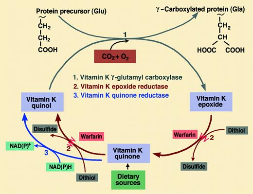
Take a hydrophilic, carboxylic amino acid and a highly hydrophobic isoprenoid naphthoquinone (that can only be synthesized by plants and bacteria), add carbon dioxide and oxygen, and we have the main ingredients of arguably the most unusual of all posttranslational modifications to a protein structure. This modification is the VKD conversion of glutamate (Glu) to γ-carboxyglutamate (Gla) catalyzed by the enzyme γ-glutamyl carboxylase. When discovered in 1974, this modification was thought to be confined to just 4 VKD procoagulants (II, VII, IX, and X). Now the list includes 3 anticoagulant proteins (C, S, and Z), 2 proteins of the extracellular matrix (osteocalcin and matrix Gla protein), a ligand for the Axl/Sky/Mer tyrosine kinase receptors (Gas6), and 4 transmembrane Gla proteins.1 VKD γ-carboxylation is intimately linked to a vitamin K cycle whereby the vitamin K quinol (KH2) cofactor is recycled via its vitamin K epoxide (KO) metabolite by the warfarin-sensitive enzyme vitamin K epoxide reductase (VKOR; see figure).
Hereditary combined deficiency of VKD coagulation factors is a very rare bleeding disorder first reported in 1966. In a 15-year follow-up study of the original patient, it was suggested that the cause might reside with a defect in one of the enzymes of the vitamin K cycle.2 Mechanistic genetic studies had to await the characterization of the gene that coded for γ-carboxylase and the very recent characterization of at least one component of a possible larger VKOR complex.3 This has enabled a few cases of congenital VKD factor deficiency to be attributed to different missense point mutations: 3 for the carboxylase (L394R, W501S, and R485P) and 1 for the VKOR (R98W). In this issue of Blood, Darghouth and colleagues describe their studies of a child who remarkably had 2 heterozygous missense mutations, W157R and T591K (which, when expressed, showed almost complete lack of γ-carboxylase activity), and a third mutation, D31N, with wild-type activity. The phenotype was also characterized by a complete lack of response to vitamin K supplementation in contrast to a partial amelioration in previously reported mutations. The higher vitamin K requirement seen in the L394R and W501S mutations seems to arise from the defective binding of glutamate and propeptide substrates, respectively, which has the knock-on effect of reducing the affinity for the KH2 cofactor compared with the wild-type enzyme.4 Darghouth and colleagues make the intriguing suggestions that the highly conserved W157 residue is part of a hydrophobic vitamin K binding site, and that the W157R mutation impairs vitamin K catalysis rather than binding.FIG1
The relationship of γ-carboxylation to the enzymes of the vitamin K cycle and vitamin K metabolism. Also shown is the inhibition of the thiol-dependent vitamin K epoxide reductase activity by warfarin. In the presence of warfarin, dietary vitamin K can enter the cycle via an NAD(P)H-dependent quinone reductase that is relatively insensitive to warfarin. Illustration by Marie Dauenheimer.
The relationship of γ-carboxylation to the enzymes of the vitamin K cycle and vitamin K metabolism. Also shown is the inhibition of the thiol-dependent vitamin K epoxide reductase activity by warfarin. In the presence of warfarin, dietary vitamin K can enter the cycle via an NAD(P)H-dependent quinone reductase that is relatively insensitive to warfarin. Illustration by Marie Dauenheimer.
The possible consequences of mutational defects in the vitamin K cycle extend beyond the coagulation function of VKD proteins. Patients with the W501S mutation have high levels of undercarboxylated osteocalcin,4 which is suspected to be deleterious to bone health. Developmental skeletal defects associated with inappropriate calcium deposition, such as those seen in the well-known warfarin embryopathy, have been reported in an infant with a putative VKOR mutatation,5 but these compound heterozygous mutations of γ-carboxylase appear to be the first to be associated with skeletal abnormalities as well as other severe pathologic features. As knowledge increases of the roles of other Gla proteins, such as matrix Gla protein in the inhibition of calcification, and of Gas6 in the regulation of cell growth and survival,1 we may be able to offer explanations for these noncoagulation defects. ▪


This feature is available to Subscribers Only
Sign In or Create an Account Close Modal