Abstract
The mechanisms by which multiple myeloma (MM) cells migrate and home to the bone marrow are not well understood. In this study, we sought to determine the effect of the chemokine SDF-1 (CXCL12) and its receptor CXCR4 on the migration and homing of MM cells. We demonstrated that CXCR4 is differentially expressed at high levels in the peripheral blood and is down-regulated in the bone marrow in response to high levels of SDF-1. SDF-1 induced motility, internalization, and cytoskeletal rearrangement in MM cells evidenced by confocal microscopy. The specific CXCR4 inhibitor AMD3100 and the anti-CXCR4 antibody MAB171 inhibited the migration of MM cells in vitro. CXCR4 knockdown experiments demonstrated that SDF-1–dependent migration was regulated by the PI3K and ERK/MAPK pathways but not by p38 MAPK. In addition, we demonstrated that AMD3100 inhibited the homing of MM cells to the bone marrow niches using in vivo flow cytometry, in vivo confocal microscopy, and whole body bioluminescence imaging. This study, therefore, demonstrates that SDF-1/CXCR4 is a critical regulator of MM homing and that it provides the framework for inhibitors of this pathway to be used in future clinical trials to abrogate MM trafficking.
Introduction
Multiple myeloma (MM) is the second most prevalent hematologic malignancy; it remains incurable, and the median survival time is 3 to 5 years.1,2 It is characterized by the presence of multiple lytic lesions and widespread involvement of the bone marrow at diagnosis, implying a continuous (re)circulation of MM cells in the peripheral blood and (re)entrance into the bone marrow.1 Studies have demonstrated the presence of circulating malignant plasma cells in more than 70% of patients diagnosed with MM.3,4 Migration of cells through the blood to the bone marrow niches requires active navigation, a process termed homing.
Chemokines are small chemoattractant cytokines that bind to specific G-protein–coupled 7-span transmembrane receptors present on the plasma membranes of target cells.5-7 Chemokines play a central role in lymphocyte trafficking and homing.8-11 One of the most extensively studied chemokines in migration is SDF-1 and its receptor, CXCR4.12,13 SDF-1 is primarily produced by stromal cells. CXCR4 is expressed on the surfaces of normal cells such as hematopoietic stem cells and T and B lymphocytes and on malignant cells such as breast cancer cells and lymphoid malignancies.6,11,14-16
To date, the role of CXCR4 in homing of MM cells to the bone marrow has not been fully elucidated. Inhibitors of CXCR4, such as AMD3100 (AnorMED, Toronto, ON, Canada), have been shown to induce the mobilization of stem cells.17,18 AMD3100 (AnorMED) is a bicyclam molecule that reversibly blocks the binding of CXCR4 with SDF-1.19 Because SDF-1/CXCR4–dependent signaling differs between cell types and between malignant and normal counterparts,20 it is critical to investigate the unique role of CXCR4/SDF-1 in MM. In this study, we sought to determine the effect of CXCR4 and its specific inhibitor, AMD3100, on the migration and in vivo homing of MM cells.
Materials and methods
MM-derived cell lines
Dexamethasone (Dex)–sensitive human MM cell line MM.1S was kindly provided by Dr Steven Rosen (Northwestern University, Chicago, IL). The KAS 6/1 human MM cell line was kindly provided by Dr Diane Jelinek (Mayo Clinic, Rochester, MN). The U266 human MM cell line was purchased from the American Tissue Type Culture Collection (Manassas, VA), and the OPM2 cell line was kindly provided by Dr Alan Lichtenstein (University of California at Los Angeles, CA). All MM cells lines were cultured in RPMI-1640 media (Sigma Chemical, St Louis, MO) containing 10% fetal bovine serum, 100 U/mL penicillin, and 100 μg/mL streptomycin.
Informed consent was obtained from all patients in accordance with the Declaration of Helsinki protocol. Approval of these studies was obtained by the Mayo Clinic College of Medicine and the University of Pittsburgh institutional review boards.
Reagents
The following inhibitors were used: the CXCR4 inhibitor AMD3100 (Sigma Chemical), the specific anti-CXCR4 antibody MAB171 (R&D Systems, Minneapolis, MN), the Gi protein inhibitor pertussis toxin (PTX; Sigma Aldrich, St Louis, MO), the PI3K inhibitor LY294002 (EMD Biosciences, San Diego, CA), the mTOR inhibitor downstream of PI3K, rapamycin (LC Laboratories, Woburn, MA), the ERK/MAPK inhibitor PD098059 (Alexis Biochemicals, San Diego, CA), and the p38 MAPK inhibitor SB203580 (Calbiochem, La Jolla, CA).
Expression of CXCR4-YFP in MM cells
To determine the effect of SDF-1 on cytoskeletal reorganization, we transiently transfected pCI-CXCR4-YFP into MM cell lines and analyzed SDF-1–dependent motility through confocal microscopy, as described in “Confocal microscopy.” Fusion of an expression vector encoding a human CXCR4 protein with a modified green fluorescent protein called the yellow fluorescent protein (YFP) added to the C-terminal of CXCR4 was performed as previously described.21 Transfection of pCI-CXCR4-YFP was performed using electroporation, as previously described.22
Lentivirus shRNA vector construction and CXCR4 gene transduction
To further determine the role of CXCR4 in the migration and regulation of downstream signaling pathways in MM, we established a CXCR4 knockout MM.1S cell line using the lentivirus system.23,24 The sequence for construction of CXCR4 shRNA was GCTGCCTTACTACATTGGGAT. pLKO.1 construct with target sequence CXCR4 shRNA or pLKO.1 control construct were cotransfected with pVSV-G and p8.9 plasmids into 293T packaging cells with Lipofectamine 2000 (Invitrogen Life Technologies, Carlsbad, CA). MM.1S cells were then transduced with the culture supernatants containing the released virus mixed with an equal amount of reconstituted RPMI 10% after 24 hours and 48 hours. Two days after infection, cells were treated with 250 ng/mL puromycin (InvivoGen, San Diego, CA) for 1 week. Stable transfection was confirmed by immunoblotting for CXCR4 expression.
Confocal microscopy
Fixed-cell method.
YFP-CXCR4–transfected MM.1S cells were stimulated with SDF-1 in a Lab-Tek chamber coverglass (Fisher Scientific, Pittsburgh, PA). The slides were then fixed, and the nuclei were counterstained with DRAQ5 (Alexis Biochemical) and analyzed with a TCS-SL confocal microscope (63× oil objective, NA 1.40; Leica Microsystems, Wetzlar, Germany) with appropriate emission filter sets—520 nm for YFP-CXCR4 and 670 nm for nuclei staining. Nuclei were pseudocolored to blue. Images were merged to localize the surface or intracellular expression of CXCR4. Cells to be assayed were first determined to be transfected and alive through their YFP fluorescence and appropriate nuclear shape.
Live-cell method.
We used glass chambers (Fisher Scientific, Pittsburgh, PA) coated with fibronectin 10 μg/mL (Gibco, Grand Island, NY) to allow the cells to adhere for imaging with confocal microscopy while they were submerged in a bath kept at 37°C. Confocal stacked images were taken using a 100× objective on an inverted laser scanning confocal microscope (model 510; Carl Zeiss, Göttingen, Germany) with 2-μm z-sections. The same cell was imaged before and after SDF-1 stimulation. We were able to take five 3-dimensional images of one cell over a period of 1 hour without significantly bleaching CXCR4-YFP fluorescence.
FACS analysis of surface and intracellular (cytoplasmic) CXCR4 expression in MM samples
Fluorescence-activated cell sorting (FACS) analysis was performed as previously described.15 For each sample, 20 × 106 whole bone marrow cells or peripheral blood cells were washed in phosphate-buffered saline (PBS) and lysed in ammonium chloride buffer (ACK). Then 1 × 10⁶ cells were analyzed using 3-color flow cytometry for CXCR4 (sCD184) CD184-PE (555974; BD Biosciences, San Diego, CA), CD38-APC (340677; BD Biosciences), and CD45-PERCP (340665; BD Biosciences). G1-PE 340761 (BD Biosciences) was included as an isotype control for CXCR4 expression. Data were analyzed using BD FACSCalibur (BD Biosciences). The percentage of expression of the chemokine receptor was determined on the CD38+ and CD45− plasma cells. For measurement of cytoplasmic CD184 (cyCD184), 100 μL medium A (Fix and Perm Gas001s; Caltag, Burlingame, CA) was added for 15 minutes. After 2 washes with PBS, 10 μL CD184–PE or isotype control was added, followed by 100 μL Medium B (Fix and Perm Gas002s; Caltag) for 15 minutes. Cells were washed twice with PBS and centrifuged for 5 minutes at 300g. Pellets were resuspended in 0.5 mL of 1% paraformaldehyde and stored at 4°C until analysis on the BD FACSCalibur.
SDF-1 ELISA
Quantitative ELISA (Quantikine; R&D Systems) was used to determine the level of SDF-1 in the peripheral blood and bone marrow of MM patients and healthy controls. Samples and standards were pipetted into wells precoated with a monoclonal antibody specific for SDF-1. After washing, an enzyme-linked polyclonal antibody specific for SDF-1 was added. Optical density was measured using a microplate reader set to 450 nm with correction at 540 nm.
Transwell migration assay
We performed transwell migration assay (Costar; Corning, Acton, MA) using MM cell lines (MM.1S, Kas 6/1, OPM2, and U266) in the presence of 0 to 100 nM SDF-1. In brief, cells were suspended in 1% FCS media and were placed (2 × 10⁵ cells) in the upper chambers of the transwell plates with serial concentrations of SDF-1 (0-100 nM) in the lower chambers in 1 mL of 1% FCS media. After 4 hours at 37°C, cells that migrated to the lower chambers were counted. Triplicates of each concentration were performed, and the means and standard deviations were calculated. Migration of primary CD138+ cells was tested in a similar fashion, with 30 nM SDF-1 in the lower chambers.
Immunoblotting
Immunoblotting was performed as previously described.25 MM cell lines (MM.1S and OPM2) were incubated with specific inhibitors, with or without SDF-1 stimulation (30 nM). After stimulation, the cells were rapidly harvested, centrifuged, and lysed in a phosphorylation lysis buffer. Cell lysates were analyzed by SDS-PAGE. Antibodies used for immunoblotting included total CXCR4 (Affinity Bioreagents, Golden, CO), anti–pan-p-PKC (Ser660), anti–p-p85PI3K, anti–p-PDK1 (Ser241), anti–p-AKT (Ser473), anti–p-ERK1/2 (Thr202/Tyr204), and anti–p-p38 MAPK (Cell Signaling Technology, Beverly, MA). Blots were then probed with anti–α-tubulin (Santa Cruz Biotechnology, Santa Cruz, CA) or antiactin antibodies (Sigma) to ensure equivalent protein loading. Similarly, CXCR4 knockdown MM.1S and mock-infected cells were stimulated with SDF-1 and analyzed using immunoblotting.
Growth inhibition assay
Growth inhibition assay was performed as previously described.22 Briefly, MM cell lines were cultured for 48 hours in media alone or with varying concentrations of AMD3100 (0-150 μM). Cells (5 × 10⁵) from 48-hour cultures were pulsed with 10 μL of 5 mg/mL 3-(4,5-dimethylthiazol-2-yl)-2,5-diphenyl tetrasodium bromide (MTT; Sigma) added to each well. The 96-well plates were incubated at 37°C for 4 hours; this was followed by the addition of 100 μL isopropanol containing 0.04 HCl. Absorbance readings at wavelengths of 570 nm (with correction readings at 630 nm) were taken on a spectrophotometer (Molecular Devices, Sunnyvale, CA), and results were verified with a standard curve.
In vivo flow cytometry
In vivo flow cytometry is a new technology for real-time continuous monitoring of fluorescent cells in the circulation of live animals without the need to draw blood samples.26 The effect of AMD3100 on homing in vivo was tested using Balb/c mice with in vivo flow cytometry, as previously described.27 In brief, MM.1S cells (2 × 10⁶/mL) were incubated in 50 μM AMD3100 or control PBS for 2 hours at 37°C. The cells were then fluorescently labeled by incubation with dialkyl carbocyanine membrane dye (DiD; Molecular Probes) at a concentration of 1 μM for 30 minutes at 37°C. Cells were then centrifuged to concentrate them to 200 to 300 μL for intravenous injection in mice. An appropriate artery for obtaining measurements was chosen in the ear. Fluorescence signal on the circulating MM cells was excited as the labeled cells passed through a slit of light (from a 632-nm HeNe laser) focused across the vessel. Signal was detected by a photomultiplier tube through a 695 ± 27.5-nm bandpass filter and was digitized for analysis with Matlab software. Cell counts were obtained every 5 minutes for 1 hour from the time of injection.
In vivo video rate confocal microscopy and 2-photon microscopy
MM cell homing to bone marrow vasculature of the skull was analyzed using fluorescence confocal microscopy, as previously described.27 A small incision was made in the scalp of the mice to expose the underlying dorsal skull surface. Images were captured after injection of the cells for 30 minutes to 1 hour per session. High-resolution images with cellular details were obtained through the intact mouse skull at a depths of up to 250 μm from the surface of the skull using a 30×/0.9 NA water-immersion objective lens. Multiple depths were obtained, and maximum intensity z-stacking (Image J) was performed to merge the images. DiD was excited with a 635-nm helium neon laser and detected with a photomultiplier tube through a 695 ± 27.5 nm bandpass filter (Omega Optical, Brattleboro, VT). Quantitative evaluation was made by dividing the bone marrow into predetermined quadrants (areas 1-4) and counting numbers of fluorescent cells per fields 3 and 4 in at least 3 different experiments. All experiments were repeated at least 3 times.
In vivo real-time bioluminescence imaging
To confirm our results with in vivo flow cytometry, we used another MM model that detects the dissemination of MM cells, as previously described,28,29 using a luciferase+ (luc+) MM.1S cell line. MM.1S Luc cells (2 × 10⁶) suspended in a volume of 100 μL saline solution were injected intravenously in the tail veins of severe combined immunodeficient/nonobese diabetic mice (SCID/NOD) mice that underwent irradiation. Cells were incubated in vitro before injection in 50 μM AMD3100 or control PBS for 4 hours at 37°C. Whole body real-time bioluminescence imaging was performed as previously described.28,29 Imaging was performed every 5 minutes for 90 minutes after injection.
Statistical analysis
Results are reported as the mean ± SD for typical experiments performed in 3 replicate samples and were compared with the Student t test. Results were considered significantly different for P values less than .05. All experiments were performed at least 3 times.
Results
CXCR4 surface expression of plasma cells was higher in the peripheral blood than in the bone marrow
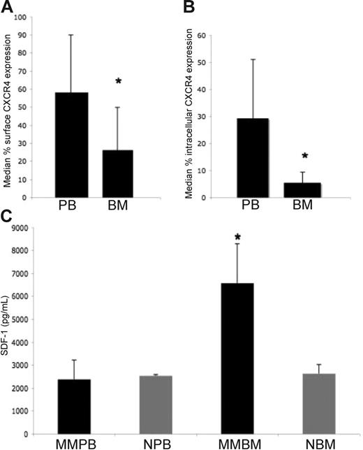
To test our hypothesis of dynamic regulation of the CXCR4 receptor as the cells move from the peripheral blood compartment to the bone marrow compartment, we used flow cytometry to analyze CXCR4 expression on the surfaces of plasma cells obtained from the peripheral blood (n = 14) or the bone marrow (n = 46) of patients in whom MM had been diagnosed. The median percentage of expression of CXCR4 in the peripheral blood and in bone marrow plasma cells in patients with MM and in patients with the early disease precursor monoclonal gammopathy of undermined significance (MGUS) is outlined in Table 1. The median percentage expression of CXCR4 in the bone marrow was lower in patients with MGUS than in those with MM, with a median expression of 58.4% (range, 6%-90%) in MGUS compared with 26.4% (range, 1%-81%) in MM (P = .005). In addition, the median percentage expression of CXCR4 in plasma cells in the peripheral blood was 60% (range, 9%-96%) compared with 26.4% (range, 1%-81%) in the bone marrow of patients with active MM (P = .001; Figure 1A). We then tested matched bone marrow and peripheral blood samples of 7 patients with MM for CXCR4 expression. The median percentage of surface CXCR4 expression in the peripheral blood was 56.3% (range, 5%-82%) compared with 34.3% (range, 17%-98%) in the bone marrow samples. Interestingly, intracellular expression was significantly higher in the peripheral blood samples than in the bone marrow samples, with a median percentage expression of 31.6% (range, 12%-71%) in peripheral blood plasma cells compared with 5% (range, 0%-13.6%) in bone marrow plasma cells; P = .001; Figure 1B.
Surface and intracellular (cytoplasmic) expression of CXCR4 on MM cells in BM and PB
| Cell type, location . | Samples, no. . | Median expression of CXCR4, % (range) . |
|---|---|---|
| BM-MGUS | 11 | 58.4 (6-90) |
| BM-MM surface | 46 | 26.4 (1-81) |
| PB-MM surface | 14 | 60 (9-96) |
| BM-MM cytoplasmic | 26 | 1.5 (0-17) |
| BM-MM surface matched | 7 | 34.3 (17-98) |
| PB-MM surface matched | 7 | 56.3 (5-82) |
| BM-MM cytoplasmic matched | 7 | 5.0 (0-13.6) |
| PB-MM cytoplasmic matched | 7 | 31.6 (12-71) |
CXCR4 and SDF1 expression in patients with MM. (A) Surface expression of CXCR4 on plasma cells in the peripheral blood (PB) and bone marrow (BM) of patients with MM. Median percentage expression of CXCR4 in plasma cells in the PB was 60% (range, 9%-96%) compared with 26.4% (range, 1%-81%) in the BM of patients with active MM (P = .001). (B) Intracellular expression of CXCR4 on plasma cells in the PB and BM of 7 matched samples. Intracellular expression was significantly higher in all PB samples compared with BM samples, with a median percentage expression of 31.6% (range, 12%-71%) in PB plasma cells compared with 5% (range, 0%-13.6%) in BM plasma cells (P = .001). (C) SDF-1 expression as measured by ELISA. SDF-1 level was markedly increased in the BM of MM patients (MM-BM; average, 6571 pg/mL) compared with healthy controls (CTRL-BM; average, 2632 pg/mL) (P < .001). In addition, the SDF-1 level in BM was significantly elevated compared with the SDF-1 level in PB of patients with MM (average, 2382.46 pg/mL) and in healthy control (MM-PB and CTRL-PB, respectively; P = .001). Error bars represent standard deviation; asterisks, statistically significant value.
CXCR4 and SDF1 expression in patients with MM. (A) Surface expression of CXCR4 on plasma cells in the peripheral blood (PB) and bone marrow (BM) of patients with MM. Median percentage expression of CXCR4 in plasma cells in the PB was 60% (range, 9%-96%) compared with 26.4% (range, 1%-81%) in the BM of patients with active MM (P = .001). (B) Intracellular expression of CXCR4 on plasma cells in the PB and BM of 7 matched samples. Intracellular expression was significantly higher in all PB samples compared with BM samples, with a median percentage expression of 31.6% (range, 12%-71%) in PB plasma cells compared with 5% (range, 0%-13.6%) in BM plasma cells (P = .001). (C) SDF-1 expression as measured by ELISA. SDF-1 level was markedly increased in the BM of MM patients (MM-BM; average, 6571 pg/mL) compared with healthy controls (CTRL-BM; average, 2632 pg/mL) (P < .001). In addition, the SDF-1 level in BM was significantly elevated compared with the SDF-1 level in PB of patients with MM (average, 2382.46 pg/mL) and in healthy control (MM-PB and CTRL-PB, respectively; P = .001). Error bars represent standard deviation; asterisks, statistically significant value.
SDF-1 levels were elevated in the bone marrow of patients with MM compared with peripheral blood samples and bone marrow samples of healthy controls
We hypothesized that low CXCR4 surface expression on MM cells in the bone marrow resulted from high bone marrow SDF-1 levels, leading to internalization/degradation of CXCR4. Therefore, we determined the level of SDF-1 in the peripheral blood and bone marrow of MM patients compared with healthy controls. As shown in Figure 1C, the level of SDF-1 was markedly increased in the bone marrow of MM patients (average, 6571 pg/mL) compared with healthy controls (average, 2632 pg/mL; P < .001). In addition, the level of SDF-1 in the MM bone marrow was significantly elevated compared with the level of SDF-1 in the peripheral blood (average, 2382.46 pg/mL; P = .001).
SDF-1 induced cytoskeletal reorganization and internalization of CXCR4 in MM cells
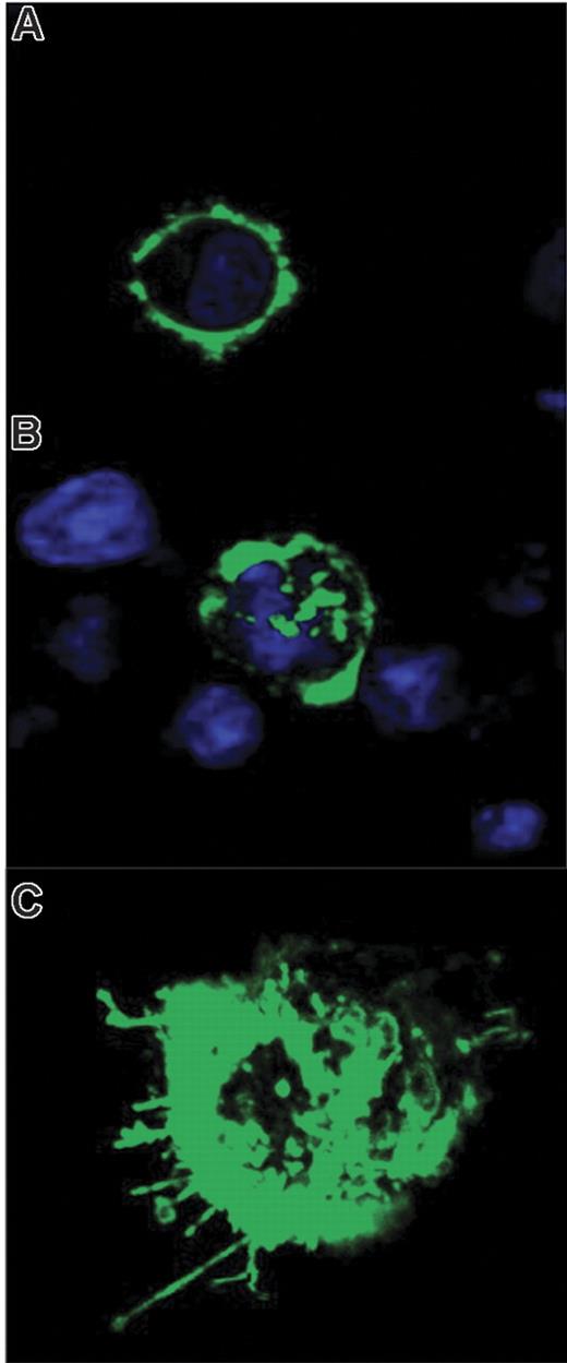
To investigate the effect of SDF-1 on CXCR4 expression in MM, we analyzed CXCR4 expression of MM.1S cells in the presence or absence of 30 nM SDF-1 for 30 minutes. We investigated the effect of SDF-1 on the cytoskeletal localization of CXCR4. MM cells (MM.1S, Kas 6/1) were transiently transfected with YFP-CXCR4. Cells were imaged in the presence or absence of 30 nM SDF-1 for 30 minutes using fixed or live cell methods. In the absence of SDF-1, CXCR4 was expressed on the surfaces of the cells (Figure 2A), whereas CXCR4 was internalized into cells in the presence of SDF-1, indicating that SDF-1 induced CXCR4 internalization (Figure 2B). With the use of live confocal microscopy and 3D reconstruction, we demonstrated that treatment with SDF-1 induced relocalization and polarization of CXCR4 and pseudopodia formation of MM cells (Figure 2C).
YFP-CXCR4 localization in response to SDF-1. (A) YFP-CXCR4 expression on the surfaces of MM.1S cells in unstimulated cells as determined by the fixed cell method. (B) Internalization of the CXCR4 receptor in response to SDF-1 (30 nM) stimulation for 1 hour. (C) Live cell method with 3D reconstruction demonstrating CXCR4 cytoskeletal rearrangement, capping, and pseudopodia formation in response to SDF-1.
YFP-CXCR4 localization in response to SDF-1. (A) YFP-CXCR4 expression on the surfaces of MM.1S cells in unstimulated cells as determined by the fixed cell method. (B) Internalization of the CXCR4 receptor in response to SDF-1 (30 nM) stimulation for 1 hour. (C) Live cell method with 3D reconstruction demonstrating CXCR4 cytoskeletal rearrangement, capping, and pseudopodia formation in response to SDF-1.
SDF-1 induced motility and migration in MM cells
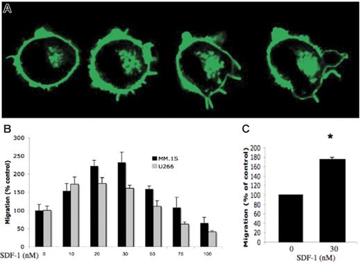
To determine the effect of SDF-1 on the motility of MM cells, we used live confocal microscopy with continuous imaging in a single MM cell treated with 30 nM SDF-1. As shown in Figure 3A, motility and pseudopodia formation increased in response to SDF-1. We hypothesized that MM cells migrated in response to SDF-1, but high levels of bone marrow SDF-1 levels inhibited migration to confine the cells to the bone marrow. Figure 3B shows transwell migration of MM.1S and U266 MM cell lines in response to serial concentrations of SDF-1, with the highest migration of MM cells at 30 nM, whereas SDF-1 concentrations of 75 to 100 nM inhibited migration. We then confirmed migration using CD138+ purified primary MM cells from 2 patients with maximal migration in response to 30 nM SDF-1, as shown in Figure 3C.
Migration and motility in response to SDF-1. (A) Increased motility of plasma cells in response to SDF-1 with pseudopodia formation. (B) Transwell migration assay demonstrating the migration of MM.1S and U266 MM cell lines in response to serial concentrations of SDF-1 (0-100 nM). SDF-1 induced maximum migration at doses of 20 to 30 nM and decreased migration at doses of 75 to 100 nM. (C) Transwell migration assay demonstrating the migration of CD138+ primary plasma cells in response to SDF-1.
Migration and motility in response to SDF-1. (A) Increased motility of plasma cells in response to SDF-1 with pseudopodia formation. (B) Transwell migration assay demonstrating the migration of MM.1S and U266 MM cell lines in response to serial concentrations of SDF-1 (0-100 nM). SDF-1 induced maximum migration at doses of 20 to 30 nM and decreased migration at doses of 75 to 100 nM. (C) Transwell migration assay demonstrating the migration of CD138+ primary plasma cells in response to SDF-1.
Molecular mechanisms regulating MM migration in response to CXCR4/SDF-1
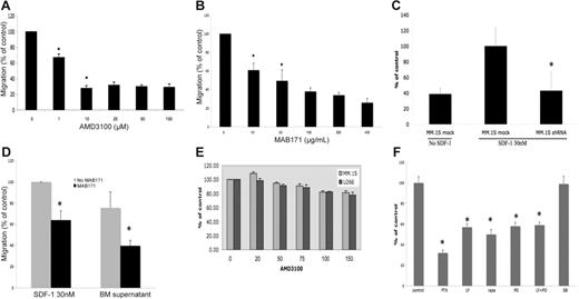
We tested the effect of the selective CXCR4 inhibitor AMD3100 (0-100 μM for 16 hours) on the migration of MM cell lines (MM.1S, U266, and Kas 6/1) using the transwell migration assay. As shown in Figure 4A, AMD3100 induced the dose-dependent inhibition of migration. AMD3100 10 μM inhibited migration by 70% compared with control (P = .03), whereas higher doses of AMD3100 did not induce further inhibition of migration. Similarly, we tested the effect of the anti-CXCR4 antibody MAB171 (10-800 μM) for 16 hours or its IgG2a isotype control on the in vitro migration of MM.1S cells. As shown in Figure 4B, anti-CXCR4 neutralizing antibody demonstrated a dose-dependent inhibition of migration. Anti–CXCR4 MAB171 inhibited 10 μM migration to 53% and 200 μM migration to 35% compared with control (P = .007). Finally, we tested the CXCR4 knockdown MM.1S cell line and demonstrated that CXCR4 knockdown cells migrated only to 43% of control, similar to cells not exposed to SDF-1 (ie, 60% reduction in migration compared with control mock cells treated with 30 nM SDF-1; Figure 4C).
Regulation of migration by inhibitors of the CXCR4 signaling pathway. (A) Migration assay using the CXCR4 inhibitor AMD3100 (0-100 μM). AMD3100 (10 μM) induced 70% inhibition of migration compared with control (P = .03). All wells contained 20 nM SDF-1 in the lower chambers. (B) Migration assay using the anti-CXCR4 antibody MAB171. Serial concentrations of MAB171 (0-400 μg/mL) inhibited migration in a dose-dependent fashion. Anti–CXCR4 MAB171 (10 μM) inhibited migration to 53%, and anti-CXCR4 MAB171 (200 μM) inhibited migration to 35% compared with control (P = .007). IgG control antibody (400 μg/mL) was used in the control well. (C) Transwell migration assay of MM.1S mock and MM.1S infected with CXCR4 shRNA (CXCR4 knockdown cells) in the presence or absence of SDF-1 (30 nM). CXCR4 knockdown cells migrated only to 43% of control, similar to cells not exposed to SDF-1 (ie, 60% reduction in migration compared with control mock cells treated with SDF-1). Control was mock cells treated with SDF-1. (D) Transwell migration assay of MM.1S in the presence or absence of the anti-CXCR4 antibody MAB171 (200 μg/mL). SDF-1 (30 nM) was placed in the lower chamber in the control wells. Bone marrow supernatant from patients with MM (2 patients) was placed in the lower chambers of the other wells. The BM supernatant bar represents the mean percentage migration of MM.1S compared with control. MM.1S migrated in response to BM supernatant (75.6%) compared with control. MAB171 resulted in 36% inhibition of migration in the SDF-1 chambers and 52.5% inhibition of migration in the chambers with BM supernatant. (E) MTT growth inhibition assay using MM cell lines treated with serial concentrations of AMD3100. AMD3100 did not inhibit survival compared with control. (F) Migration assay in MM.1S treated with inhibitors of pathways downstream of CXCR4: PTX, LY294002, rapamycin, PD098059, combination LY 294002 and PD098059, and p38 MAPK inhibitor SB203580. SDF-1 (30 nM) was placed in the lower chambers. PTX (50 ng/mL) significantly inhibited migration to 30% compared with control in the presence of 30 nM SDF-1 (P = .004). The PI3K inhibitor LY294002 and the MEK inhibitor PD098059 inhibited migration by 57% and 58%, respectively. Rapamycin downstream of PI3K demonstrated results similar to those of LY294002. The combination of the PI3K and ERK/MAPK inhibitors was not additive (59%), indicating that both signal through the same pathway. The p38 MAPK inhibitor SB203580 (SB) did not inhibit migration in response to SDF-1.
Regulation of migration by inhibitors of the CXCR4 signaling pathway. (A) Migration assay using the CXCR4 inhibitor AMD3100 (0-100 μM). AMD3100 (10 μM) induced 70% inhibition of migration compared with control (P = .03). All wells contained 20 nM SDF-1 in the lower chambers. (B) Migration assay using the anti-CXCR4 antibody MAB171. Serial concentrations of MAB171 (0-400 μg/mL) inhibited migration in a dose-dependent fashion. Anti–CXCR4 MAB171 (10 μM) inhibited migration to 53%, and anti-CXCR4 MAB171 (200 μM) inhibited migration to 35% compared with control (P = .007). IgG control antibody (400 μg/mL) was used in the control well. (C) Transwell migration assay of MM.1S mock and MM.1S infected with CXCR4 shRNA (CXCR4 knockdown cells) in the presence or absence of SDF-1 (30 nM). CXCR4 knockdown cells migrated only to 43% of control, similar to cells not exposed to SDF-1 (ie, 60% reduction in migration compared with control mock cells treated with SDF-1). Control was mock cells treated with SDF-1. (D) Transwell migration assay of MM.1S in the presence or absence of the anti-CXCR4 antibody MAB171 (200 μg/mL). SDF-1 (30 nM) was placed in the lower chamber in the control wells. Bone marrow supernatant from patients with MM (2 patients) was placed in the lower chambers of the other wells. The BM supernatant bar represents the mean percentage migration of MM.1S compared with control. MM.1S migrated in response to BM supernatant (75.6%) compared with control. MAB171 resulted in 36% inhibition of migration in the SDF-1 chambers and 52.5% inhibition of migration in the chambers with BM supernatant. (E) MTT growth inhibition assay using MM cell lines treated with serial concentrations of AMD3100. AMD3100 did not inhibit survival compared with control. (F) Migration assay in MM.1S treated with inhibitors of pathways downstream of CXCR4: PTX, LY294002, rapamycin, PD098059, combination LY 294002 and PD098059, and p38 MAPK inhibitor SB203580. SDF-1 (30 nM) was placed in the lower chambers. PTX (50 ng/mL) significantly inhibited migration to 30% compared with control in the presence of 30 nM SDF-1 (P = .004). The PI3K inhibitor LY294002 and the MEK inhibitor PD098059 inhibited migration by 57% and 58%, respectively. Rapamycin downstream of PI3K demonstrated results similar to those of LY294002. The combination of the PI3K and ERK/MAPK inhibitors was not additive (59%), indicating that both signal through the same pathway. The p38 MAPK inhibitor SB203580 (SB) did not inhibit migration in response to SDF-1.
The bone marrow milieu contains many chemokines and cytokines that induce the migration of MM cells other than SDF-1.30 To determine whether the inhibition of CXCR4 significantly inhibited the migration of MM cells in the bone marrow milieu, we tested the effect of MAB171 on the migration of MM.1S cells in response to bone marrow supernatant. Supernatant from the bone marrow of patients with MM was obtained by centrifugation of the bone marrow aspirates and was placed in the lower chambers of the transwell migration assay. SDF-1 (30 nM) was placed in the lower chamber of the control experiment. As demonstrated in Figure 4D, anti-CXCR4 antibody MAB171 (200 μM) inhibited the migration of MM.1S cells in response to bone marrow supernatant. MAB171 resulted in 36% inhibition of migration in the SDF-1 chambers and 52.5% inhibition of migration in the chambers containing bone marrow supernatant.
To determine whether AMD3100 specifically inhibited migration and did not affect the survival or proliferation of MM cells, we next investigated the effect of this agent on proliferation and apoptosis in MM cell lines. As shown in Figure 4E, serial concentrations of AMD3100 (0-150 μM) did not induce cytotoxicity in MM cell lines at 48 hours. Similar results were obtained in assays for apoptosis (data not shown).
We then sought to determine the downstream signaling pathways regulating SDF-1–dependent migration, specifically examining the role of the PI3K and MAPK pathways in SDF-1–dependent migration in MM. We tested the effect of the Gi protein inhibitor PTX (50-100 ng/mL) for 16 hours, the PI3K inhibitor LY294002 (25-50 μM) for 20 minutes, the mTOR inhibitor downstream of PI3K rapamycin (20-50 nM) for 16 hours, the ERK/MAPK inhibitor PD098059 (25-50 μM) for 90 minutes, and the p38 MAPK inhibitor SB203580 (10 μM) for 16 hours. All tested doses and durations of these inhibitors did not induce apoptosis in the MM cells (data not shown). As demonstrated in Figure 4F, PTX (50 ng/mL) significantly inhibited migration to 30% compared with control in the presence of 30 nM SDF-1 (P = .004). The PI3K inhibitor LY294002 and the MEK inhibitor PD098059 decreased migration by 57% and 58%, respectively. Rapamycin downstream of PI3K demonstrated results similar to those of LY294002. In contrast, the p38 MAPK inhibitor did not affect migration. The combination of the PI3K and the ERK/MAPK inhibitors was not additive (59%), indicating that both signal through the same pathway.
Signaling pathway regulated by SDF-1/CXCR4 in MM
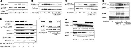
We next determined the effect of SDF-1 on activation of the PI3K and ERK/MAPK pathways. MM cells were cultured with serial concentrations of SDF-1 and for different durations. SDF-1 led to a rapid activation of pERK1/2 and pAKT downstream of PI3K. pAKT activation occurred within 30 seconds and peaked at 5 minutes, followed by a decrease at 10 minutes. pERK1/2 activation occurred within 30 seconds and peaked at 3 minutes, followed by a decrease at 5 minutes. Figure 5A shows the activation of PERK and pAKT in response to 30 nM SDF-1 at 1, 3, and 5 minutes of stimulation. We then investigated the effect of SDF-1 and the CXCR4 inhibitor AMD3100 on CXCR4 protein level and downstream proteins, including p-p85 PI3K, pAKT, pERK, and pPKC. As shown in Figure 5B, CXCR4 expression was up-regulated by 30 and 100 nM SDF-1 and was inhibited by 30 μM AMD3100 even in the presence of SDF-1 stimulation. Figure 5C shows that SDF-1 30 nM and 100 nM led to the activation of p85-PI3K at 5 minutes of stimulation. This activation was abrogated by 30 μM AMD3100 even in the presence of SDF-1, thereby confirming that SDF-1 activates PI3K through CXCR4. We also demonstrated that SDF-1 activates pPKC and that AMD3100 30 μM leads to a decrease in pPKC, pAKT, and pERK1/2 proteins, as shown in Figure 5D.
Signaling pathways regulated by SDF1/CXCR4 in MM. (A) Immunoblotting for pERK and pAKT, demonstrating rapid activation in response to SDF-1 30 nM in a time-dependent fashion at 1, 3, and 5 minutes. (B) Immunoblotting for total CXCR4 demonstrating up-regulation of CXCR4 by SDF-1 (30 and 100 nM for 5-minute incubation) and inhibition by AMD3100 (100 μM for 16-hour incubation), even in the presence of SDF-1 stimulation. (C) Immunoblotting for pPI3K (p85) demonstrating activation in response to SDF-1 in a dose-dependent fashion with maximum activation at 100 nM at 5 minutes. This effect was abrogated by AMD3100 (30-100 μM for 16-hour incubation), confirming that SDF-1 activates PI3K through CXCR4. (D) Immunoblotting for PKC, pAKT, and pERK1/2 in the presence or absence of AMD3100 (30 μM for 16-hour incubation). SDF-1 led to a rapid up-regulation of pPKC, pAKT, and pERK1/2 at 1 minute and 5 minutes. AMD3100 inhibited the expression of pPKC, pAKT, and pERK1/2. (E) Immunoblotting with CXCR4 knockdown MM.1S (lanes 3-4) and mock-infected MM.1S (lanes 1-2), with or without stimulation with 30 nM SDF-1 for 1 minute. CXCR4 knockdown with shRNA led to the inhibition of CXCR4, p-PDK-1, pAKT, and pERK1/2, but not p-p38 MAPK. (F) Immunoblotting for pERK and pAKT in the presence of 50 ng/mL pertussis toxin (PTX) for 90 minutes. SDF-1 (30 nM) induced pERK and pAKT as a control in lane 1. PTX inhibited pERK and pAKT even in the presence of SDF-1, indicating that activation of these pathways by SDF-1 is Gi dependent. (G) Immunoblotting for pAKT and pERK in the presence of the PI3K inhibitor LY294002 (25 μM for 20 minutes) or the MEK inhibitor PD098059 (25 μM for 90 minutes) with or without SDF-1. LY294002 inhibited pAKT even in the presence of SDF-1, whereas PD294002 did not affect AKT activity. LY294002 inhibited pERK1/2, indicating that ERK/MAPK is downstream of PI3K. PD294002 inhibited pERK activity even in the presence of SDF-1.
Signaling pathways regulated by SDF1/CXCR4 in MM. (A) Immunoblotting for pERK and pAKT, demonstrating rapid activation in response to SDF-1 30 nM in a time-dependent fashion at 1, 3, and 5 minutes. (B) Immunoblotting for total CXCR4 demonstrating up-regulation of CXCR4 by SDF-1 (30 and 100 nM for 5-minute incubation) and inhibition by AMD3100 (100 μM for 16-hour incubation), even in the presence of SDF-1 stimulation. (C) Immunoblotting for pPI3K (p85) demonstrating activation in response to SDF-1 in a dose-dependent fashion with maximum activation at 100 nM at 5 minutes. This effect was abrogated by AMD3100 (30-100 μM for 16-hour incubation), confirming that SDF-1 activates PI3K through CXCR4. (D) Immunoblotting for PKC, pAKT, and pERK1/2 in the presence or absence of AMD3100 (30 μM for 16-hour incubation). SDF-1 led to a rapid up-regulation of pPKC, pAKT, and pERK1/2 at 1 minute and 5 minutes. AMD3100 inhibited the expression of pPKC, pAKT, and pERK1/2. (E) Immunoblotting with CXCR4 knockdown MM.1S (lanes 3-4) and mock-infected MM.1S (lanes 1-2), with or without stimulation with 30 nM SDF-1 for 1 minute. CXCR4 knockdown with shRNA led to the inhibition of CXCR4, p-PDK-1, pAKT, and pERK1/2, but not p-p38 MAPK. (F) Immunoblotting for pERK and pAKT in the presence of 50 ng/mL pertussis toxin (PTX) for 90 minutes. SDF-1 (30 nM) induced pERK and pAKT as a control in lane 1. PTX inhibited pERK and pAKT even in the presence of SDF-1, indicating that activation of these pathways by SDF-1 is Gi dependent. (G) Immunoblotting for pAKT and pERK in the presence of the PI3K inhibitor LY294002 (25 μM for 20 minutes) or the MEK inhibitor PD098059 (25 μM for 90 minutes) with or without SDF-1. LY294002 inhibited pAKT even in the presence of SDF-1, whereas PD294002 did not affect AKT activity. LY294002 inhibited pERK1/2, indicating that ERK/MAPK is downstream of PI3K. PD294002 inhibited pERK activity even in the presence of SDF-1.
We next examined the effect of CXCR4 knockdown on signaling in MM cells. As shown in Figure 5E, CXCR4 inhibition with shRNA resulted in the inhibition of CXCR4 protein and signaling pathways downstream of PI3K, including pPDK1 and pAKT. In addition, we explored the effect of CXCR4 knockdown on MAPK signaling and demonstrated that pERK1/2 was inhibited in response to CXCR4 knockdown but not to p38 MAPK.
To further explore the mechanism of regulation of the PI3K and ERK pathways in response to SDF-1, we performed immunoblotting in the presence of the Gi protein inhibitor PTX (50 ng/mL for 90 minutes), the PI3K inhibitor LY294002 (25 μM for 20 minutes), or the MEK inhibitor PD098059 (25 μM for 90 minutes), with or without SDF-1. As shown in Figure 5F, PTX led to the inhibition of pERK and pAKT activation even in the presence of SDF-1 (30 nM), indicating these signaling pathways are dependent on the Gi protein of the G-protein–coupled receptor (GPCR) CXCR4. In Figure 5G, LY294002 abrogated pAKT stimulation in response to SDF-1. Interestingly, LY294002 also inhibited pERK1/2, indicating that ERK/MAPK is downstream of PI3K. Similar results were obtained with pPKC, indicating that pPKC is downstream of PI3K (data not shown). PD098059 abrogated phosopho-ERK1/2, consistent with its specific effect on MEK, but had no effect on pAKT.
In vivo effect of AMD3100 on the homing of MM cells
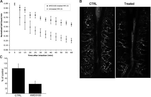
We next determined the in vivo effect of AMD3100 on MM cell homing. DID-labeled MM.1S cells treated with AMD3100 or control (PBS) were injected in the tail veins of mice, followed by in vivo flow cytometry every 5 minutes for 1 hour after injection. As shown in Figure 6A, the number of cells decreased dramatically (86% decrease) after 1 hour in the control experiment, indicating exit out of the circulation or homing, whereas only a 47% reduction in cells occurred in the AMD3100-treated cohort at 1 hour (P = .002). Similarly, we demonstrated that the number of cells present in the perivascular bone marrow niches of the skull was significantly higher in control mice than in the AMD3100-treated group at 1 hour after injection. Figure 6B shows areas 3 and 4 of the perivascular bone marrow niches and demonstrates that the number of cells that homed and adhered to these areas was lower in the AMD3100-treated mice than in the control group. The mean cell count in the AMD3100-treated mice decreased to 38% compared with that in controls (P = .01; Figure 6C).
In vivo flow cytometry and confocal microscopy imaging of MM.1S in the presence or absence of AMD3100. (A) In vivo confocal flow cytometry. DiD-labeled cells (treated with 50 μM AMD3100 for 2 hours or untreated control) were injected in the tail veins of 2 BALB/c mice. Cells were counted every 5 minutes for 1 hour. The cell count decreased by 86% in the control and by 47% in the AMD3100-treated mouse (P = .002). (B) In vivo confocal imaging of 4 quadrants of the skulls of mice showing BM niches on each side of the sagittal sinus (center of each picture). Fluorescent cells homed to the parasagittal vascular segments in the untreated control mouse; the number of cells that homed to the BM was significantly lower in the AMD3100-treated mouse. (C) Mean cell count of fluorescent cells that homed to BM niches in areas 3 and 4 in 3 experiments of the untreated (CTRL) and treated AMD3100 mice. The cell count in the AMD3100-treated mice decreased to 38% compared with control (P = .01).
In vivo flow cytometry and confocal microscopy imaging of MM.1S in the presence or absence of AMD3100. (A) In vivo confocal flow cytometry. DiD-labeled cells (treated with 50 μM AMD3100 for 2 hours or untreated control) were injected in the tail veins of 2 BALB/c mice. Cells were counted every 5 minutes for 1 hour. The cell count decreased by 86% in the control and by 47% in the AMD3100-treated mouse (P = .002). (B) In vivo confocal imaging of 4 quadrants of the skulls of mice showing BM niches on each side of the sagittal sinus (center of each picture). Fluorescent cells homed to the parasagittal vascular segments in the untreated control mouse; the number of cells that homed to the BM was significantly lower in the AMD3100-treated mouse. (C) Mean cell count of fluorescent cells that homed to BM niches in areas 3 and 4 in 3 experiments of the untreated (CTRL) and treated AMD3100 mice. The cell count in the AMD3100-treated mice decreased to 38% compared with control (P = .01).
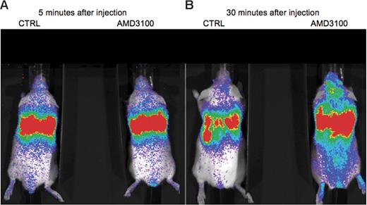
Figure 7 shows that with the use of the bioluminescence imaging Luc+ MM model, a significant difference resulted in the distribution of bioluminescence between AMD3100-treated and untreated mice. Bioluminescence was maximal at the cardiopulmonary area, indicating circulation in both groups at the beginning of treatment. This intensity diminished rapidly in the untreated mice at 20 minutes after injection, indicating exit from the circulation or homing. In the AMD3100-treated mice, bioluminescence remained more intense in the cardiopulmonary area through the 90 minutes of imaging compared with the control group. This result was consistent with the in vivo flow cytometry data (prolonged circulation time for AMD3100-treated group).
Bioluminescence imaging of Luc+ MM.1S cells injected intravenously into the tail veins of SCID/NOD mice. Cells were incubated in vitro before injection in 50 μM AMD3100 or control PBS for 4 hours at 37°C. Whole body real-time bioluminescence imaging was performed every 5 minutes for 90 minutes after injection. (A) Equal distribution of bioluminescence in the control (CTRL) and treated (AMD3100) mice 5 minutes after injection. (B) Thirty minutes after injection, fluorescence intensity diminished in the cardiopulmonary area, indicating exit from the circulation in the CTRL mice. In AMD3100-treated mice, bioluminescence remained high in the cardiopulmonary area.
Bioluminescence imaging of Luc+ MM.1S cells injected intravenously into the tail veins of SCID/NOD mice. Cells were incubated in vitro before injection in 50 μM AMD3100 or control PBS for 4 hours at 37°C. Whole body real-time bioluminescence imaging was performed every 5 minutes for 90 minutes after injection. (A) Equal distribution of bioluminescence in the control (CTRL) and treated (AMD3100) mice 5 minutes after injection. (B) Thirty minutes after injection, fluorescence intensity diminished in the cardiopulmonary area, indicating exit from the circulation in the CTRL mice. In AMD3100-treated mice, bioluminescence remained high in the cardiopulmonary area.
Discussion
In this study, we demonstrated that MM cells from patients with active MM expressed high levels of CXCR4 in the peripheral blood but that the expression level of CXCR4 on the surfaces of the MM cells decreased dramatically in the bone marrow, where levels of SDF-1 are high, potentially to confine the cells to the marrow and to prevent further trafficking of the cells. Studies to identify the expression of chemokine receptors in MM have demonstrated controversial results.31,32 Large variations were reported in CXCR4 expression, ranging from 10% to 100%,32 and in vitro migration of MM cells directly correlated with the expression level of CXCR4 in MM.31,32 In addition, CXCR4 expression on MM cells inversely correlated with disease activity in one study,33 consistent with the hypothesis that high bone marrow SDF-1 levels in patients with higher disease activity resulted in lower CXCR4 expression on the surfaces of MM cells. This is further confirmed by our data showing that CXCR4 expression level was lower in MM cells from the bone marrow of patients with MM than in those with MGUS.
We then demonstrated that SDF-1 induced cytoskeletal rearrangement, pseudopodia formation, and internalization of the CXCR4 receptor in MM cells. These results showed for the first time the cytoskeletal changes that occur in response to SDF-1 in MM cells. In addition, we demonstrated the translocation of surface CXCR4 to the intracellular compartment in response to SDF-1. Previous studies have demonstrated that GPCR heteromultimerized with receptor tyrosine kinases translocated to the endosome and promoted the activation of endosomal Ras-ERK pathways in other cell types.34 It is, therefore, possible that the subcellular localization of CXCR4 in MM mediates an enhanced activation of signaling pathways. In addition, CXCR4 has been shown to heteromultimerize with other receptors, including tyrosine kinase receptor35 and T-cell receptor, in other cell types.36 Therefore, it is possible that CXCR4 interacts with other receptors before internalization and activation of downstream signaling pathways.
We next demonstrated that the CXCR4 inhibitor AMD3100 and the anti-CXCR4 antibody MAB171 blocked migration in response to SDF-1. These data were confirmed using a CXCR4 knockdown MM cell line. We then determined the role of downstream signaling pathways in the regulation of SDF-1–dependent migration in MM. Specifically, we demonstrated that SDF-1–dependent migration is Gi dependent and that the PI3K and ERK/MAPK pathways are important regulators of migration in MM. We further demonstrated the inhibition of SDF-1–dependent signaling in response to the Gi protein inhibitor PTX. Gi protein signaling stimulated the activity of the ERK1/2 and PI3K pathways in many cell types.37-39 These data, therefore, indicated that SDF-1 stimulation of the PI3K and ERK pathways was dependent on the Gi protein of CXCR4 in MM. Previous studies in hematopoietic stem cells demonstrated that, in contrast to MM, the ERK/MAPK pathway did not regulate migration.40 The mechanisms of migration and SDF-1–dependent signaling differed between cell types and between malignant cells and normal cells.14,20,41 Speigel et al20 demonstrate that the migration and homing of human precursor-B acute lymphoblastic leukemia (ALL) cells differ in comparison with normal CD34+ in response to SDF-1. Similarly, studies in chronic myelogenous leukemia (CML) demonstrate lower migration of CML cells to SDF-1 compared with normal CD34+ cells.41,42 These data indicate that SDF-1 signaling differs between cell types, highlighting the importance of defining specific signaling pathways in MM. Similarly, we demonstrated that ERK/MAPK was downstream of PI3K in MM and that p38 MAPK did not regulate migration in MM. Previous studies in ALL have demonstrated that p38 MAPK is a critical regulator of migration,43 again highlighting the differences in signaling between malignant cells types. In addition, SDF-1 may also activate another newly discovered receptor, CXCR7 or RDC-1.44,45 We are exploring the function of CXCR7 in MM and the possible interactions of this receptor with CXCR4 and downstream signaling.
Finally, we determined the effect of CXCR4 inhibition on the homing of MM cells in vivo. We demonstrated that AMD3100 and PTX resulted in a significant inhibition of homing of MM cells to bone marrow niches. In vivo flow cytometry indicated the rapid exit of circulating MM cells within 20 minutes of injection in control mice, whereas in AMD3100-treated mice, the cells continued to circulate in the peripheral blood. We then confirmed—by demonstrating a significant difference in the number of MM cells that homed to the perivascular bone marrow niches—that the decrease in circulating cells was indeed attributed to homing. We confirmed those results using another MM model with in vivo whole body bioluminescence imaging.
The SDF-1/CXCR4 axis not only regulates migration and homing in MM, it also regulates adhesion, invasion, and possibly egression or mobilization of MM cells out of the bone marrow. In MM, SDF-1 rapidly and transiently up-regulates VLA-4–mediated MM cell adhesion to fibronectin and VCAM-146,47 and increases invasion and MMP secretion in MM.47,48 In addition, studies using G-CSF for mobilization have demonstrated a significant decrease in surface expression of CXCR4 and VLA-4 in the mobilized MM cells compared with cells obtained from the bone marrow, indicating a potential role of CXCR4 and VLA-4 in the mobilization/egression of MM cells.49 In addition, recent studies have demonstrated in an in vivo MM model that blocking CXCR4 led to a 20% reduction in bone marrow tumor load.50
Understanding the pathways that regulate MM cell trafficking is critical to rationally design future therapeutic trials that specifically target these mechanisms. Inhibition of the SDF-1/CXCR4 axis may have positive effects in regulating tumor metastasis and growth, but this may also negate immunologic responses through dysregulated lymphocyte trafficking and may contribute to the disruption of hematopoiesis. As with any therapy, the usefulness of this type of intervention will require a balance between its positive effect on disease outcome and its deleterious effects on normal physiological functions. Greater understanding of the role of SDF-1 and CXCR4 in the body will allow greater manipulation of this important biological axis to affect disease outcome. In summary, we demonstrated that SDF-1/CXCR4 is a critical regulator of migration and homing of MM and that it provides the preclinical framework for clinical trials of specific inhibitors of CXCR4 to improve patient outcomes.
Authorship
Contribution: G.D.R., T.E.W., T.H., K.C.A., C.P.L., and I.M.G. designed the research; Y.A., H.N., U.K.S., J.R., X.L., C.M.P., J.A.S., T.K., J.M.G., G.L., X.J., M.T., A.K., D.C., I.V., and I.M.G. performed the research; K.E.H. contributed a vital reagent; K.E.H. and C.P.L. contributed vital new analytical tools; A.K. contributed vital cell lines and an animal model; T.H., G.D.R., and I.M.G. analyzed the data; and Y.A., J.R., X.L., and I.M.G. wrote the paper.
Conflict-of-interest disclosure: The authors declare no competing financial interests.
Y.A. and H.N. contributed equally to this study.
Correspondence: Irene M. Ghobrial, Dana Farber Cancer Institute, 44 Binney St, Mayer 548A, Boston, MA 02115; e-mail: irene_ghobrial@dfci.harvard.edu.
The publication costs of this article were defrayed in part by page charge payment. Therefore, and solely to indicate this fact, this article is hereby marked “advertisement” in accordance with 18 USC section 1734.
Acknowledgments
This work was supported in part by National Institutes of Health grant EB000664, the Leukemia and Lymphoma Research Foundation, the Multiple Myeloma Research Foundation, and an ASH Scholar Award. I.M.G. is a Lymphoma Research Foundation Scholar.

This feature is available to Subscribers Only
Sign In or Create an Account Close Modal