In the century since the discovery of the ABO blood groups, numerous associations between ABO groups and disease have been noted. However, the selection pressures defining the ABO distributions remain uncertain. We review published information on Plasmodium falciparum infection and ABO blood groups. DNA sequence information dates the emergence and development of the group O allele to a period of evolution before human migration out of Africa, concomitant with P falciparum's activity. The current geographic distribution of group O is also consistent with a selection pressure by P falciparum in favor of group O individuals in malaria-endemic regions. We critically review clinical reports of ABO and P falciparum infection, documenting a correlation between disease severity and ABO group. Finally, we review published data on the pathogenesis of P falciparum infection, and propose a biologic model to summarize the role of ABO blood groups in cytoadherence biology. Such ABO-related mechanisms also point to a new hypothesis to account for selection of the Le(a−b−) phenotype. Taken together, a broad range of available evidence suggests that the origin, distribution, and relative proportion of ABO blood groups in humans may have been directly influenced by selective genetic pressure from P falciparum infection.
Introduction
The ABO blood group system is arguably the best known, and yet the most functionally mysterious, genetic polymorphism in humans. In clinical practice, ABO is the most important system for blood group compatibility. In the century since their discovery, ABO antigen associations with infections and other diseases have been the subject of hundreds of publications.1,2 Some reports found unexpected associations, such as the susceptibility of group A individuals to salivary or gastric cancers.3 However, associations with diseases affecting humans after reproduction are not expected to exert any genetic selection. Thus, despite a large body of literature, the evolutionary basis for the origin and diversity of ABO blood group antigens remains uncertain. Much new information has emerged since a relationship between ABO and malaria was first suggested more than 40 years ago.4 We review literature in support of the hypothesis that Plasmodium falciparum malaria has shaped the distribution of ABO blood groups in humans.5
We offer 4 arguments in support of this hypothesis. First, we review evidence that P falciparum was present at the time when ABO polymorphisms arose. Second, we note that the current worldwide distribution of ABO groups is consistent with an effect from P falciparum. Third, we critically review studies examining clinical outcomes during P falciparum infection. Fourth, we offer proposed biologic mechanisms relating host ABO group to the pathophysiology and lethality of P falciparum malaria.
P falciparum: the strongest force in the recent history of the human genome
P falciparum has been called “the strongest known force for evolutionary selection in the recent history of the human genome.”6 The signature of P falciparum has been its enormous toll on human life, especially children. Infectious diseases that kill children select for survival genes and effectively prevent transmission of genotypes unfavorable to survival. In the case of P falciparum, untreated children have a 20-fold higher case fatality rate than adults. Even today, the worldwide loss of human life is staggering, with almost all of the 1 million to 2.7 million annual deaths, one every 12 seconds, occurring among children younger than age 5.7 The ability of P falciparum to kill before reproduction has given it the capacity to select emerging polymorphisms as rapidly as can be witnessed in evolutionary time.
The coevolution of humans and P falciparum
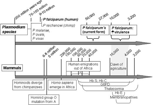
Figure 1 shows the parallel coevolution of P falciparum and its human host. The 4 species of malaria that infect humans today (P vivax, P ovale, P malariae, and P falciparum) arose from an ancestral form 200 million years ago.8 P falciparum's closest genetic relative, P reichenowi, which infects chimpanzees, diverged from P falciparum 9 million to 10 million years ago, around the time that prehuman species diverged from chimpanzees and great apes.8 Thus, P falciparum long predated the development of modern humans.
Modern humans, Homo sapiens, began to emerge in Africa approximately 200 000 years ago9,10 and developed during a period overlapping with the current genetic form of P falciparum.11 Humans first emigrated out of Africa between 100 000 and 40 000 years ago to colonize Asia, Europe, and Oceania,12 and carried with them blood group polymorphisms that had developed under selection pressure of P falciparum.
During the period from 70 000 to 4000 years ago, multiple human erythrocyte mutations associated with a survival advantage in P falciparum infection are thought to have developed. These include structural hemoglobinopathies (Hb S, Hb C),13 quantitative hemoglobinopathies (the thalassemias),14 membrane mutations (spherocytosis, elliptocytosis, ovalocytosis),15 and enzymopathies (glucose-6-phosphate dehydrogenase [G6PD] deficiency).16 The prevalence of these malaria-selected mutations, as well as the pressure P falciparum exerted on the distribution of blood groups, may have further increased 10 000 years ago when the death toll from P falciparum is thought to have increased substantially.17 This increase in prevalence probably resulted from agriculture, forest-clearing, and animal domestication, all of which promoted success of the Anopheles mosquito. As a result, the most intense malarial selection pressures were effectively applied to the human genome in the relatively compressed period of the past few millennia, coincident with the observed development of multiple adaptive erythrocyte mutations.
P falciparum was present during genetic selection of group O alleles
The evolutionary story of the ABO genes sits squarely within this malarial timeline. Among modern humans, the most common allele for group O (termed O01) is identical to the group A allele (A01) for the first 261 nucleotides, at which point a guanosine base is deleted, resulting in a frame-shift mutation that produces a premature stop codon and failure to produce a functional A or B transferase. This single base deletion, from what would otherwise have been a functional group A gene, implies that group O individuals with allele O01 arose as a mutation from A01.18 The second most common allele for group O (termed O02) is considered to be an even more ancient O allele, and also shares the 261 deletion. Other less common group O alleles have been discovered18 and nearly all share deletion 261, leading Roubinet et al18 to conclude that virtually all group O alleles arose at a single time point in human evolution.
Because deletion 261 is found in all populations worldwide, it presumably arose during evolution in Africa before the outward migrations of early humans. Thus, whatever selection pressure favored the group O phenotype must have been active in Africa earlier than 50 000 to 100 000 years ago. P falciparum's established presence in Africa at the origin of modern humans makes it a plausible candidate to influence human blood groups. In fact, during the 150 000 years from the origin of H sapiens to the migration out of Africa, even a slight selective advantage of group O during P falciparum infection would have resulted in a gradual but persistent increase in the prevalence of group O individuals. After the migration, group O genes may have been further enriched among humans inhabiting malaria regions outside of Africa through the introduction of independent group O mutations at position 261. In summary, any infectious disease driving the distribution of ABO groups must be an agent that was active in Africa before the early migration of humans and must be lethal before or during the human reproductive years. P falciparum, considered to be one of the strongest forces in evolutionary history, was in the right place at the right time.
The worldwide prevalence of group O parallels malarial regions
Our hypothesis is that during infection with P falciparum, group O offers a survival advantage, group A confers a disadvantage, and group B has an intermediate effect. Given this hypothesis, one would expect to find that the ratio of group O to A is higher in geographic regions where malaria is currently, or was previously, endemic. This indeed appears to be the case (Table 1).19,,–22 An especially high prevalence of group O coupled with a low prevalence of group A is found throughout subSaharan Africa, where P falciparum persists to this day. In the Western hemisphere, the distribution of group A and group O generally matches malaria's tropical distribution.19 From the tropical regions of Central and South America southward, the indigenous peoples are almost exclusively group O. In Asia, the prevalence of group O rises among peoples who live closer to the equator. For example, in Beijing, China (a cold weather zone), group O is 29% and group A 27%, but in Canton, China (a more tropical zone), group O is 46% and group A is 23%. Group O is the most common blood group in Turkey and Persia.19 In contrast, group A is the predominant blood group in the colder regions of the Earth, where malaria has not been endemic (Table 1). In fact, group A is found in highest frequency in Scandinavia, Greenland, and the subarctic regions of Europe and North America. Thus, if survival from malaria is associated with group O, and mortality is associated with group A, then the worldwide distribution of ABO groups is consistent with selective pressure from malaria.
| Country/people . | Group O, % . | Group A, % . | Non–group O, % . | Ratio O to A . | Malaria area, current or past . |
|---|---|---|---|---|---|
| Southeast Nigeria | 87 | 8 | 13 | 11.0 | Yes |
| Sudan | 62 | 16 | 38 | 3.86 | Yes |
| Kenya/Kikuyu | 60 | 19 | 40 | 3.16 | Yes |
| African Bushmen | 56 | 34 | 44 | 1.65 | Yes |
| Central African Republic | 44 | 28 | 56 | 1.57 | Yes |
| Congo | 52 | 24 | 48 | 2.17 | Yes |
| Eritrea/Somalia | 60 | 22 | 40 | 2.73 | Yes |
| Ghana | 47 | 23 | 53 | 2.04 | Yes |
| Namibia | 47 | 10 | 53 | 4.70 | Yes |
| Central America; Amazon basin | 90 | < 10 | 10 | > 9 | Yes |
| North American Indians/Navajo | 73 | 27 | 27 | 2.70 | Yes |
| USA, general/Indians | 79 | 16 | 21 | 4.94 | Yes |
| Persia | 38 | 33 | 62 | 1.15 | Yes |
| Turkey | 43 | 34 | 57 | 1.26 | Yes |
| Philippines | 45 | 22 | 55 | 2.04 | Yes |
| Vietnam | 42 | 22 | 58 | 1.91 | Yes |
| Papua New Guinea | 41 | 27 | 59 | 1.52 | Yes |
| Finland | 34 | 41 | 66 | .83 | No |
| Austria & Hungary | 36 | 44 | 64 | .82 | No |
| Sweden | 38 | 47 | 62 | .81 | No |
| Switzerland | 40 | 50 | 60 | .80 | No |
| Norway | 39 | 50 | 61 | .78 | No |
| Bulgaria | 32 | 44 | 68 | .73 | No |
| Czech Republic | 30 | 44 | 70 | .68 | No |
| Portugal | 35 | 53 | 65 | .66 | No |
Relationship between ABO groups and the severity of malaria: a critical review of the literature
We critically reviewed literature published during the past 28 years that investigated the relationship between P falciparum infection and ABO groups. Studies in humans published through January 2007 were identified using Medline searches, textbook citations, and the reference lists from retrieved articles. Search terms included “Plasmodium falciparum,” “malaria,” and “ABO.” Related articles for relevant publications were also queried.
Flawed reports exploring associations
Although we found 22 reports that explored an association between ABO group and P falciparum malaria,23,,,,,,,,,,,,,,,,,,,–43 there was no definitive study with adequate sample size investigating ABO group and survival from P falciparum infection. We focused, therefore, on studies that examined ABO group and severity of disease. The sample size of 12 studies was too small to ascertain variations in the distribution of the ABO blood groups.23,,,,,,,,,,–34 Eleven studies were flawed by absent or inappropriate control groups.21,23,28,,,–32,34,–36,38 Several publications reported data on individuals unlikely to die of P falciparum, including 15 studies of individuals with asymptomatic infection/incidental parasitemia, mild infection, or infection of unspecified severity.21,26,,,,,,,,,,,,–39 Finally, many studies failed to report outcomes among the most informative cohort, children younger than 5 years old, who have the highest morbidity and mortality. Given the small sample sizes, the flaws in study design, and unspecified or absent focus on clinical severity, it is not surprising that these studies failed to identify any association between ABO groups and outcomes from P falciparum infection.
Well-designed clinical studies of ABO and clinical severity of P falciparum infection
In contrast to these reports, Table 2 lists those controlled studies40,,–43 that examined ABO distributions and clinical severity of P falciparum infection. Clinical severity, rather than incidence or prevalence of detectable parasitemia, is a more relevant outcome to assess ABO group and survival. Studies reporting clinical features such as cerebral malaria carry more weight than those reporting only laboratory markers such as percent parasitemia, because the latter does not always predict survival. For example, a low percentage of circulating parasitized red cells may either reflect less severe infection (good prognosis), or a greater degree of adhesion of parasitized cells to the vascular endothelium (bad prognosis).44,–46 Conversely, among those with a well-developed humoral immunity, there is little correlation between high circulating parasitemia and severity of illness.47,48
| Author/year/locale . | Sample size . | Focus . | Findings . |
|---|---|---|---|
| Fischer et al40 /1998/Zimbabwe | 209 mild malaria outpatients vs 280 severe malaria inpatients | Coma and anemia severity by blood group | Coma in 3% of group non-A vs 9% of group A, P = .008 |
| Lell et al41 /1999/Gabon | 100 mild malaria cases vs 100 severe malaria cases | Proportion of severe cases among ABO blood groups | Group O: 46% severe group A: 71% severe, P < .01 |
| Pathirana et al42 /2005/Sri Lanka | 243 malaria cases (163 mild, 80 severe) and 65 nonmalarial controls with other infections | Blood group distributions among the mild vs severe, odds ratio for severe malaria if group O vs. non-O | Blood group, mild/severe: group O, 48%/24%; group B, 23%/28%; group A, 25%/33%; group AB, 5%/16%; P < .001; odds ratio (severe malaria:controls) for group O status, 0.37; P < .001 |
| Loscertales et al43 /2006/The Gambia | 41 primiparae with active placental infection. | Maternal and infant hemoglobin; infant weight and size; placental weight; placental parasite count | Placental weight group O, 2893 g; non–group O, 2639 g; P = .04 |
In 1998, Fischer et al40 reported favorable outcomes for group O individuals compared with group A among 489 patients in Zimbabwe with P falciparum malaria. They studied 209 outpatients and 280 severely ill inpatients. Coma was 3-times more common among group A individuals compared with non-A persons (9 of 104 group A versus 11 of 385 with non-A blood, χ2 = 7.0; p = .008; odds ratio, 3.6). Because patients with coma are at a higher risk for death, this study supports the hypothesis that group O individuals may have a survival advantage in severe malaria. However, the sample size was insufficient to observe an effect of ABO group on survival.
Lell et al41 compared 100 cases of severe P falciparum malaria with 100 cases of mild malaria in Gabon. Severe malaria was defined as either hyperparasitemia with more than 0.25 × 10¹² infected red blood cells (RBCs) per L or more than 10% parasitemia; or severe anemia with Hb less than 50 g/L; or clinical signs of severe malaria. Mild malaria cases had minimal laboratory abnormalities and were treated as outpatients. The ratio of group A to group O in patients with severe malaria was 0.50, but was only 0.17 among those with mild malaria (3-fold relative risk). Among all group A individuals, 71% had severe malaria and only 29% had mild malaria (P < .01). In contrast, among all group O cases 46% had severe malaria and 54% had mild malaria (P = .21).
In Sri Lanka, Pathirana et al42 assessed 243 adult cases (mean age, 29.8 years) of P falciparum malaria (163 mild, 80 severe) compared with 65 control patients with other infections. Severe malaria was defined as the presence of cerebral malaria, severe malarial anemia, or multiple organ dysfunction syndromes. The proportion of group O in mild malaria cases was 48%, but was only 24% in severe malaria cases. In striking contrast, the proportion of group A in mild cases was 25%, but was 33% in severe cases (O-to-A ratio: 1.92 mild, .73 severe). The distribution of ABO groups was highly statistically different in severe malaria syndromes compared with uncomplicated malaria or the control population (χ2 = 28.66; P < .001). Once again, a case of severe malaria was nearly 3-times as likely to be group A as O (P = .005). This study provided the strongest statistical evidence of an association between ABO and disease severity in P falciparum infection.
Last year, Loscertales et al43 reported data collected before the HIV pandemic on pregnancy outcomes among women infected with P falciparum in The Gambia. Subjects of interest were pregnant women with no evidence of malaria infection of the placenta, placental malaria pigment without parasites (evidence of cleared placental infection), or active placental infection. Among 89 primiparae, 41 had active placental parasite infection. In this subset, there was a significantly higher birth weight of children born to group O mothers compared with non-group O mothers (2893 ± 362 g vs 2639 ± 323 g; P = .04). Infant length and placental weight were greater, and placental parasite count was lower among group O mothers compared with non-group O, although these values did not reach statistical significance, perhaps because of the small sample size. A larger study is needed to confirm any association between birth outcomes and ABO among women with P falciparum infection during pregnancy. Nevertheless, this initial study is intriguing because it suggests that a group O survival advantage in P falciparum infection may begin in utero during fetal development.
The definitive study to assess ABO groups and survival from infection with P falciparum has not yet been conducted. It will need to examine mortality, especially among pediatric patients, before the age of reproduction. In addition, because even a slight survival advantage for any blood group would accumulate over many generations, it should have a sufficient sample size to detect a small effect, analyze outcomes separately for A, B, O and AB, and control for other phenotypes known to be associated with malaria survival. Currently, the best available studies,40,,–43 listed in Table 2, consistently suggest that group O hosts of P falciparum malaria tend to have milder disease and less severe clinical outcomes than group A hosts. These reports represent substantial, although not yet definitive, evidence for a survival advantage for group O individuals with P falciparum infection.
Possible mechanisms to explain P falciparum's selective genetic pressure in favor of group O and against group A
The adherence of parasitized RBCs to other cells is central to the pathophysiology of severe malaria syndromes including cerebral malaria, respiratory failure, multiorgan failure, and death.49,50 Parasitized RBCs adhere to the vasculature through a process termed “sequestration,” closely mimicking inflammatory leukocyte attachment.51 Furthermore, half of infected RBC isolates52 form occlusive intravascular aggregates, which consist not only of infected RBCs bound to each other (“autoagglutinates”)53 but also of infected RBCs bound to uninfected RBCs (“homotypic RBC rosettes”)54 and/or to platelets (“heterotypic RBC rosettes”).55 Sequestration and rosette formation impair blood flow, causing tissue ischemia and cell death.56 Not surprisingly, in vitro rosetting is more pronounced in parasite strains derived from patients with severe disease, particularly in cases of cerebral malaria.57,,–60
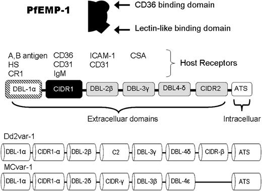
P falciparum is unique among malaria species in that infected erythrocytes express an adhesive determinant, termed P falciparum erythrocyte membrane protein-1 (PfEMP-1). PfEMP-1 is encoded by the parasite genome and is expressed on the outer surface of infected RBCs. PfEMP-1 binds to a variety of target molecules found on RBCs, platelets, and vascular endothelial cells.49,50 The structure of PfEMP-1 includes variable numbers of 2 types of adhesive binding domains: Duffy binding-like (DBL) regions and cysteine-rich interdomain regions (Figure 2).61 DBL-1α demonstrates lectin-like properties, causing it to bind primarily to cells bearing A and B blood group oligosaccharides, and to other glycosylated targets, such as the glycoprotein CD35 (CR1), and heparan sulfate–like glycosaminoglycan.62,63 Cysteine-rich interdomain region-1, however, binds principally to CD36 (platelet glycoprotein IV), thus targeting platelets and endothelium. We summarize literature here on the relationship between ABO, rosetting, and vascular cytoadherence. In Figures 3 and 4, we highlight known and proposed mechanisms by which ABO exerts its influence.
The PfEMP-1 molecule, protein domains, and gene structure.112 The cartoon at the top indicates that PfEMP-1 contains both a CD36-binding domain and a lectin-like binding domain. This depiction is used in Figures 3 and 4. A schematic of the expressed PfEMP-1 protein is shown in the center. DBL-1α, far left in stripe binds to blood group A or B determinants. The CD36-binding region (cysteine-rich interdomain region-1) is shown in black. Two different examples of var genes encoding PfEMP-1 are shown at the bottom. For each parasite, a single PfEMP-1 protein, selected from among 50 to 60 different var genes, is expressed on the RBC surface.112 HS, heparan sulfate; CR1, complement receptor 1; ICAM-1, intracellular adhesion molecule 1; CSA, chondroitin sulfate A; ATS, acid terminal segment.
The PfEMP-1 molecule, protein domains, and gene structure.112 The cartoon at the top indicates that PfEMP-1 contains both a CD36-binding domain and a lectin-like binding domain. This depiction is used in Figures 3 and 4. A schematic of the expressed PfEMP-1 protein is shown in the center. DBL-1α, far left in stripe binds to blood group A or B determinants. The CD36-binding region (cysteine-rich interdomain region-1) is shown in black. Two different examples of var genes encoding PfEMP-1 are shown at the bottom. For each parasite, a single PfEMP-1 protein, selected from among 50 to 60 different var genes, is expressed on the RBC surface.112 HS, heparan sulfate; CR1, complement receptor 1; ICAM-1, intracellular adhesion molecule 1; CSA, chondroitin sulfate A; ATS, acid terminal segment.
Lectin-like binding of parasitized erythrocytes with non-O blood group antigens
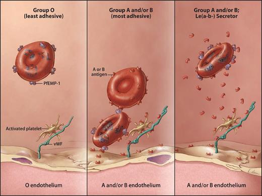
Figure 3 shows a hypothetical model to explain binding of parasitized RBCs to group A or B antigens. Three lines of evidence suggest a direct role for group A or B antigens in cytoadherence as measured by rosette formation: (1) higher rosette rates and larger rosette sizes among non–group O compared with group O RBCs; (2) rosette disruption by soluble group A or group B oligosaccharides; and (3) correlation of rosette formation with transcription of the lectin-specific binding domain of PfEMP-1. ABO effects on the frequency, size, and strength of rosette formation in vitro were first reported by Carlson et al64 in 1992. When RBCs from 52 donors of different ABO groups were incubated with O RBCs infected with P falciparum strain R + PA1, they found rosetting was greatest with group A/AB RBCs, lowest with O RBCs, and intermediate with B RBCs.
Hypothetical model for cytoadhesion of parasitized RBCs to blood group A or group B structures. Infected RBCs expressing PfEMP-1 can bind to group A (or to a lesser extent, group B) determinants on other cells. In the left panel, cytoadhesion from lectin-binding fails to occur because of the absence of group A or group B antigens. In the center panel, an infected RBC adheres to an uninfected RBC (homotypic adherence) via A or B antigens. The infected cell in turn adheres to endothelium either by binding to blood group antigen on endothelial cells or by binding to blood group antigens on platelets or VWF (heterotypic adherence). In the right panel, cytoadherence is blocked by soluble blood group substance present in the plasma of group A and/or B individuals with the Le(a−b−) Secretor phenotype. Copyright Kimberly Main Knoper; used with permission.
Hypothetical model for cytoadhesion of parasitized RBCs to blood group A or group B structures. Infected RBCs expressing PfEMP-1 can bind to group A (or to a lesser extent, group B) determinants on other cells. In the left panel, cytoadhesion from lectin-binding fails to occur because of the absence of group A or group B antigens. In the center panel, an infected RBC adheres to an uninfected RBC (homotypic adherence) via A or B antigens. The infected cell in turn adheres to endothelium either by binding to blood group antigen on endothelial cells or by binding to blood group antigens on platelets or VWF (heterotypic adherence). In the right panel, cytoadherence is blocked by soluble blood group substance present in the plasma of group A and/or B individuals with the Le(a−b−) Secretor phenotype. Copyright Kimberly Main Knoper; used with permission.
Udomsangpetch et al65 showed a strong association between rosette formation and ABO blood group, with group A and group B RBCs (A > B) forming rosettes more than group O cells in each of 8 tested strains (P < .001). Rowe et al60 confirmed that from among 154 isolates, RBCs from group O patients rosetted less (median rosette frequency, 2%; range, 0%-45%) than those from group A (median, 7%; range, 0%-82%; P < .01) or group AB (median, 11%; range, 0%-94%; P < .03). Chotivanich et al54 found the highest relative rosette ratios in vitro among RBCs from healthy donors who were group A (2.7 ± 1.4) and B (2.4 ± 1.1) compared with group O (1.6 ± 0.7; P = .05). Most recently, Barragan et al confirmed that group A targets formed the strongest rosettes. Furthermore, they reported that group A RBCs enzymatically converted to group O RBCs, and Bombay RBCs rosetted minimally and to the same degree.66 Thus, potentiation of rosetting appears specific (but not exclusive) to the A and B antigens.
The binding of parasitized RBCs to groups A and B determinants on endothelial cells67 has not yet been demonstrated experimentally but would be expected to contribute to cytoadherence. As proposed in Figure 3, such adherence may involve not only blood group antigens on endothelial cells but also antigens present on platelets or von Willebrand factor (VWF). Whether P falciparum has selected for individuals with weak subgroups of A or B and whether such individuals have improved survival during P falciparum infection is unknown at this time and deserves further research.
Lectin-like binding by parasitized RBCs is not limited to A and B antigens alone, as other heteropolysaccharides, such as the glycosaminoglycans, are also well-known targets. These include chondroitin sulfate A68,69 and the complement receptor CR1, which depends heavily for its function on extensive N-glycosylation.70,71 CR1 polymorphisms form the basis of the Knops blood group system.72 Knops-null cells fail to rosette. Mutations with reduced CR1 expression, such as the Sl(a−) (KN7) phenotype found in Africans or a phenotype found in 80% of Papua New Guineans, are associated with reduced rosetting and lower risk of cerebral malaria.73,74
Disrupting rosettes with competitors of the lectin-binding domain of infected RBCs
Further evidence supporting a role for group A or B antigens in rosetting comes from inhibition experiments. Trisaccharides corresponding to the blood group A or B determinants reduced rosette formation from parasitized group A or B RBCs.65,66 In contrast, the monosaccharides were unable to disrupt rosettes. Given that soluble forms of group A and B antigen in plasma might reduce or disrupt rosette formation, higher plasma concentrations of these antigens might carry a selective advantage in P falciparum malaria (Figure 3). The action of 2 independent fucosyltransferases, the Secretor gene transferase and the Lewis gene transferase, directly influence the quantity of free A and B antigen found in the plasma of A and B individuals. The highest levels are seen among those with the combination of Secretor-positive and Le(a−b−) phenotypes.75,76 For example, sera from Le(a−b−) Secretor-positive group A individuals contained levels of group A glycoprotein over 200 ng/mL in 5 of 9 sera, which was an order of magnitude higher than the levels seen in Le(a + b−) or Le(a−b+) subjects.75
Recent evidence has shown that the selective pressure in favor of the Secretor-negative phenotype probably results from protection afforded against viral gastrointestinal infections, which are known to cause lethal diarrhea in children.77 In contrast, no explanation has yet emerged to account for a selective genetic pressure in favor of the Lewis-negative phenotype. Although the Le(b) antigen has previously been a suggested binding site for Helicobacter pylori,78 it is difficult to attribute any strong selective genetic pressure to this nonlethal bacterial infection. However, given the high levels of soluble blood group substance found in the plasma of Le(a−b−) Secretor-positive individuals and given the in vitro evidence that soluble blood group substances can disrupt malaria rosette formation, we offer the new hypothesis that P falciparum may account for the 3-fold higher prevalence of the Le(a−b−) phenotype among people of African ancestry.79
DBL-α domain's selective expression correlates with rosettes and disease severity
Recent analysis of P falciparum var gene expression suggests that host ABO blood group is relevant to disease severity. Var genes are the highly variable sequences that encode the polymorphic PfEMP-1 protein, including its outermost DBL-α domain (Figure 2). DBL-α domains fall into 2 groups: DBL-α1 and DBL-α0; only the former binds to A and B antigens. Each mature parasite clone within a generation selects for expression only one var gene80 from among 50-60 found across all 14 chromosomes.63 If group A or B expression is indeed pathogenically relevant, then DBL-α1 expression should be associated with severe, cytoadhesive complications. To investigate this, Kyriacou et al81 sequenced var gene tags found in parasite isolates obtained from 9 children with cerebral malaria compared with 8 children with an equally high parasite burden without cerebral symptoms. Eighty percent of the isolates from patients with cerebral malaria transcribed var genes coding for the DBL-α1 domain. In contrast, only 25% of isolates of children with equivalent degrees of parasitemia but without cerebral malaria contained DBL-α1 var genes (P < .001, χ2). Furthermore, parasite rosette frequency was higher among children with cerebral malaria, with a significant correlation between rosette formation and DBL-α1 gene expression (P = .009, Spearman rank correlation.) This study links cerebral malaria to DBL-α1 expression, rosette formation, and adhesion to group A or group B structures (Figure 3).
Despite these intriguing reports, the overall importance of ABO antigens in pathologic adhesion may thus far have been underestimated for various reasons. Previous studies have almost uniformly been conducted using O RBCs. It is also difficult to dissociate the role played by ABO sugars from the contribution of other glycosylated adhesion molecules (CD36,82 ICAM,83 CR170 ). In addition, host ABO group may affect other aspects of malaria pathogenesis. For example, Chung et al84 recently reported that group A1 RBCs were more likely to be invaded in vitro by P falciparum than group A2, O, or B RBCs. Group A1 cells were more easily invaded by both sialic acid–dependent and sialic acid–independent parasite strains. Invasion of A1 RBCs was specifically reduced by enzymatic conversion of group A to group O cells. These findings, if confirmed and supported by in vivo observations, would lend additional support to the hypothesis that P falciparum has affected the distribution of ABO blood groups.
Recently, Fernandez et al53 identified other rosette-forming surface molecules, termed “rosettins,” with a tropism for the abundant carbohydrate antigens on RBCs. The non-PfEMP1 rosettins, known as “rifins,” encompass the diverse protein products of approximately 200 different rif genes.85,–87 Currently, little is known about the relationship between rifins and ABO blood group antigens, and more research is needed to understand their contribution to the lectin-binding properties of malaria-parasitized RBCs.
P falciparum, CD36, platelets, and VWF
Throughout most of the vasculature, parasitized RBCs expressing cysteine-rich interdomain region-1 can tether and roll on endothelial adhesion molecules, with final direct adhesion to CD36. In addition to direct endothelial binding, infected RBCs may also bind to platelets, in turn adhering to endothelial cells. Platelet-mediated RBC adhesion may be especially relevant to cerebral malaria because CD36 is not strongly expressed on cerebral endothelial cells.88 Combes and Wassmer reviewed previous research on cerebral malaria and concluded that platelets “serve as a bridge” between parasitized RBCs and endothelium. In one set of experiments, Wassmer et al89 co-incubated infected RBCs with CD36-deficient brain microvascular endothelial cells grown to confluence in vitro. Adherence of infected RBCs to endothelial cells under both static and flow conditions depended on the presence of platelets. The adhesion of infected RBCs to platelets was mediated by platelet CD36, and could be blocked by anti-CD36. Electron microscopy showed platelets localized between the infected RBCs and the endothelium.89 Thrombocytopenia is common in severe malaria and platelet localization to brain endothelium was found to be higher in children who died from cerebral malaria compared with uninfected coma controls.90
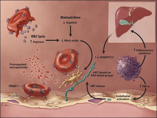
The mechanism by which platelets promote adhesion of parasitized RBCs is not fully understood. A mechanism is proposed in Figure 4. Central is the role of VWF. VWF function is directly related to ADAMTS13 activity and the VWF of group A individuals is more resistant to proteolysis by ADAMTS13 than that of group O individuals.91 The lower levels of VWF found in group O individuals compared with non-O92 may contribute to the apparent genetic advantage of group O in malaria.
Proposed model for cytoadhesion of parasitized RBCs to CD36. Infected RBCs expressing PfEMP-1 can bind to CD36 (platelet glycoprotein IV) determinants on other cells. On the left, an infected cell binds to endothelial CD36 (direct sequestration). In the center, an infected cell binds to CD36 expressed on activated platelets, which in turn bind to endothelial cells via VWF. The lower levels of VWF in group O patients may confer a survival advantage. Inflammation, endothelial activation, NO depletion, reduced ADAMTS13 activity, and procoagulant microparticles may contribute to cytoadhesion. TNF-α, tumor necrosis factor α; ADAMTS13, a disintegrin-like and metalloprotease with thrombospondin type 1 motifs. Copyright Kimberly Main Knoper; used with permission.
Proposed model for cytoadhesion of parasitized RBCs to CD36. Infected RBCs expressing PfEMP-1 can bind to CD36 (platelet glycoprotein IV) determinants on other cells. On the left, an infected cell binds to endothelial CD36 (direct sequestration). In the center, an infected cell binds to CD36 expressed on activated platelets, which in turn bind to endothelial cells via VWF. The lower levels of VWF in group O patients may confer a survival advantage. Inflammation, endothelial activation, NO depletion, reduced ADAMTS13 activity, and procoagulant microparticles may contribute to cytoadhesion. TNF-α, tumor necrosis factor α; ADAMTS13, a disintegrin-like and metalloprotease with thrombospondin type 1 motifs. Copyright Kimberly Main Knoper; used with permission.
Evidence for an important role of VWF in malaria is emerging. Hollestelle et al93 have shown that cerebral malaria is associated with increased levels of VWF propeptide. VWF propeptide is released from endothelial storage sites in response to endothelial damage or activation by cytokines found in children with malaria.94,95 Although ADAMTS13 levels have not been measured in malaria, reduced hepatic release resulting from inflammation96 would further enhance VWF-mediated platelet adhesion. In addition, reduced levels of nitric oxide (NO) probably contribute to the adhesive pathophysiology of malaria. Reduced NO promotes platelet activation and VWF-mediated adhesion,97 and worsened survival in a rodent model of cerebral malaria.98 Moreover, NO antagonists were observed to increase cytoadherence of parasitized cells to human endothelial cells under flow conditions in vitro.99 Low levels of NO would be expected to occur in malaria as a result of arginase release during hemolysis and arginine deficiency from malnutrition. In fact, low arginine and low nitrite levels (an indirect marker of NO availability) were associated with the development of cerebral malaria in Tanzanian children.100 Finally, multiple classes of cellular microparticles, including endothelial microparticles resulting from cell injury, platelet microparticles resulting from platelet activation, and RBC microparticles resulting from hemolysis, probably exert a pro-adhesive effect in the setting of active P falciparum infection.101 Recently, microparticles have been described in malaria and to correlate with disease severity.102,103 Microparticles may worsen clinical outcomes in malaria not only by acting as small adhesive intermediates of heterotypic cellular adhesion, but also through tissue factor expression and procoagulant activity that generates thrombin, resulting in additional platelet activation.104 Thus, multiple lines of evidence suggest that VWF may be central to heterotypic cell adhesion in malaria. As a result, the consistently lower levels of VWF found in group O subjects may provide an additional selection advantage among group O individuals infected with P falciparum.
Conclusions
We have proposed that human and P falciparum coevolution has shaped the proportions of ABO antigens observed in humans across geographic regions. However, we recognize that the distribution of ABO has also been influenced by other events,105,106 including the migration of peoples with founder effects, population splitting from wars and famine, and other lethal pediatric diseases in which survival may be associated with a specific ABO antigen. For example, the possibility that the isohemagglutinins found in group O individuals provide broader immunity against various pathogens has been previously suggested.107,108
Our review of published data serves to emphasize the role of ABO in cytoadherence, rather than P falciparum invasion of RBCs, which depends instead on glycophorin A. P falciparum thus contrasts with P vivax, in which another particular blood group (Duffy) is essential to invasion.
Although our review highlights the reduced cytoadherence of P falciparum among group O individuals, survival after infection is known to depend on a highly complex interaction between both host and parasite genes.41,109,110 Indeed, the full extent of the selective pressure exerted by P falciparum on the human genome has yet to be realized, and new examples of host survival genes continue to be discovered. Examples include recent evidence that a point mutation in the gene for NO synthetase provides as much protection against severe malaria as sickle cell trait,111 or the discovery that the abca-1 gene deletion protects against cerebral malaria through reduced procoagulant microparticle generation.112 Furthermore, because cell adhesion and malarial lethality also depend on parasite gene expression, the evolution of parasite genes has direct bearing on the selective pressure favoring different blood groups. Thus, the influence of P falciparum infection on the relative proportion of ABO antigens is mutually complex. Malaria is known to have affected many erythrocyte genes, including those concerned with globin synthesis, membrane proteins, and RBC enzymes. Given the importance of RBCs in malaria, an influence on genes encoding the most abundant antigens on the RBC membrane is not surprising. Our review suggests that P falciparum was in the right position during evolutionary history to affect the origin and relative proportion of ABO antigens; that the geographic distribution of ABO antigens worldwide is consistent with a survival advantage in malaria among group O individuals; that clinical studies provide supporting evidence in favor of an effect of ABO group on disease severity; and that recent discoveries suggest biologic mechanisms linking disease pathogenesis to ABO antigen expression. Taken together, these findings suggest that the evolution and distribution of ABO blood groups is linked to P falciparum infection.
Acknowledgments
The authors express their appreciation to Dr Christopher Stowell for his critical reading of the manuscript.
This work was supported in part by a grant from the Evelyn and Robert Luick Memorial Fund.
Authorship
Contribution: C.C. and W.D. contributed equally to the intellectual content, research, graphics design, and text.
Conflict-of-interest disclosure: The authors declare no competing financial interests.
Correspondence: Christine M. Cserti, MD, University Health Network, Toronto General Hospital (Blood Transfusion Laboratory), 200 Elizabeth Street, 3EC-306, Toronto ON M5G-2C4; e-mail: christine.cserti@uhn.on.ca.


