Abstract
Toll-like receptor (TLR) 9 recognizes unmethylated microorganismal cytosine guanine dinucleotide (CpG) DNA and elicits innate immune responses. However, the regulatory mechanisms of the TLR signaling remain elusive. We recently reported that Ly49Q, an immunoreceptor tyrosine-based inhibitory motif–bearing inhibitory receptor belonging to the natural killer receptor family, is crucial for TLR9-mediated type I interferon production by plasmacytoid dendritic cells. Ly49Q is expressed in plasmacytoid dendritic cells, macrophages, and neutrophils, but not natural killer cells. In this study, we showed that Ly49Q regulates TLR9 signaling by affecting endosome/lysosome behavior. Ly49Q colocalized with CpG in endosome/lysosome compartments. Cells lacking Ly49Q showed a disturbed redistribution of TLR9 and CpG. In particular, CpG-induced tubular endolysosomal extension was impaired in the absence of Ly49Q. Consistent with these findings, cells lacking Ly49Q showed impaired cytokine production in response to CpG-oligodeoxynucleotide. Our data highlight a novel mechanism by which TLR9 signaling is controlled through the spatiotemporal regulation of membrane trafficking by the immunoreceptor tyrosine-based inhibitory motif–bearing receptor Ly49Q.
Introduction
Toll-like receptors (TLRs) recognize molecular patterns unique to microorganisms, and then elicit host immune responses against the pathogens. Among the TLR family members, TLR3, 4, 7, and 9 can elicit type I interferon (IFN) production in response to microbial components.1 TLR3, 7, and 9 reside in intracellular compartments such as endosomes and detect microbial nucleic acids. The intracellular localization of such TLRs might be necessary to prevent the recognition of self nucleotides while facilitating access to microbial ones.2 TLR9 is a sensor of unmethylated cytosine guanine dinucleotide (CpG) DNA and resides in the endoplasmic reticulum.2,3 TLR9's recognition of CpG DNA is accompanied by changes in membrane dynamics and trafficking, resulting in a strict spatiotemporal compartmentalization of the TLR9 and CpG DNA. The CpG oligonucleotide (CpG) moves into early endosomes and is subsequently transported to a tubular endolysosomal compartment, and in both of these structures it is colocalized with TLR9.3,4 In endosomes, TLR9 forms a complex with myeloid differentiating factor 88 (MyD88) and IFN regulatory factor 7, and activates MyD88-IFR7–dependent type I IFN induction.5 Interestingly, the manner of CpG internalization and the retention time of CpG in endosomes differ between CpG-A and CpG-B, and the retention of CpG/TLR9 complex in the endosomes is the primary determinant of TLR9 signaling.6 After the TLR9 signaling complexes form in endosomes, they translocate to the juxtanuclear area, in tubular endolysosomal structures that extend toward the cell periphery and plasma membrane.3,4 However, the molecular mechanisms underlying the intracellular trafficking of TLR9 are largely unknown
Ly49Q is an immunoreceptor tyrosine-based inhibitory motif (ITIM)–bearing inhibitory receptor belonging to the lectin-type natural killer (NK) receptor family.7,8 However, it possesses unique distinguishing features. Ly49Q is expressed neither on NK nor NKT cells, but on plasmacytoid dendritic cells (DCs), macrophages, and neutrophils.7,9,10 The expression of Ly49Q appears to be regulated during the maturation of these cells and is significantly up-regulated by IFN-γ treatment.7,10,11 Ly49Q can associate with both Src homology 2-containing protein tyrosine phosphatase (SHP)-1 and SHP-2 in a tyrosine phosphorylation-dependent manner.7 We recently reported that Ly49Q is crucial for TLR9-mediated type I IFN production by pDCs.12 pDCs in Ly49Q-deficient mice show impaired CpG-triggered IFN-α and interleukin (IL)-12 production; consequently, TLR9-dependent antiviral responses are diminished in Ly49Q-deficient mice. To gain insight into how Ly49Q regulates TLR9 signaling, we focused on the fact that Ly49Q localizes to endosome-like vesicular compartments. We found that Ly49Q was crucial for the efficient development of the tubular endolysosomes during the intracellular trafficking of CpG and TLR9. Our results reveal a novel mechanism by which TLR signaling is controlled through the spatiotemporal regulation of membrane trafficking by the receptor Ly49Q.
Methods
Mice
Mice (6-7 weeks old) were purchased from CLEA Japan. Experiments were performed according to the Guidelines for Animal Use and Experimentation as set by the International Medical Center of Japan, and were approved by the International Medical Center of Japan. Ly49Q knockout mice were described previously.
Antibodies and reagents
The preparation of the anti-Ly49Q antibody was described previously.7 The following monoclonal antibodies were purchased from BD Pharmingen: phycoerythrin (PE)-conjugated anti–mouse Mac-1 (M1/70); streptavidin-conjugated allophycocyanin and PE; control rat immunoglobulin (Ig)G2a and IgG2b; and anti–mouse CD18 (M18/2). Biotin-conjugated and purified anti-FLAG M2 antibodies were purchased from Upstate Biotechnology. PE-conjugated H-2Kb tetramer was purchased from MBL. Alexa Fluor 594-conjugated anti–rat IgG, Alexa Fluor 594-conjugated anti–mouse IgG, and Alexa Fluor 488-conjugated anti–rat IgG were purchased from Molecular Probes. Antibodies for phospho-c-Jun N-terminal kinase (JNK) and JNK were purchased from Santa Cruz Biotechnology. Antibodies for phospho-p38 and p38 were purchased from Dai-ichi Pure Chemicals. Antibody for Glu-tubulin was purchased from Chemicon International. Recombinant IFN-γ was purchased from PeproTech. CpG-oligodeoxynucleotide1668 and rhodamine-conjugated CpG-oligodeoxynucleotide1668 were purchased from Hokkaido System Science.
Cell culture
The murine macrophage cell line RAW264.7 was purchased from American Type Culture Collection. Cells were cultured in complete RPMI 1640 medium supplemented with 10% fetal calf serum, 10 mM HEPES (N-2-hydroxyethylpiperazine-N′-2-ethanesulfonic acid), 2 mM l-glutamine, 1 mM sodium pyruvate, 50 μM 2-mercaptoethanol, 1% (vol/vol) nonessential amino acids, 100 U/mL penicillin, and 100 μg/mL streptomycin. Macrophages and pDCs were treated with CpG-N-[1-(2,3-Dioleoyloxy)propyl]-N,N,N-trimethylanmonium methylsulfate (DOTAP; Roche Diagnostics), prepared following the manufacturer's instructions. Peritoneal exudation macrophages were prepared by injecting 2 mL of 4% thioglycolate medium intraperitoneally into mice. Cells infiltrating the peritoneal cavity were collected 4 days after the injection.
Flow cytometric analysis
Acid treatment and immunofluorescence analysis were performed, as previously described.13 Stained cells were analyzed with a FACSCalibur (BD Biosciences).
Vectors and cDNA transfection
The expression vector for TLR9-green fluorescent protein (GFP; pCAGGS-TLR9-GFP) was a gift of Dr K. Miyake (Tokyo University).14,15 Expression vector for Rab5-DsRed was a gift of Dr C. R. Roy (Yale University). WEHI3 transfectants expressing wild-type Ly49Q (Ly49Q-WT), the ITIM-less mutant, or containing the mock vector were previously described.7 The vectors for the Ly49Q knockdown were a gift of Dr Takayanagi (Tokyo Medical and Dental University). RAW264.7 cells were transfected by electroporation using a Microporator MP-100 (Digital Bio; NanoEnTek), following the manufacturer's instructions.
Immunohistochemical staining
Cells adhering to glass coverslips were fixed with 3.7% formalin in phosphate-buffered saline (PBS) at room temperature for 15 minutes, and then treated with 0.1% Triton X-100 in PBS for 20 minutes. After being washed with PBS containing 0.05% bovine serum albumin, the cells were treated with 3% bovine serum albumin in PBS to prevent nonspecific protein binding. The cells were then stained with the indicated antibodies or reagents, mounted, and analyzed by confocal (Zeiss) or fluorescence (Olympus) microscopy.
Western blot analyses
The cells were lysed in radioimmunoprecipitation assay buffer consisting of 10 mM Tris-HCl, pH 7.5, 150 mM NaCl, 1 mM EGTA (ethyleneglycoltetraacetic acid), 1.5 mM MgCl2, 10 mM NaF, 1 mM Na3VO4, and complete EDTA (ethylenediaminetetraacetic acid)-free protease inhibitor mixture (Roche Diagnostics). The lysates were resolved by 5% to 20% sodium dodecyl sulfate (SDS)–polyacrylamide gel electrophoresis and transferred to polyvinylidene difluoride membranes. The proteins were detected by incubating the membranes with the indicated antibodies and visualized using SuperSignal West Dura Extended Duration Substrate (Pierce). In some experiments, SuperSignal West Femto Extended Duration Substrate was used for detection (Pierce). For quantification of the precipitated proteins, a LAS-3000 (Fuji Photo Film) was used.
Reverse transcription–polymerase chain reaction and quantification of mRNA
For RNA preparations, Isogen was used according to the manufacturer's instructions (Wako Pure Chemical). cDNA synthesis was performed according to standard protocols using oligo(dT) and random hexamer oligonucleotides. For semiquantitative reverse transcription–polymerase chain reaction (RT-PCR), gene-specific fragments were obtained by linear phase PCR amplification, and standardized using the β-actin or hypoxanthine guanine phosphoribosyl transferase 1 level. Quantitative differences in mRNA levels were determined by real-time RT-PCR using SYBR Green PCR Master Mix (Applied Biosystems) and a thermal cycler controlled by the 7900HT Fast Real Time PCR system (Applied Biosystems). Primers used for PCR analyses are shown in supplemental Table 1 (available on the Blood website; see the Supplemental Materials link at the top of the online article).
RNA interference
Double-stranded RNA for Ly49Q interference (UGAGGACAAUCAAGGGUCAAGAGAA) was purchased from Hokkaido System Science. To introduce the small interfering RNA (siRNA) into RAW264 cells, a Microporator MP-100 (Digital Bio; NanoEnTek) was used according to the manufacturer's instructions. Transfection efficiency was estimated by using a fluorescein isothiocyanate–conjugated control siRNA-A (Santa Cruz Biotechnology).
Statistical analysis
The statistical significance of differences in values for the migration, invasion, adhesion, and spreading of neutrophils was determined with the 2-tailed Student t test. Differences with a P value of less than .05 were considered significant.
Results
Colocalization of Ly49Q with CpG in lysosomal compartments
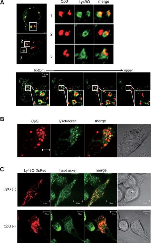
We recently reported that, in Ly49Q-deficient mice, the TLR9-triggered production of cytokines such as IFN-α and IL-12 is severely impaired.12 To investigate how Ly49Q affects TLR9 signaling, we first examined whether Ly49Q colocalizes with CpG-containing endosomes. Because Ly49Q is internalized and localizes to endocytic compartments, we hypothesized that Ly49Q colocalizes with CpG/TLR9 in endosomal compartments and affects TLR9 signaling. To facilitate the immunohistochemical analyses, peritoneal macrophages expressing FLAG-tagged Ly49Q were obtained from Ly49Q-expressing transgenic (Tg) mice (supplemental Figure 1). Ly49Q was detected not only at the cell surface, but also in annular-shaped vesicular compartments (Figure 1A). The CpG fluorescence partially overlapped with the Ly49Q-associated vesicles, and some of the CpG-containing endosomes were closely encircled by Ly49Q+ vesicular structures, suggesting that the Ly49Q-associated vesicles had fused with the CpG-containing endosomes. Z-axis sections revealed that the Ly49Q-containing compartments were intricately intertwined with the CpG-containing endosomes. Similar results were obtained using the RAW264 macrophage cell line (data not shown). As previously demonstrated, the CpG-containing endosomes subsequently acquired lysosome-like features along with acidification, which was detectable with an acidic organelle-targeting fluorescent dye (Figure 1B).16,17 To clarify whether Ly49Q also localized to the lysosome-like compartments, a Ly49Q-DsRed fusion protein was expressed in RAW264 cells. The Ly49Q localized to lysotracker-targeted compartments in the steady state (Figure 1C). CpG stimulation caused the Ly49Q to be redistributed to tubular vesicular compartments, which were previously described as tubular endolysosomal structures.
Ly49Q colocalized with CpG in endosome/lysosome compartments. (A) Colocalization of Ly49Q with CpG in endosomes. Peritoneal exudate macrophages were prepared from Ly49Q Tg mice. The cells were incubated with rhodamine-conjugated CpG (0.3 μM) for 15 minutes, fixed with 4% formalin in PBS, then stained with an anti-FLAG antibody and analyzed by confocal microscopy. The bottom 4 photographs show serial Z-axis–sectioned patterns after 60-minute incubation with rhodamine-conjugated CpG. Squares indicate the region shown in higher magnification. (B) Localization of CpG to late endosomes/lysosomes. RAW264 cells were incubated with rhodamine-conjugated CpG for 60 minutes. To visualize the late endosomes/lysosomes, the cells were incubated with lysotracker for the last 30 minutes. (C) Localization of Ly49Q in the late endosomes/lysosomes. A Ly49Q-DsRed fusion construct was introduced into RAW264 cells. Twenty-four hours after transfection, the cells were cultured with lysotracker at 37°C for 30 minutes, and the intracellular localization of Ly49Q was examined by confocal microscopy. Ly49Q localized to the late endosomes/lysosomes. In the presence of CpG stimulation, Ly49Q-containing compartments showed a tubular structure.
Ly49Q colocalized with CpG in endosome/lysosome compartments. (A) Colocalization of Ly49Q with CpG in endosomes. Peritoneal exudate macrophages were prepared from Ly49Q Tg mice. The cells were incubated with rhodamine-conjugated CpG (0.3 μM) for 15 minutes, fixed with 4% formalin in PBS, then stained with an anti-FLAG antibody and analyzed by confocal microscopy. The bottom 4 photographs show serial Z-axis–sectioned patterns after 60-minute incubation with rhodamine-conjugated CpG. Squares indicate the region shown in higher magnification. (B) Localization of CpG to late endosomes/lysosomes. RAW264 cells were incubated with rhodamine-conjugated CpG for 60 minutes. To visualize the late endosomes/lysosomes, the cells were incubated with lysotracker for the last 30 minutes. (C) Localization of Ly49Q in the late endosomes/lysosomes. A Ly49Q-DsRed fusion construct was introduced into RAW264 cells. Twenty-four hours after transfection, the cells were cultured with lysotracker at 37°C for 30 minutes, and the intracellular localization of Ly49Q was examined by confocal microscopy. Ly49Q localized to the late endosomes/lysosomes. In the presence of CpG stimulation, Ly49Q-containing compartments showed a tubular structure.
Defective CpG redistribution and endolysosome extension in Ly49Q− pDCs and macrophages
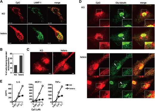
Previous studies demonstrated that the intracellular trafficking of CpG is spatiotemporally regulated, and that the behavior of CpG endosomes affects the quality of TLR9 signaling.5,6 Therefore, we next investigated whether Ly49Q is involved in CpG trafficking using Ly49Q−/− pDCs and macrophages. The internalization of CpG and its subsequent redistribution to tubular endolysosomes were observed in bone marrow-derived pDCs prepared from a Ly49Q+/− mouse (Figure 2A). The tubular endolysosomal structures radiated from the perinuclear region, extending toward the cell periphery. Lysosomal-associated membrane protein-1 (LAMP-1)+ late endosomes were localized to the perinuclear region, and some of the CpG colocalized with LAMP-1. These results are consistent with a previous report showing that internalized CpG is transported to the perinuclear region through late endosomes and subsequently distributed into tubular endolysosomes.3
Formation of tubular endolysosomal structures was impaired in pDCs and macrophages derived from Ly49Q knockout mice. (A) Bone marrow–derived pDCs were prepared by culturing bone marrow cells with Flt3L, and they were then incubated with rhodamine-conjugated CpG (0.3 μM) for 60 minutes. The cells were fixed and then stained with an anti–LAMP-1 antibody. Confocal images of representative cells are shown. (B) The number of cells showing the directionally extending CpG-including tubular endolysosomal structures was counted, and the proportion of total cells was calculated. (C) Peritoneal exudate macrophages were prepared from Ly49Q knockout and control littermate mice and incubated with rhodamine-conjugated CpG (0.3 μM) for 24 hours. The intracellular distribution of CpG was examined by confocal microscopy. (D) The peritoneal exudate macrophages were treated with rhodamine-conjugated CpG (0.3 μM) for 24 hours. The cells were fixed and then stained with anti–detyrosinated tubulin (Glu-tubulin) antibody. Arrowheads indicate MTOC. (E) Cytokine production by peritoneal exudate macrophages was compared between Ly49Q-deficient and control littermate mice. Macrophages were stimulated with CpG, and the amount of cytokine in the culture supernatant was estimated by cytokine bead array 24 hours later.
Formation of tubular endolysosomal structures was impaired in pDCs and macrophages derived from Ly49Q knockout mice. (A) Bone marrow–derived pDCs were prepared by culturing bone marrow cells with Flt3L, and they were then incubated with rhodamine-conjugated CpG (0.3 μM) for 60 minutes. The cells were fixed and then stained with an anti–LAMP-1 antibody. Confocal images of representative cells are shown. (B) The number of cells showing the directionally extending CpG-including tubular endolysosomal structures was counted, and the proportion of total cells was calculated. (C) Peritoneal exudate macrophages were prepared from Ly49Q knockout and control littermate mice and incubated with rhodamine-conjugated CpG (0.3 μM) for 24 hours. The intracellular distribution of CpG was examined by confocal microscopy. (D) The peritoneal exudate macrophages were treated with rhodamine-conjugated CpG (0.3 μM) for 24 hours. The cells were fixed and then stained with anti–detyrosinated tubulin (Glu-tubulin) antibody. Arrowheads indicate MTOC. (E) Cytokine production by peritoneal exudate macrophages was compared between Ly49Q-deficient and control littermate mice. Macrophages were stimulated with CpG, and the amount of cytokine in the culture supernatant was estimated by cytokine bead array 24 hours later.
In contrast, Ly49Q−/− pDCs showed fractionated and undirected CpG-containing compartments. Only a small portion of the CpG compartments colocalized with the late endosomes, and the perinuclear localization of the late endosomes was not obvious in the Ly49Q−/− pDCs. The frequency of cells possessing radiating and elongating endolysosome structures was decreased in the Ly49Q−/− pDCs compared with the Ly49Q+/− pDCs (Figure 2B). A similar defect in the redistribution of CpG-containing endolysosomes was observed in Ly49Q−/− peritoneal exudate macrophages (Figure 2C). The directional extension of CpG-containing endolysosomes was not observed in the Ly49Q+/− macrophages, whereas numerous directional CpG-containing endolysosomes elongating from the perinuclear region were observed in the Ly49Q+/− macrophages. The tubular endolysosome elongation has been shown to be MyD88 independent, but microtubule dependent.3,18 Stabilized microtubules are enriched with posttranslationally modified tubulins such as detyrosinated tubulin (Glu-tubulin), in which the C-terminal tyrosine of α-tubulin is removed by tubulin carboxypeptidase.19 Immunohistochemical staining of Glu-tubulin clearly demonstrated that, in Ly49Q+/− macrophages, Glu microtubules colocalized with the tubular endolysosomes, indicating that stabilized microtubules exist along the endolysosomes (Figure 2D). In addition, the Glu-tubulin at the microtubule organizing center (MTOC) appeared to be connected to the tubular endolysosomes in Ly49Q+/− macrophages. In contrast, in Ly49Q−/− macrophages, Glu-tubulin did not colocalize with the endolysosomes, and no connection between Glu-tubulin and the endosomes at the MTOC was observed. Thus, the impairment of endolysosome elongation in the Ly49Q−/− macrophages was associated with decreased stability of the microtubules. Consistent with these observations, in Ly49Q−/− macrophages, the CpG-triggered production of cytokines, including IL-6, tumor necrosis factor α, and monocyte chemoattractant protein-1, was severely diminished (Figure 2E). Thus, the absence of Ly49Q upset the CpG trafficking in both pDCs and macrophages, in close correlation with the failure of cytokine production in both pDCs and macrophages.
Importance of Ly49Q in TLR9-mediated IFN-β and IL-6 production in RAW264 cells
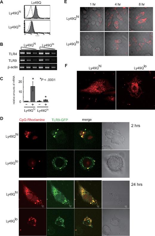
Next, we tried to investigate the intracellular trafficking of CpG and TLR9 in the presence or absence of Ly49Q using the mouse macrophage cell line RAW264, which expresses TLR9 and readily permits the introduction of various expression constructs. To do this, we established Ly49Qhi and Ly49Qlo RAW264 clones to examine the relevance of Ly49Q in CpG/TLR9 trafficking, because the expression level of Ly49Q in RAW264 cells was heterogeneous in the steady state (Figure 3A). Four clones each of Ly49Qhi or Ly49Qlo RAW264 cells were analyzed, and the same results were obtained (data not shown). Ly49Qhi and Ly49Qlo RAW264 clones showed comparable expression levels of CD11b, F4/80, TLR4, and TLR9 (supplemental Figures 2A, 3B). To confirm the importance of Ly49Q in TLR9 signaling in RAW264 cells, Ly49Qhi or Ly49Qlo RAW264 cells were treated with CpG, and the TLR9-triggered cytokine production was examined. When the cells were stimulated with CpG, the Ly49Qhi RAW264 cells produced IFN-β and IL-6, and the Ly49Qlo RAW264 cells produced little, if any (Figure 3C and supplemental Figure 2B). These results were consistent with our findings that pDCs in Ly49Q−/− mice show impaired TLR9-mediated type I IFN production and that Ly49Q− pDCs from bone marrow are less potent producers of IFN-β and IL-6 (Figure 2E).10 In addition, the Ly49Qhi and Ly49Qlo RAW264 cells were morphologically different in their spreading and adhesion properties and in their formation of cytoplasmic vacuolar structures in the presence of CpG (supplemental Figure 2C). When exogenous Ly49Q-WT was expressed in Ly49Qlo RAW264 cells, the CpG-induced IL-6 production recovered (supplemental Figure 2D-E). However, the ITIM-less mutant (Ly49Q-YF) did not rescue the IL-6 production efficiently, indicating that the ITIM is important for TLR9 signaling.
Inefficient uptake of CpG and retarded redistribution of TLR9 in Ly49Qlo cells. (A) Expression of Ly49Q in RAW264 clones. Ly49Qhi or Ly49Qlo RAW264 clones were established from bulk RAW264 cells by limiting dilution. Four clones of each type were analyzed, and similar results were obtained. Data from representative clones are shown. (B) No difference in TLR9 and TLR4 expression in RAW264 cells. Semiquantitative RT-PCR was carried out using RNA prepared from the indicated cells. The presence or absence of Ly49Q had no effect on the transcription of these TLRs. In the photographs, the PCR templates were sequentially diluted by 5-fold. (C) Quantitative analysis of IFN-β transcription in RAW264 cells in response to CpG. Ly49Qhi or Ly49Qlo RAW264 cells were stimulated with CpG1668 (0.3 μM) for 4 hours, and quantitative RT-PCR analyses were performed. The histograms show the relative amounts of IFN-β mRNA evaluated by real-time PCR. (D) Intracellular redistribution of CpG and TLR9. TLR9-GFP expression plasmids were introduced into RAW264 cells. Twenty-four hours after transfection, the cells were incubated with rhodamine-conjugated CpG (0.3 μM) for the indicated periods. Impaired CpG/TLR9 redistribution was observed in the Ly49Qlo RAW264 cells. (E) Internalization and distribution of CpG in RAW264 cells. RAW264 cells were incubated with rhodamine-conjugated CpG (0.3 μM) and fixed at the indicated time points. (F) Intracellular distribution of rhodamine-conjugated CpG. After 24 hours of incubation with CpG, tubular endolysosomal structures were observed in Ly49Qhi RAW264, but not in Ly49Qlo RAW264 cells.
Inefficient uptake of CpG and retarded redistribution of TLR9 in Ly49Qlo cells. (A) Expression of Ly49Q in RAW264 clones. Ly49Qhi or Ly49Qlo RAW264 clones were established from bulk RAW264 cells by limiting dilution. Four clones of each type were analyzed, and similar results were obtained. Data from representative clones are shown. (B) No difference in TLR9 and TLR4 expression in RAW264 cells. Semiquantitative RT-PCR was carried out using RNA prepared from the indicated cells. The presence or absence of Ly49Q had no effect on the transcription of these TLRs. In the photographs, the PCR templates were sequentially diluted by 5-fold. (C) Quantitative analysis of IFN-β transcription in RAW264 cells in response to CpG. Ly49Qhi or Ly49Qlo RAW264 cells were stimulated with CpG1668 (0.3 μM) for 4 hours, and quantitative RT-PCR analyses were performed. The histograms show the relative amounts of IFN-β mRNA evaluated by real-time PCR. (D) Intracellular redistribution of CpG and TLR9. TLR9-GFP expression plasmids were introduced into RAW264 cells. Twenty-four hours after transfection, the cells were incubated with rhodamine-conjugated CpG (0.3 μM) for the indicated periods. Impaired CpG/TLR9 redistribution was observed in the Ly49Qlo RAW264 cells. (E) Internalization and distribution of CpG in RAW264 cells. RAW264 cells were incubated with rhodamine-conjugated CpG (0.3 μM) and fixed at the indicated time points. (F) Intracellular distribution of rhodamine-conjugated CpG. After 24 hours of incubation with CpG, tubular endolysosomal structures were observed in Ly49Qhi RAW264, but not in Ly49Qlo RAW264 cells.
To further confirm that the effects in the Ly49Qlo RAW264 cells were due to the decreased Ly49Q and not to some other difference, we performed siRNA knockdown experiments. Ly49Q expression in the Ly49Qhi RAW264 cells was down-modulated by introducing Ly49Q antisense RNA, and the CpG-induced IL-6 production was examined. The expression of Ly49Q short hairpin RNA in the Ly49Qhi RAW264 cells resulted in diminished IL-6 production in response to CpG (supplemental Figure 3A-B). The down-modulation of Ly49Q expression also caused a decrease in lysosome-like vesicular structures after CpG stimulation, implying a functional correlation between Ly49Q and lysosome and/or vesicle trafficking (supplemental Figure 3C). In addition, Ly49Q expression was induced in Ly49Qlo RAW264 cells by IFN-γ treatment, as we reported previously (supplemental Figure 3D). In association with the increased Ly49Q expression, the IL-6 production and vacuolar formation by CpG stimulation were recovered in these originally Ly49Qlo RAW cells (supplemental Figure 3E-F). Furthermore, the inhibition of Ly49Q expression using Ly49Q-specific short hairpin RNA in IFN-γ-treated RAW264 cells diminished the CpG-induced IL-6 production and vacuolar formation, indicating that Ly49Q is important for the IL-6 production triggered by TLR9 (supplemental Figure 3G-H). Therefore, TLR9-mediated cytokine production in RAW264 cells was also dependent on Ly49Q.
Defective TLR9 trafficking in the absence of Ly49Q
Therefore, we next investigated the intracellular trafficking of a TLR9-GFP fusion protein using the Ly49Qhi and Ly49Qlo RAW264 cells. Two hours after CpG stimulation, internalized CpG was colocalized with TLR9 in endosomes in both the Ly49Qhi and Ly49Qlo RAW264 cells (Figure 3D). However, there were great differences in the diameter, number, and cytoplasmic localizations of the CpG/TLR9 endosomes between the Ly49Qhi and Ly49Qlo RAW264 cells. In the Ly49Qlo RAW264 cells, the diameters of the TLR9+ vesicles appeared smaller than in the Ly49Qhi RAW264 cells (Figure 3D). In addition, several TLR9+ vesicles were observed scattered throughout the cytoplasm in the Ly49Qlo RAW264 cells. After 24 hours of stimulation, the difference in TLR9 distribution between the Ly49Qlo and Ly49Qhi RAW264 cells was remarkable. TLR9 in the Ly49Qhi RAW264 cells was localized along tubular endosomal structures and distributed at the edges of protrusions that were focal adhesion-like attachment sites. In contrast, in the Ly49Qlo RAW264 cells, even though the TLR9+ vesicles colocalized with the CpG-containing endosomes, they remained scattered in the cytoplasm, and no vesicular fusion or elongation was observed. These tubular structures did not colocalize with either Rab11 or the transferrin receptor (data not shown).
Defects in tubular endolysosome extension, as observed in the Ly49Q knockout pDCs and macrophages, were also observed in Ly49Qlo RAW264 cells (Figure 3E-F). Kinetic analyses of CpG trafficking demonstrated that both Ly49Qhi and Ly49Qlo RAW264 cells internalized CpG, although the amount of internalized CpG in Ly49Qlo RAW264 cells seemed slightly lower than in Ly49Qhi RAW264 cells at the early time point (1 hour; Figure 3E). After 8 hours of CpG stimulation, in the Ly49Qhi RAW264 cells, CpG-containing endosomes appeared to spread or diffuse through the cytoplasm, in contrast to Ly49Qlo RAW264 cells, which showed no diffuse distribution in CpG fluorescence in the cytoplasm. In addition, no obvious change of distribution pattern was observed in Ly49Qlo RAW264 cells from 4 to 8 hours. A remarkable difference was also observed after 24 hours of CpG stimulation in the extension of tubular endolysosomal structures (Figure 3F). These results strongly suggest that the CpG trafficking in RAW264 cells was also regulated by Ly49Q.
Mitogen-activated protein kinase activation, but not NF-κB-related transcription factor expression, was affected by Ly49Q
Next, we examined which signaling pathways could be affected by Ly49Q. RT-PCR analyses revealed no great differences in the expression of NF-κB–related transcription factors between the Ly49Qhi and Ly49Qlo RAW264 cells (supplemental Figure 4; data not shown). However, the TLR9-triggered activation of p38 was severely impaired in the Ly49Qlo RAW264 cells (Figure 4A). In addition, CpG-induced JNK activation was dysregulated in the Ly49Qlo RAW264 cells. The activation of JNK in Ly49Qhi RAW264 cells was sustained between 4 and 7 hours after CpG stimulation, but in the Ly49Qlo RAW264 cells, the level of phospho-JNK decreased during this time (Figure 4B). In addition, immunohistochemical analyses clearly showed that Ly49Q colocalized with phosphorylated p38 in late endosome/lysosome compartments after CpG stimulation. A portion of the phosphorylated JNK also colocalized with LAMP-1+ late endosome compartments (Figure 4C). These results strongly suggest that Ly49Q influences TLR9-mediated p38 and JNK activation in the late endosome/lysosome compartments.
Ly49Q is involved in the regulation of MAP kinase activation after CpG stimulation. (A) Activation kinetics of p38 and JNK. RAW264 cells were treated with CpG (0.3 μM) for the indicated periods, and the total cell lysates were prepared by directly adding SDS sample buffer. The total lysates were separated by SDS–polyacrylamide gel electrophoresis, and the activation of p38 and JNK was examined by Western blotting. (B) The signal intensity (pixel numbers) of phosphorylated MAP kinases was normalized to that of each total MAP kinase, and semiquantitative values for the MAP kinase activation are shown. (C) Intracellular distribution of phosphorylated p38 and JNK in the presence of CpG stimulation. FLAG-tagged Ly49Q expression plasmids were introduced into Ly49Qhi RAW264 cells, and 24 hours after transfection, the cells were incubated with unlabeled CpG (0.3 μM) for 2 hours. The cells were then fixed and stained with antibodies against FLAG and phosphorylated p38. To analyze the distribution of phosphorylated JNK, Ly49Qhi RAW264 cells were treated with CpG for 2 hours and stained with antibodies against phospho-JNK and LAMP-1.
Ly49Q is involved in the regulation of MAP kinase activation after CpG stimulation. (A) Activation kinetics of p38 and JNK. RAW264 cells were treated with CpG (0.3 μM) for the indicated periods, and the total cell lysates were prepared by directly adding SDS sample buffer. The total lysates were separated by SDS–polyacrylamide gel electrophoresis, and the activation of p38 and JNK was examined by Western blotting. (B) The signal intensity (pixel numbers) of phosphorylated MAP kinases was normalized to that of each total MAP kinase, and semiquantitative values for the MAP kinase activation are shown. (C) Intracellular distribution of phosphorylated p38 and JNK in the presence of CpG stimulation. FLAG-tagged Ly49Q expression plasmids were introduced into Ly49Qhi RAW264 cells, and 24 hours after transfection, the cells were incubated with unlabeled CpG (0.3 μM) for 2 hours. The cells were then fixed and stained with antibodies against FLAG and phosphorylated p38. To analyze the distribution of phosphorylated JNK, Ly49Qhi RAW264 cells were treated with CpG for 2 hours and stained with antibodies against phospho-JNK and LAMP-1.
Ly49Q was internalized and recycled through an ITIM-mediated mechanism
To obtain insights into a mechanism of the Ly49Q-mediated TLR-containing vesicular trafficking, we analyzed trafficking of Ly49Q and its ligand, major histocompatibility complex (MHC) class I. We previously demonstrated that Ly49Q associates with MHC class I in cis at the cell surface.20 By confocal microscopic analyses, we found that Ly49Q was colocalized with H-2Kb in peritoneal exudate macrophages (Figure 5A). This colocalization occurred not only at the cell surface, but also in cytoplasmic vesicles, suggesting that Ly49Q was internalized together with H-2Kb. We next tested whether the removal of β2-microglobulin (β2m) from the cell surface by acid treatment would elicit binding of the H-2Kb tetramer to Ly49Q, as shown for Ly49A.13 As we previously reported, Ly49Q on the pDCs in C57BL/6 mice showed low levels of H-2Kb binding (Figure 5B).20 C57BL/6 β2m−/− pDCs showed increased H-2Kb binding, which was completely inhibited by an anti-Ly49Q antibody. These results indicate that Ly49Q associates with H-2Kb in a cis configuration. The acid treatment of pDCs did not affect the H-2Kb binding, even though β2m was successfully removed from the cell surface (Figure 5C-D; data not shown). Furthermore, neither the cell viability nor the Ly49Q expression levels on the cell surface were affected by acid treatment (data not shown). These results strongly suggested that Ly49Q's association with H-2Kb is β2m independent and stable under acidic conditions; therefore, this interaction should be sustained in intracellular acidic compartments, such as endosomes/lysosomes.
Ly49Q was internalized with MHC class I, and its localization was regulated in an ITIM- and tyrosine phosphatase-dependent manner. (A) Colocalization of Ly49Q with H-2Kb in intracellular vesicular compartments. Peritoneal exudation macrophages were prepared from Ly49Q Tg mice and examined for Ly49Q (red) and MHC class I (green) by immunohistochemical analyses with a confocal microscope. FLAG-tagged Ly49Q in Tg mice was detected with an anti-FLAG antibody. (B) Binding of H-2Kb tetramer to pDCs. Cells enriched in bone marrow pDCs were obtained by AutoMACS using an anti–plasmacyloid DC Ag-1 antibody, and the binding of PE-conjugated H-2Kb tetramer was examined by flow cytometry. In the absence of β2m, binding of the H-2Kb tetramer in trans was detectable due to loss of the cis interaction. The tetramer binding was abrogated by an anti-Ly49Q antibody. (C) Removal of β2m from the cell surface by acid treatment. Bone marrow cells were treated with citrate buffer (0.133 M citric acid and 0.066 M Na2HPO4, pH 3.3) at 20°C for the indicated periods. Removal of β2m from the cell surface was confirmed by a decreased fluorescence intensity of anti-β2m antibody staining. CD11c+ cells were gated and analyzed. (D) Binding of H-2Kb tetramer before and after acid treatment. H-2Kb tetramer bound in trans to Ly49Q after the removal of β2m from the cell surface, indicating that the cis interaction between Ly49Q and H-2Kb was still maintained, and the interaction was β2m independent and acid resistant. (E) ITIM and tyrosine phosphatase dependence of Ly49Q redistribution. WEHI3 transfectants expressing Ly49Q-WT or Ly49Q-YF were incubated at 37°C in the presence or absence of the indicated inhibitors of membrane trafficking. The cells were then fixed and stained with an anti-FLAG antibody to visualize Ly49Q.
Ly49Q was internalized with MHC class I, and its localization was regulated in an ITIM- and tyrosine phosphatase-dependent manner. (A) Colocalization of Ly49Q with H-2Kb in intracellular vesicular compartments. Peritoneal exudation macrophages were prepared from Ly49Q Tg mice and examined for Ly49Q (red) and MHC class I (green) by immunohistochemical analyses with a confocal microscope. FLAG-tagged Ly49Q in Tg mice was detected with an anti-FLAG antibody. (B) Binding of H-2Kb tetramer to pDCs. Cells enriched in bone marrow pDCs were obtained by AutoMACS using an anti–plasmacyloid DC Ag-1 antibody, and the binding of PE-conjugated H-2Kb tetramer was examined by flow cytometry. In the absence of β2m, binding of the H-2Kb tetramer in trans was detectable due to loss of the cis interaction. The tetramer binding was abrogated by an anti-Ly49Q antibody. (C) Removal of β2m from the cell surface by acid treatment. Bone marrow cells were treated with citrate buffer (0.133 M citric acid and 0.066 M Na2HPO4, pH 3.3) at 20°C for the indicated periods. Removal of β2m from the cell surface was confirmed by a decreased fluorescence intensity of anti-β2m antibody staining. CD11c+ cells were gated and analyzed. (D) Binding of H-2Kb tetramer before and after acid treatment. H-2Kb tetramer bound in trans to Ly49Q after the removal of β2m from the cell surface, indicating that the cis interaction between Ly49Q and H-2Kb was still maintained, and the interaction was β2m independent and acid resistant. (E) ITIM and tyrosine phosphatase dependence of Ly49Q redistribution. WEHI3 transfectants expressing Ly49Q-WT or Ly49Q-YF were incubated at 37°C in the presence or absence of the indicated inhibitors of membrane trafficking. The cells were then fixed and stained with an anti-FLAG antibody to visualize Ly49Q.
Next, we asked whether the ITIM of Ly49Q was involved in the internalization of Ly49Q, because the tyrosine motifs (Yxxφ) in ITIMs have been suggested to function as an internalization signal.21 We first investigated Ly49Q endocytosis in the presence of various inhibitors of membrane trafficking, using Ly49Q-null myeloid lineage WEHI3 cells transduced with Ly49Q-WT or an ITIM-less mutant (Ly49Q-YF). In the absence of inhibitors, Ly49Q-WT was largely observed at the cell surface, but Ly49Q-YF inhabited perinuclear intracellular granules (Figure 5E). Methyl-β-cyclodextrin, which inhibits raft-dependent endocytosis by depleting cholesterol from the plasma membrane,22 abrogated both the perinuclear distribution of Ly49Q-YF and the juxtamembranous endosomal distribution of Ly49Q-WT. Chlorpromazine, an inhibitor of clathrin-dependent endocytosis,23 did not inhibit the internalization of either Ly49Q-YF or Ly49-WT. These results strongly suggest that Ly49Q is internalized via lipid raft-mediated endocytosis, and that the ITIM is not necessary for endocytosis itself, but important for the retention of Ly49Q at the cell surface.
We also found that the internalized endosomes were transported along microtubules, because nocodazole treatment strikingly diminished the accumulation of Ly49Q-YF in the perinuclear regions.24 Importantly, treatment with a phosphatase inhibitor, sodium vanadate,25 caused Ly49Q-WT to be redistributed to the perinuclear region in the same pattern as Ly49Q-YF. Given that Ly49Q-WT can associate with tyrosine phosphatases via its ITIM, these results suggest that the ITIM-associated phosphatase is important for regulating the intracellular distribution of Ly49Q. Because MHC class I recycles between the cell surface and endosomes, this finding also suggests that Ly49Q recycles together with MHC class I along microtubules in the steady state.
Discussion
In this study, we demonstrated that an inhibitory receptor, Ly49Q, plays an important role in the signaling of TLR9 by controlling the intracellular trafficking of TLR9 and CpG. The spatiotemporal regulation of the vesicular compartments containing TLR9 and CpG and their associated adaptor proteins is crucial for TLR9 signaling.5,6 Ly49Q itself was internalized and appeared to move along microtubules in the steady state. The observation that Ly49Q associated with MHC class I in a cis configuration, even in an acidic environment, strongly suggests that Ly49Q recycles together with MHC class I. Importantly, the Ly49Q movement was regulated by an ITIM- and tyrosine phosphatase-dependent mechanism. Because Ly49Q itself can recruit tyrosine phosphatases such as SHP-1 and SHP-2 to the ITIM,7 it is possible that Ly49Q-associated phosphatases play a role in the trafficking of Ly49Q itself.
How Ly49Q influences TLR9/CpG trafficking in addition to its own recycling process is an important question. Not all the CpG-containing endosomes included Ly49Q, and some endosomes contained only Ly49Q. This observation suggests that Ly49Q-containing and CpG-containing endosomes fused after these molecules were internalized separately. In endosomes/lysosomes, where the pH ranges from 5.0 to 6.5 depending on the type of compartment, some receptor-ligand interactions are disrupted due to the acidic environment. However, internalized Ly49Q might maintain an association with MHC class I in endosomes, because the Ly49Q-MHC class I association was β2m independent and acid resistant. In fact, Ly49Q and MHC class I were still colocalized in the endosomes/lysosomes after being internalized. Therefore, Ly49Q may influence endosome behavior and TLR9 signaling events in such vesicular compartments through an interaction with MHC class I, which may contribute to the sustained activation of mitogen-activated protein (MAP) kinases that are colocalized with Ly49Q at the endosomes. Further investigations focusing on the roles of this inhibitory receptor not only at the plasma membrane, but also at intracellular vesicular compartments, may help us in understanding the fine-tuning of immune responses.
In this study, we found that the TLR9-triggered extension of tubular endolysosome structures was severely impaired in both Ly49Qlo RAW264 cells and Ly49Q knockout pDCs and macrophages. Tubular endolysosomal structures have attracted attention for their functional associations not only with TLR signaling, but also with various cellular activities, including cytokine secretion and phagocytosis, and antigen presentation.3,18,26-30 Although neither the regulatory mechanisms that govern the formation and trafficking of these compartments nor their nature are fully understood, it has been established that the dynamic movement and reconstitution of the TLR9-associated endoplasmic reticulum and endolysosomes provide a site where TLR9-associated signal complexes containing the receptor, the ligand, MyD88, and IFN regulatory factor 7 can be assembled.3,5 In addition, the location and retention of these molecules in endosomes are the key factors controlling the nature of TLR9 responses. These observations together lead to a novel concept that the trafficking of TLR signaling complexes to the correct membrane compartment with appropriate timing may be important for eliciting effective and controlled immune responses. In light of our present findings, we propose that Ly49Q is involved in the optimization of TLR9 signaling by controlling the redistribution of endolysosome compartments spatiotemporally.
In addition to the impaired formation of tubular endolysosomal structures, Ly49Qlo RAW264 cells contained smaller TLR9/CpG endosomes than Ly49Qhi cells. This observation suggests that Ly49Q plays a role not only in the intracellular movement of endosomes/lysosomes, but also in the endocytic process itself. Given that the CpG-containing endosomes were smaller in Ly49Qlo than in Ly49Qhi RAW264 cells, Ly49Q may affect the dimension of the endocytic cups. It has been proposed that the Yxxφ (where φ is a hydrophobic amino acid) of the ITIM sequence functions as the internalization signal for membrane protein trafficking.21 Tyrosine-mediated internalization has been demonstrated for inhibitory receptors such as cytotoxic T lymphocyte-associated antigen-1 and CD33.31,32 In these receptors, tyrosine phosphorylation of the ITIM is essential for the recruitment of the internalization machinery, including the μ2 or suppressor of cytokine signaling 3-E3 ligase complex, and the subsequent internalization and degradation of the receptors. However, Ly49Q internalization seems to be mediated by a different mechanism from that reported for cytotoxic T lymphocyte-associated antigen-1 and CD33, because the ITIM is essential for the maintenance of Ly49Q at the cell surface, and Ly49Q-YF, whose mutant ITIM lacked tyrosine, was internalized. Alternatively, Ly49Q may be involved in the directional transport of recycling endosomes from the perinuclear region to the plasma membrane along microtubules. The accumulation of Ly49Q-YF at the perinuclear region may reflect stagnating endolysosomes that ought to have been added to the tubular endolysosomal structures to extend them. Importantly, the improper trafficking of TLR9/CpG vesicular compartments in Ly49Q− and Ly49Qlo cells was closely related to the impaired production of cytokines such as IFN-β and IL-6. Although the precise molecular basis for Ly49Q's regulation of endosome/lysosome trafficking is still largely unknown, our data indicate that Ly49Q is important for the physical positioning of TLR9 signaling within a cell.
The mechanistic linkages among the tubular endolysosome extension, MAP kinase activation, and cytokine production still need to be elucidated. We showed that p38 activation was impaired, and JNK activation was temporally dysregulated in Ly49Qlo cells during CpG stimulation. We also demonstrated that phospho-p38 and phospho-JNK distributed to Ly49Q-containing endosome/lysosome compartments. Therefore, TLR9, CpG, pp38, pJNK, and Ly49Q might be colocalized in the same endosomes/lysosomes and functionally associated. The JNK pathway is required for the formation of the immunologic synapse between NK and their target cells: the expression of dominant-negative JNK or its siRNA knockdown blocks the cytotoxic granule movement along microtubules to the interface between the NK and its target cells, preventing NK cell polarization.33 The cytotoxic granules have been suggested to share a common biogenesis with lysosomes.34 Therefore, it is possible that an impairment of sustained JNK activation disrupts the directional movement of TLR9/CpG-riding endolysosomes along microtubules. In addition to JNK, p38 modulates endocytosis, by regulating a guanosine diphosphate dissociation inhibitor in the cytosolic cycle of Rab5, a key regulator of endocytic membrane traffic.35,36 Further detailed analyses will be necessary to examine these possibilities.
Our present study established that Ly49Q is important for the regulation of TLR trafficking, which is associated with temporally regulated MAP kinase activation and cytokine production. The inappropriate positioning and timing of TLR9 signaling complexes may be caused by the lack of Ly49Q, resulting in impaired cytokine responses. Therefore, the fine-tuning of the intracellular trafficking of TLR9/CpG compartments by such an inhibitory receptor might be crucial for optimizing the responses to various infectious microbes. It is intriguing that the inflammatory cell-specific inhibitory receptor, Ly49Q, is a key regulator for the correct positioning of the TLR9 signaling complex. Understanding the multiple functions of this inhibitory receptor will help reveal the molecular bases of TLR9 receptor functioning as well as that of the NK receptors, and will shed light on the origin and role of inhibitory receptors recognizing MHC class I in the regulation of immune cells ranging from macrophages to NK cells.
The online version of this article contains a data supplement.
The publication costs of this article were defrayed in part by page charge payment. Therefore, and solely to indicate this fact, this article is hereby marked “advertisement” in accordance with 18 USC section 1734.
Acknowledgments
We thank Dr T. Kitamura for providing the pMxs-IRES-GFP plasmid; Dr K. Miyake for the TLR9-GFP expression plasmid; Drs C. R. Roy and A. Hubber for the Rab5-DsRed expression plasmid; Dr Takayanagi for the Ly49Q knockdown plasmids; Drs L. A. Miglietta and G. E. Gray for critical reading of this manuscript; Drs T. Okamura and M. Goto, S. Takahashi, and other members of Sankyo Lab Inc of Japan for animal care; and M. Nakasuji for technical support. We also thank Drs H. Sorimachi and K. Nishikawa and the staff of our laboratory for helpful discussion.
This work was supported by grants-in-aid for scientific research from the Ministry of Education, Science, Sports, and Culture of Japan (17590445 and 19590507 to N.T.-S.; 1839012 to K.I.), and grants from the Ministry of Health and Welfare and the Naito Memorial Foundation.
Authorship
Contribution: M.Y., A.T., E.K., S.S., and K.I. performed experiments; T.D., K.I., T.S., and A.P.M. analyzed results; and N.T.-S. designed the research and wrote the paper.
Conflict-of-interest disclosure: The authors declare no competing financial interests.
Correspondence: Noriko Toyama-Sorimachi, Department of Gastroenterology, Research Institute, International Medical Center of Japan, Toyama 1-21-1, Shinjuku-ku, Tokyo 162-8655, Japan; e-mail: nsorima@ri.imcj.go.jp.
References
Author notes
*M.Y., A.T., and E.K. contributed equally to this study.


This feature is available to Subscribers Only
Sign In or Create an Account Close Modal