Abstract
Ectopically expressed, human B-domainless (hB) factor 8 (F8) in platelets improves hemostasis in hemophilia A mice in several injury models. However, in both a cuticular bleeding model and a cremaster laser arteriole/venule injury model, there were limitations to platelet-derived (p) hBF8 efficacy, including increased clot embolization. We now address whether variants of F8 with enhanced activity, inactivation resistant F8 (IR8) and canine (c) BF8, would improve clotting efficacy. In both transgenic and lentiviral murine model approaches, pIR8 expressed at comparable levels to phBF8, but pcBF8 expressed at only approximately 30%. Both variants were more effective than hBF8 in cuticular bleeding and FeCl3 carotid artery models. However, in the cremaster injury model, only pcBF8 was more effective, markedly decreasing clot embolization. Because inhibitors of F8 are stored in platelet granules and IR8 is not protected by binding to von Willebrand factor, we also tested whether pIR8 was effective in the face of inhibitors and found that pIR8 is protected from the inhibitors. In summary, pF8 variants with high specific activity are more effective in controlling bleeding, but this improved efficacy was inconsistent between bleeding models, perhaps reflecting the underlying mechanism(s) for the increased specific activity of the studied F8 variants.
Introduction
Hemophilia A is an X-chromosome–linked bleeding disorder due to a deficiency in clotting factor VIII (F8), affecting approximately 1:5000 males.1,2 In this country, significant hemophilia A bleeding episodes are primarily treated by infusions of recombinant F8; however, limitations result from F8's short half-life,3 the high cost of the replacement factor,4 and clinically relevant inhibitor development to F8 in 20%-30% of patients.5 Several studies have focused on modulating F8 hemostasis using therapeutic strategies such as the attachment of recombinant F8 to pegylated liposomes.6 Bypass products of either activated prothrombin complex concentrates or activated recombinant F8 have been successfully used in patients with inhibitors.7-9 These alternate approaches to F8 therapy do not provide continuous coverage, and may not always be effective.8,9
Gene therapy for F8 replacement is attractive as there is a wide therapeutic window for F8 corrective plasma levels.10 Past gene transfer studies have focused on liver expression of hF8; however, sustained high F8 expression levels have proven difficult to achieve in these studies.11-13 One approach to improve outcome in these studies has been to increase the efficacy of F8 by designing variants of F8 with improved in vitro activity.14 Inactivation resistant F8 (IR8) is one such variant and has increased resistance to thrombin and activated protein C inactivation in vitro.15 However, this variant also has a decreased von Willebrand factor (VWF) binding, which may limit its plasma half-life and clinical utility.16
Previous work has demonstrated that targeted delivery of ectopic hBF8 from within platelet α-granules is effective at improving clotting in F8null mice.17,18 Most importantly, platelet-derived (p) human B-domainless (hB) F8 remains markedly more effective in the presence of circulating inhibitors than plasma hBF8.19-21 This pF8 is stored in α-granules independent of VWF and can be effective when released at sites of injury in F8null mice.18,22 Despite having this advantage, we found that the efficacy of phBF8 in F8null mice varied based on the bleeding model studied. In particular, phBF8 was not as effective in the arteriole/venule cuticular bleeding model.17 Of greater concern was that phBF8 was associated with increased arteriole and venule embolization in the cremaster injury model. This is likely due to the observation that α-granule release begins in the base of a growing clot and then extends throughout the clot, perhaps leading to inadequate fibrin generation at key areas within the clot.23
Thus, while pF8 appears an attractive strategy for hemophilia A therapy because of its potential use in the setting of inhibitors, one also needs to carefully examine whether this nontraditional delivery system would not lead to untoward clot embolization. In this study, we tested whether using 2 F8 variants with increased F8 activity, the abovementioned IR8 and canine (c) BF8, a species of F8 that has 3- to 5-fold greater specific activity than hBF8,24,25 could decrease clot instability. We found that both F8 species were stored in murine platelets and released upon platelet activation. While pIR8 antigen reached levels comparable to phBF8, surprisingly pcBF8 was expressed at approximately 30% of the level of phBF8 using both transgenic and lentiviral expression approaches. Both of these F8s were more effective than phBF8 in the FeCl3 and cuticular model; however, only pcBF8 was more effective than phBF8 and pIR8 in the cremaster model. This improved efficacy by pcBF8 was associated with a marked decrease in embolization rate to almost that in wild-type (WT) mice. Additional observations on the efficacy of pF8 in the F8null mice were also made with regard to the benefit of concurrent low-level plasma F8 on pF8 efficacy, and on the importance of VWF binding of F8 on the protection of pF8 from circulating inhibitors. The biological and clinical implications of these findings are discussed.
Methods
pF8 transgenic mice construction and characterization
cDNAs for IR814 and cBF826 (kindly provided by Dr Rita Sarkar and Dr Haig Kazazian, University of Pennsylvania), with Not1 restriction sites immediately upstream and downstream of the coding sequence, were subcloned into a similarly prepared, previously described27 vector encoding the murine glycoprotein Ibα (GPIbα; 2.6 kb) proximal promoter (supplemental Figure 1A, available on the Blood Web site; see the Supplemental Materials link at the top of the online article). DNA sequence from the GPIbα promoter through the polyadenylation site was confirmed; the Sal1/Xho1 fragment containing the entire construct was then excised as described,27 and transgenic mice established.17 Positive transgenic lines were defined using genomic polymerase chain reaction (PCR) ampli-fication as previously described.17 Murine studies were done with Children's Hospital of Philadelphia Institutional Animal Utilization Committee approval.
Positive founder lines were tested for ectopic F8 message in platelets as described.17 Reverse-transcription (RT)–PCR amplification was done using a GPIbα exon 1 (5′-TCTGTTCCTCCAAAGGACTG-3′) sense primer and an hBF8 coding region (5′-CACAGGCAGCTCACCGAGATC-3′) antisense primer, yielding a 320-bp cDNA fragment for IR8. For cBF8 transgenic message, the antisense primer (5′-CAGCACCTTCAGAAGCTTTC-3′) was used instead to detect a 600-bp cDNA fragment. Platelet specific control RT-PCR primers for mouse platelet factor 4 (PF4) message were sense 5′-GTAGAACTTTATCTTTGGGT-3′ and antisense 5′-AATTTTCCTCCCATTCTTCA-3′, with an expected size of 300 bp.
Transgenic lines expressing different levels of pF8 were crossed onto the previously described28 F8null mice (provided by Dr Kazazian), which has a targeted disruption of Exon 16. Genomic confirmation was done using PCR amplification, primer pairs, and amplification conditions previously described.28 For most studies, F8null male mice that were transgenic for the pF8 constructs were compared with sibling male F8null mice that were pF8 nontransgenic.
Lentivirus vector construction and corrected F8null mice
The self-inactivating (SIN) lentivirus based on the HIV type 1 (HIV-1) has been previously described29 and has a ubiquitin (FUGW) promoter driving eukaryotic green fluorescent protein (eGFP) expression.30 The 1.2-kb ubiquitin promoter was replaced by 1.1 kb of murine PF4 promoter,31 and the eGFP was replaced with F8 cDNAs for hBF8, IR8 and cBF8 (supplemental Figure 1B). Infectious stocks of vectors were produced by transient transfection of human embryonic kidney (HEK) 293T cells by Ca3(PO4)2 precipitation as described30 with 3 plasmids: the appropriate lentiviral plasmid, the cytomegalovirus Δ8.2 (CMVΔ8.2) packaging vector, and the vesicular stomatitis virus glycoprotein (VSVG) envelope vector.32 Transfections were performed with 11 μg/mL F8 plasmid DNA, 7 μg/mL CMVΔ8.2, and 3.9 μg/mL VSVG in Dulbecco modified Eagle medium (DMEM) supplemented with 10% fetal bovine serum (FBS), 1% of penicillin and streptomycin (PS), and 1% l-glutamine (LG; all GIBCO BRL). Viral supernatant was collected 48 hours posttransfection, and concentrated by ultracentrifugation at 30 000g for 2 hours. Viral pellets were resuspended in DMEM and stored at −80°C until used. Viral titers were quantified for both infectivity (transforming units [TFU] per milliliter), and total RNA content. For TFU per milliliter, 3T3 fibroblast cells were infected with serial 10-fold dilutions of the lentiviral supernatant. Percent positive cells for hF8 were determined using a sheep polyclonal anti-hF8 fluorescein isothiocyanate (FITC)–labeled antibody (Affinity Biologicals) and flow cytometry after intracellular fixation and permeabilization. For total RNA content, virus particle (VP) per milliliter was measured using a quick titer lentiviral kit (Cell Biolab).
These lentiviral stocks were subsequently used to transfect murine marrow. Whole bone marrow was collected from femurs and tibias of donor F8null mice and cultured for 48 hours at 37°C in 5% CO2 in DMEM containing 15% FBS, 1.25% PS, 1.25% LG, 6 ng/mL recombinant murine (rm) interleukin-3 (IL-3), 10 ng/mL rmIL-6, and 100 ng/mL rm stem cell factor (rmSCF; all Pepro-Tech). Cells were transduced at 5 to 10 × 106 cells per well of a 24-well nontissue culture-treated plate (Falcon-Becton Dickinson) coated with 10 to 20 μg/cm2 Retronectin (PanVera) by spin infection at 2000g for 1.5 hours. Multiplicity of infection was optimized for each lentiviral stock by in vitro marrow transduction and growth of megakaryocytes as described.18
On the day of bone marrow transplantation (BMT), 6- to 8-week-old recipient F8null mice expressing human αIIb/mouse β3 on their platelets33 received a lethal dose of 1000 cGy total body irradiation in a split dose. Three hours postirradiation, 2-5 × 10⁶ virally transduced BM cells in a volume of 150-200 μL of phosphate-buffered saline (PBS) was injected via the tail vein. Platelet count recovery was monitored starting at 3 weeks posttransplantation, and the degree of chimerism was determined by flow cytometry using a phycoerythrin (PE)–conjugated rat anti–mouse αIIb antibody (BD Pharmingen) and a FITC-conjugated anti–human αIIb antibody (eBioscience).
Determination of F8 antigenic and functional levels
Platelets and plasma were obtained as described.17 Levels of hBF8 and IR8 in plasma and platelets of both transgenic and bone marrow reconstituted mice were determined using an anti–human specific enzyme linked immunosorbent assay (ELISA) kit (Affinity Biologicals) compared with recombinant full-length hF8 protein (Baxter Health Corporation). cBF8 levels were detected using a cF8-specific ELISA as described24 compared with purified recombinant cBF8 protein.24 Total pF8 determinations were done on platelet releasate after 3 cycles of freeze/thaw at −80°C/37°C of the platelet pellet. For platelet-releasate F8 determination, platelets were incubated at 37°C with thrombin (final concentration, 1 U/mL; Sigma-Aldrich) for 10 minutes. Both total and releasate samples were then centrifuged at 600g for 15 minutes, and the supernatant frozen at −80°C until assayed for F8 antigen.
For transgenic mice, functional levels of F8 were determined by a 2-stage clotting Coatest assay using a Coamatic Factor 8 kit (DiaPharma Group) as described by the manufacturer. The 2-stage Coatest assay was done after activation of platelets with phorbol 12-myristate 13-acetate (PMA; final concentration, 500nM) for 15 minutes at 37°C. Full-length recombinant hF8 protein and normal pooled canine plasma24 were used as standards for the quantification of F8 activity.
Cuticular blood loss studies
Cuticular bleeding time study was done using the middle hind digit of 6- to 10-week old mice as described.17 Before the removal of the cuticle, blood was collected by retro-orbital puncture to determine initial hemoglobin levels. At the end of 6 hours, another blood count was determined before euthanizing the animal.
FeCl3 carotid artery studies
For transgenic mice studies, FeCl3-induced carotid arterial injuries were performed as described17,31,34 using 6- to 8-week-old animals. Briefly, the right common carotid artery was exposed, and a miniature Doppler flow probe (Model 0.5VB; Transonic Systems) was positioned around the artery. A 1 × 2-mm2 strip of 1M Whatman filter paper, soaked in 10%, 15%, or 20% FeCl3, was applied to the advential surface of the artery for 2 minutes. The area was then flushed with PBS, and blood flow through the artery was then monitored for 30 minutes.
Cremaster laser injury thrombus and embolization studies
F8null pF8 transgenic mice were intraperitoneally injected with sodium pentobarbital (11 mg/kg, Abbott Laboratories), and maintained under anesthesia with the same anesthetic delivered via catheterized jugular vein as described previously.23,35,36 The cremaster muscle was isolated and the microvessels studied using an Olympus BX61WI microscope with a 40×/0.8 numeric aperture water-immersion objective lens. Labeled antibodies for platelets and fibrin as previously described23 were injected into the jugular vein 5 minutes before injury and laser injury was induced using an SRS NL100 Nitrogen Laser system (Photonic Instruments). Data were collected over a course of 2.5 minutes at 5 frames/s. No more than 5 arteriole and 5 venule injuries were done per mice. Embolization studies were performed as described.23 All data were analyzed using Slidebook 4.2 software (Intelligent Imaging Innovations).
IR8 inhibition studies
Mice that were either phBF8.38/F8null and pIR8.5/F8null were studied in the FeCl3 carotid artery injury model in a F8 inhibitor model previously described.19 These mice were given either 1.5 μg, 3.0 μg, or 7.5 μg total of a 1:5 (μg/μg) inhibitor cocktail of monoclonal antibodies ESH8:GMA-8021 in 100 μL of PBS (American Diagnostica and Green Mountain Antibodies, respectively)19 on days 0, 3, 6, and 9 by retro-orbital injection. A control group was given control murine isotype immunoglobulin (IgG) (Innovative Research) in the same amounts over the same 9-day schedule. In a separate study, mice were given 1.0 μg or 5.0 μg total of purified IgG from F8-deficient plasma with inhibitors (George King Biomedical Inc.) In acute studies, separate hF8.38 and IR8.5 transgenic F8null mice were given a single dose of 7.5 μg total of the inhibitor mix on day 10. All injury studies were performed on day 10 with 15% FeCl3 for 2 minutes, and blood flow was measured for 30 minutes.
Statistical analysis
Differences between groups were compared using 2-sided Student t test. Statistical analyses were performed using Microsoft Excel 2008 for Macintosh. Differences were considered significant when P values were ≤ .05.
Results
pF8 antigenic and activity levels
A series of pIR8 and pcBF8 transgenic mice lines were established. Those with detectable pF8 message levels were crossed onto a F8null background. These pF8/F8null lines were then compared with the previously described phBF8/F8null line (phF8.38), wherein the highest level of releasable hBF8 was noted.17 For pIR8/F8null lines, F8 antigen was measured in platelet-lysate and releasate and in parallel plasma samples using an anti-hF8 ELISA (Figure 1A). Similar studies were done for pcBF8 using a similarly designed anti-cF8 ELISA (Figure 1B). Because expression levels and tissue distribution in transgenic lines can vary widely depending on copy number and genomic site of insertion, a second approach was also carried out to examine the ability to express phBF8, pIR8, and pcBF8. These studies were done using a BMT/lentiviral approach where viral constructs had identical promoters and anticipated levels more likely would depend on the F8 being expressed (Figure 1C-D).
F8 levels in transgenic and post-BMT mice. (A-D) Antigenic levels in platelet lysates (open bars), and releasates (gray bars), and plasma (black bars) were measured. Panels A and B were transgenic mice expressing pF8 studies and panels C and D were BMT/lentiviral pF8 studies. All mice were FVIIInull. Background measurements in FVIIInull mice were subtracted from expressed F8 levels. Mean ± 1 SD are shown. The numbers of animals studied in each arm are shown above each bar. Panel A shows the transgenic IR8 studies and panel B the pcBF8. The highest expressing phBF8 line previously studied phF8.3817 is included as positive control. Panels C and D show similar data for BMT/lentiviral pF8 studies. U indicates studies with the lentivirus driven by the ubiquitin promoter and P indicates studies with the PF4 promoter. BMT38 were FVIIInull mice reconstituted with marrow from the phF8.38 transgenic line as positive control. (E) Activity level of F8 from platelet releasate (open bars) and plasma (gray bars).
F8 levels in transgenic and post-BMT mice. (A-D) Antigenic levels in platelet lysates (open bars), and releasates (gray bars), and plasma (black bars) were measured. Panels A and B were transgenic mice expressing pF8 studies and panels C and D were BMT/lentiviral pF8 studies. All mice were FVIIInull. Background measurements in FVIIInull mice were subtracted from expressed F8 levels. Mean ± 1 SD are shown. The numbers of animals studied in each arm are shown above each bar. Panel A shows the transgenic IR8 studies and panel B the pcBF8. The highest expressing phBF8 line previously studied phF8.3817 is included as positive control. Panels C and D show similar data for BMT/lentiviral pF8 studies. U indicates studies with the lentivirus driven by the ubiquitin promoter and P indicates studies with the PF4 promoter. BMT38 were FVIIInull mice reconstituted with marrow from the phF8.38 transgenic line as positive control. (E) Activity level of F8 from platelet releasate (open bars) and plasma (gray bars).
Each transgenic line had a very different level of pF8 for both pIR8 and pcBF8 and thus allowed us to study the efficacy of these variants over a range of pF8 levels. Releasable pIR8 levels were ≥ 70% of that seen with line phF8.38 (Figure 1A). Lentiviral studies support this finding, suggesting that the level of pIR8 can be comparable to that of phBF8 (Figure 1C), and supporting our previous finding that the lack of VWF binding only minimally decreases pF8 storage.22 Based on the efficient expression of cBF8 expression in baby hamster kidney cells compared with hBF8,24 we expected that cBF8 would be highly expressed in murine megakaryocytes. Surprisingly, maximum pcBF8 antigenic levels achieved with both transgenic and lentiviral approaches was only approximately 30% of that seen with phBF8 (Figure 1B,D).
Some of our pIR8 and cBF8 transgenic lines had detectable plasma F8 as well (Figure 1A-B), an observation that was not seen in our prior studies with phBF8 transgenic lines.17 This is unlikely due to pIR8 or pcBF8 “leaking” from the platelets. The GPIbα promoter used in the transgenic studies may normally also be active at a low level in endothelial cells37 and/or random genomic insertion may have lead to F8 expression in endothelial cells or in other marrow cells, and ultimately its presence in the plasma. In addition, lentiviral studies with the ubiquitin promoter also showed the presence of plasma pIR8 and cBF8 (Figure 1C and D, respectively), but with the megakaryocyte-specific PF4 promoter, this did not occur even though the pF8 level was higher. Our observations with the ubiquitin-driven lentivirus of detectable plasma F8 are consistent with other studies using ubiquitously expressing lentiviruses, where plasma F8 was seen in similar BMT/lentiviral F8 studies.38,39
F8 activity was determined in the highest expressing pF8 transgenic lines for pIR8/F8null (pIR8.5) and pcBF8/F8null (pcF8.24) and compared with phF8.38 both in the platelet releasate and in plasma (Figure 1E). Our studies suggest that pIR8.5 platelet releasate had an approximately 1.5-fold increased activity over phF8.38 platelet releasate, consistent with prior studies that reported increased specific activity of IR8 relative to hF814,16 and the near-equal antigenic level of pIR8 in the platelets (Figure 1A). For cBF8.24, a 1.7-fold increase over line phF8.38 is also consistent with published studies on the specific activity of cBF8 relative to hBF824,25 and the antigenic level in platelets (Figure 1B). In all 3 of these transgenic lines on a F8null background, plasma F8 activity levels were undetectable, consistent with the antigenic studies.
Cuticular bleeding studies
We have previously shown that the standard tail bleed model, which involves both arteriole and venule injuries, requires very low levels of pF8 to be fully corrective.17 We developed a cuticular bleed model that also involves arterial and venule injury, but is less sensitive to low pF8 levels. The time to cessation of bleeding varied among the pF8/F8null transgenic mice: pIR8.5 and pcF8.24 had shorter times than phF8.38 (Table 1). The percentage drop in hemoglobin was the same in WT and all the pF8 transgenic/F8null mice, showing good hemostatic efficacy of all 3 pF8s in this model.
Cuticular bleeding time
| Genotype . | No. of animals studied . | Clotting time, h . | Original hemoglobin, % . |
|---|---|---|---|
| WT | 8 | 0.7 ± 0.7 | 71 ± 15 |
| F8null | 4 | > 6 | 25 ± 6 |
| phF8.38/F8null | 4 | 2.6 ± 1.2 | 65 ± 20 |
| pIR8.5/F8null | 5 | 1.6 ± 0.1 | 70 ± 14 |
| pIR8.16/F8null | 4 | 3.5 ± 0.3 | 69 ± 14 |
| pIR8.18/F8null | 4 | 2.6 ± 0.1 | 67 ± 23 |
| pcF8.24/F8null | 9 | 1.1 ± 0.3 | 66 ± 19 |
| pcF8.45/F8null | 5 | 1.2 ± 0.3 | 79 ± 14 |
Clotting time in hours and the percentage of original hemoglobin at the end of the 6-hour study. Values are shown as means ± 1 SD.
FeCl3 carotid artery injury studies
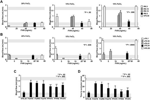
Previously, we showed that clotting in the FeCl3 carotid artery injury model followed a dose-response to infused hF8.17 In that study, we showed that phF8.38/F8null was effective at clotting after a standard 2 minutes 20% FeCl3 injury, but that other phBF8/F8null lines with lower pF8 levels did not develop occlusive clots.17 Therefore, we tested the degree of correction of the various transgenic pIR8 and pcBF8 lines on the F8null background in this system compared with phF8.38/F8null mice as positive control and F8null mice as negative control. Despite having lower pF8 levels than line phBF8, virtually all of the lines exhibited normal clotting after a standard 20% FeCl3 injury (Figure 2A-B left panels). We therefore used less chemosclerotic injuries, and showed that pIR8 and pcBF8 were more effective than phF8.38/F8null mice after these milder injuries (Figure 2A-B middle and right panels). Moreover, line IR8.18/F8null, which expresses both plasma and pIR8, was no more effective than IR8.16/F8null line that expresses pIR8 at an equivalent level but with no concurrent plasma F8, suggesting that there is no synergistic effect of having both platelet and low circulating plasma F8 in this model. These 2 findings are further supported in FeCl3 injury studies using F8null mice reconstituted with F8null marrow transduced with ubiquitin or PF4 promoter-driven phBF8, pIR8, or pcBF8 lentiviral constructs (Figure 2C-D). Here as well, pIR8 and pcBF8 improved clotting better than phBF8 though the overall efficacy by the lentiviral approach is less than in the transgenic approach as these reconstituted mice have lower pF8 levels (Figure 1), a 30% decrease in steady state platelet count (not shown) and likely have greater variability in pF8 level per platelet than the transgenic mice. Likewise, the ubiquitin promoter-driven constructs have concurrent low levels of plasma pIR8 or pcBF8 (Figure 1C-D), but were again no more effective in improving hemostasis in this injury model than PF4 promoter-driven constructs with no plasma F8.
FeCl3 carotid artery studies. Transgenic lines for pIR8 (A) and pcBF8 (B) were studied for clot development in the carotid artery injured with 20%, 15% and 10% FeCl3 for 2 minutes (left to right). Mean ± 1 SD of blood flow rate seen over 30 minutes are shown for the various transgenic lines (defined on the right) represented at their mean pF8 antigenic levels in Figure 1A and B. Gray horizontal bars indicate the mean ± SD for blood flow rate in WT (bottom bar) and F8null (top bar) mice. N-5 mice per arm. *P < .035 relative to phF8.38. Panels C and D show BMT/lentiviral F8null studies for average blood flow (C) and the average time to occlusion (D) over 30 minutes. Gray horizontal bars indicate the mean ± SD for time to occlusion in WT (bottom bar) and F8null (top bar) mice. N < 8 mice per arm. ** P < .02 and ***P < .002 for ubiquitin and PF4 promoter-driven vector mice relative to F8null mice.
FeCl3 carotid artery studies. Transgenic lines for pIR8 (A) and pcBF8 (B) were studied for clot development in the carotid artery injured with 20%, 15% and 10% FeCl3 for 2 minutes (left to right). Mean ± 1 SD of blood flow rate seen over 30 minutes are shown for the various transgenic lines (defined on the right) represented at their mean pF8 antigenic levels in Figure 1A and B. Gray horizontal bars indicate the mean ± SD for blood flow rate in WT (bottom bar) and F8null (top bar) mice. N-5 mice per arm. *P < .035 relative to phF8.38. Panels C and D show BMT/lentiviral F8null studies for average blood flow (C) and the average time to occlusion (D) over 30 minutes. Gray horizontal bars indicate the mean ± SD for time to occlusion in WT (bottom bar) and F8null (top bar) mice. N < 8 mice per arm. ** P < .02 and ***P < .002 for ubiquitin and PF4 promoter-driven vector mice relative to F8null mice.
Cremaster arteriole and venule model
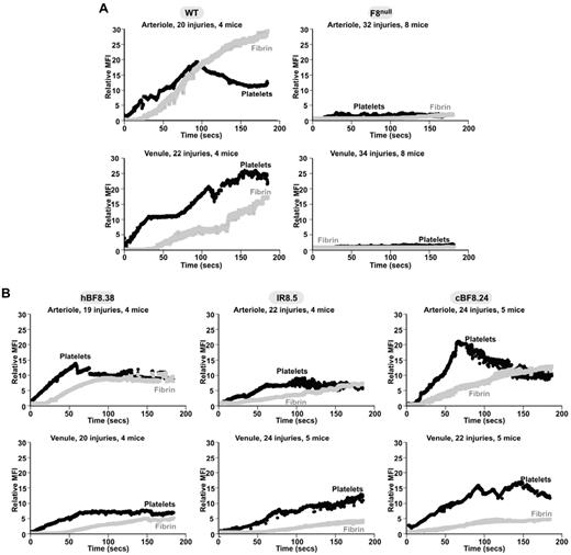
To study arteriole versus venule efficacy of pF8 variants hBF8, IR8, and cBF8, we used the in situ laser-induced cremaster arteriole/venule injury system previously described.23 Studies of arteriole and venule laser injury in WT and in F8null mice were consistent with published studies (Figure 3A).23,35,36 WT mice had an immediate accumulation of platelets in arterioles and venules, while fibrin took 20 and 40 seconds to accumulate in the arterioles and venules, respectively. F8null mice had little platelet and fibrin accumulation in both arterioles and venules.
In situ cremaster arteriole and venule studies. Average data for platelet (black) and fibrin (gray) accumulation. The number of injuries and mice studied are shown. (A) WT and F8null mice and (B) transgenic lines phF8.38, pIR8.5 and pcF8.24 on a F8null background were studied.
In situ cremaster arteriole and venule studies. Average data for platelet (black) and fibrin (gray) accumulation. The number of injuries and mice studied are shown. (A) WT and F8null mice and (B) transgenic lines phF8.38, pIR8.5 and pcF8.24 on a F8null background were studied.
Line phF8.38/F8null also showed a partial correction of platelet and fibrin accumulation, consistent with published studies.23 Initially, pIR8.5/F8null appeared not as effective at fibrin accumulation as phF8.38/F8null (Figure 3B); however, while hBF8 fibrin accumulation rapidly plateaued on the arteriole side, pIR8 fibrin curve continued to accumulate with time (supplemental Figure 2), consistent with IR8 being resistant to inactivation and showing prolonged activity in vitro. On the other hand, pcF8.24/F8null showed platelet accumulation and fibrin incorporation almost as well as WT mice (Figure 3A left panel, B right panel). Time of fibrin accumulation onset was better than the other transgenic mice (Figure 3B), especially in the venules. Thus, pcBF8 showed a further improvement in platelet and fibrin hemostasis in this bleeding model compared with the other variants despite having a 3-fold lower level of pF8.
Embolization studies
Previous studies showed that phF8.38/F8null was associated with an increased risk of clot embolization in the cremaster laser injury model. We now tested whether either pIR8 or pcBF8 had fewer emboli as an indication of greater clot stability. Studies of WT and untreated F8null mice gave similar arteriole and venule results to what had been previously reported23 (Figure 4). Our studies ofIR8 showed that pIR8.5/F8null mice had the same degree of embolization as phF8.38/F8null mice (Figure 4). In contrast, even though the pcF8.24/F8null mice had an approximately 3-fold lower pF8 levels than phF8.38/F8null mice, they had a marked decrease in both arteriole and venule embolization, especially for embolic size (Figure 4B) and total embolic mass (Figure 4C).
Embolization studies after cremaster arteriole and venule injuries. Embolization studies downstream of arteriole and venule injuries. All transgenics are on a F8null background. Open bars indicate arteriole studies and closed bars indicate venule studies. N = 18 videos per arm. Mean ± 1 SD shown. *P < .035 relative to WT. **P < .04 relative to phF8.38. (A) Number of detectable emboli. (B) Average relative embolic size determined by mean fluorescence. (C) Total embolic mass per study determined from the number and size of emboli.
Embolization studies after cremaster arteriole and venule injuries. Embolization studies downstream of arteriole and venule injuries. All transgenics are on a F8null background. Open bars indicate arteriole studies and closed bars indicate venule studies. N = 18 videos per arm. Mean ± 1 SD shown. *P < .035 relative to WT. **P < .04 relative to phF8.38. (A) Number of detectable emboli. (B) Average relative embolic size determined by mean fluorescence. (C) Total embolic mass per study determined from the number and size of emboli.
Inhibitor studies of pIR8
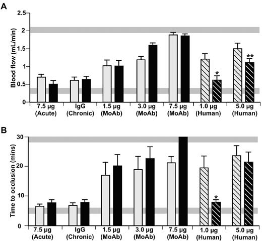
One of the greatest challenges in the care of hemophilia A is the care of patients with inhibitors. Because IR8 is not protected from inhibitors by binding to VWF,14 we examined whether pIR8 is as effective in F8null mice as phBF8 in the presence of inhibitors. We tested the pIR8.5/F8null mice in the FeCl3 system when challenged with a F8 antibody model compared with phF8.38/F8null transgenic mice. In these studies, the mice were repeatedly infused with F8 inhibitor mixture over a 9-day schedule to allow steady-state levels of the inhibitors to accumulate in the platelets.19 In the chronic mixed monoclonal inhibitor study, pIR8 was protected from the inhibitors, but only to the same extent as phBF8 (Figure 5, “MoAb”). Because ESH8 inactivation of IR8 may be complex,14,40 we also tested pIR8 and phBF8 after a similar infusion schedule using IgG purified from F8-deficient plasma with inhibitors and showed that pIR8 had greater resistance to this inhibitor than phBF8 (Figure 5, “Human”).
Inhibitor studies in pIR8.5/F8null mice. FeCl3 carotid artery injury studies in hF8.38/F8null mice (gray bars), and pIR8.5/F8null mice (black bars). In panels A and B on the left is an acute study where the animals received a one time infusion of the inhibitor mix, and in the middle are chronic studies done on day 10 of study after a murine IgG control or murine inhibitor mix infusions of F8 on days 0, 3, 6 and 9. On the right are chronic studies done using inhibitor IgG purified from F8-deficient plasma. In panels A and B, mean ± 1 SD are shown for N < 5 mice per arm. Results in panel A are expressed as average blood flow seen. Studies in panel B show the time to occlusion. *P < .025 and **P < .05 relative to phF8.38. Gray horizontal bars in panels A and B indicate the mean ± 1 SD for WT (bottom) and F8null (top) mice.
Inhibitor studies in pIR8.5/F8null mice. FeCl3 carotid artery injury studies in hF8.38/F8null mice (gray bars), and pIR8.5/F8null mice (black bars). In panels A and B on the left is an acute study where the animals received a one time infusion of the inhibitor mix, and in the middle are chronic studies done on day 10 of study after a murine IgG control or murine inhibitor mix infusions of F8 on days 0, 3, 6 and 9. On the right are chronic studies done using inhibitor IgG purified from F8-deficient plasma. In panels A and B, mean ± 1 SD are shown for N < 5 mice per arm. Results in panel A are expressed as average blood flow seen. Studies in panel B show the time to occlusion. *P < .025 and **P < .05 relative to phF8.38. Gray horizontal bars in panels A and B indicate the mean ± 1 SD for WT (bottom) and F8null (top) mice.
Discussion
Present day management of severe hemophilia A with early introduction of recombinant F8 prophylaxis has reduced the incidence of chronic complications.41 Among the remaining challenges in the care of hemophilia A is the development of inhibitors in a significant portion of these individuals.5 The recent recognition that early exposure to F8 increases one's risk of developing inhibitors has only further sharpened the need to find effective therapies for this complication beyond present-day induction of tolerance strategies.5,42,43 Shi et al first showed,20 and we confirmed,19 that pF8 is protected from circulating inhibitors. Our data suggest that pF8 is 100-fold more effective than plasma F8 in the presence of inhibitors in spite of the copresence of the inhibitors within the platelets.19
However, while ectopic expression of F8 within platelets may offer a potential advantage in the presence of inhibitors,39,44 this strategy has certain limitations. One clear disadvantage is that platelet activation needs to occur to release pF8. We have shown that the availability of the pF8 is dependent on platelet degranulation, and this process is delayed after injury, begins at the base of a growing clot, and then slowly spreads throughout the clot.19 We believe that this temporal and physical maldistribution underlies the increased rate of embolization we saw in F8null mice expressing phBF8 in the cremaster laser arteriole/venule injury model. We now tested whether this risk of embolization could be improved or eliminated using F8 variants IR8 and cBF8, each with increased specific activity.
Our data show that both variants showed promise: pIR8 levels and releasibility after activation were comparable to phBF8 by both transgenic and BMT/lentiviral approaches in spite of IR8's inability to bind VWF.14 It was significantly more effective than phBF8 in both the FeCl3 and cuticular clotting models in the F8null setting, reflecting IR8's resistance to spontaneous A2-domain dissociation and proteolytic inactivation. Surprisingly, in the cremaster laser injury system it was no better than phBF8. Consistent with this limited degree of improvement in this clotting model, pIR8/F8null mice had as much embolization as the phBF8/F8null mice. We believe that this difference in pIR8 efficacy in the various models tested may be explained by the fact that F8 normally may be sequentially activated by thrombin with R740 cleavage occurring before R372.45 The R740A mutation in IR8 may slow cleavage at R372 so that this mutation in IR8 may protect it from inactivation,14 but also may slow activation, as supported by the longer cremaster studies (supplemental Figure 2). We propose that the cuticular and FeCl3 injuries are both more diffuse injuries allowing wider zones of pIR8 release and also have endpoints only after a much longer time period than in the cremaster studies.
The pIR8 studies also provide additional insight into the biology of megakaryocyte-expressed F8, because it does not bind VWF and could potentially be poorly stored in α-granules and also more susceptible to inhibitors than phBF8. These pIR8 studies support our prior studies with VWF−/− mice that pF8 α-granule storage and release from activated platelets is mostly independent of concurrent VWF expression and storage.22 We also found that pIR8 retains its effectiveness in the presence of anti-F8 inhibitors, supporting that VWF binding is not essential for pF8 efficacy in the presence of circulating inhibitors. These findings are consistent with the evolving concept that there are separate platelet granule pools within platelets,46 and perhaps pF8 has limited intraplatelet contact with the inhibitors and may have different time or spatial release as well. These inhibition observations may also be important in large animal studies of pF8. The F8null canine model is the best-studied large animal model for hemophilia A.47 Prior studies suggest that these animals have little to no platelet VWF.48,49 We believe that our studies support the ability to use this large animal hemophilia A model to study pF8 biology, including inhibitor studies.
Canine BF8 was a potential candidate as a platelet-expressed F8 because it had both increased specific activity and higher expression levels than hBF8 when studied in baby hamster kidney cells.24 The bases for these species differences have yet to be determined. We found that pcBF8 expressed in mice demonstrated increased specific activity, but was expressed at a decreased level in megakaryocytes and platelets. Overall, there was a 1.7× increase in the pcBF8 activity relative to phBF8, roughly equal to the increase in the pIR8 transgenic mice (Figure 1E). Like pIR8, pcBF8 was significantly more effective than phBF8 in the cuticular and FeCl3 carotid artery clotting models; however, unlike pIR8, pcBF8 was also more effective in the cremaster laser injury model both on the arteriole and venules sides. This observation was supported by the embolization studies in which the cBF8 was the first platelet-delivered F8 studied that had almost normal embolization size and total mass in the F8null setting.
The molecular basis of why pcBF8 was effective in the cremaster model and pIR8 was not as effective is not apparent. Similarly, the basis for the lower level of pcBF8 relative to the levels seen with the phF8s is also unclear. This observation of low pcBF8 could be due to RNA message level, but the same cDNAs were used in the baby hamster kidney cell expression studies, where cBF8 was expressed at levels several fold higher than hBF8.24 More likely the underlying defect in pcF8 levels is related to the well-known difficulty for F8 to undergo intracellular processing, leading to intracellular accumulation and cellular apoptosis. Both the transgenic and lentivirus/BMT pcBF8 mice had normal steady-state platelet counts (data not shown). Further, time to platelet count recovery in the pcBF8 lentiviral/BMT mice was no different from the same studies using an empty lentiviral vector or one expressing an hF8 (data not shown). Because compensatory increases in thrombopoietin may mask deleterious effects of F8 on megakaryopoiesis in the mice, it is still possible that cF8 is more poorly processed during megakaryopoiesis and leads to the destruction of megakaryocytes expressing higher levels of pcBF8. In preliminary studies, we believe that this actually occurs: cBF8 and hBF8 mRNA levels are comparable, while megakaryocytes expressing cBF8 have an increase in apoptosis (data not shown). Whether the species differences in canine and human F8 that affect specific activity and intracellular processing are distinct are unknown, but future studies to pursue the basis of these properties may be worthwhile to further improve pF8 efficacy.
Finally, the possibility exists that because platelet- and plasma-delivered F8 may have different physical and temporal availability that low levels of plasma F8 may have a synergistic effect with pF8. This concept was tested in the cuticular and FeCl3 clotting models using transgenic mice with equal levels of platelet, but different levels of plasma F8 (eg, pIR8.16/F8null and pIR8.18/F8null) wherein the pIR8.18 mice have plasma as well as platelet F8. In both models, pIR8.18/F8null was at best slightly more effective than pIR8.16/F8null. These data are supportive that pF8 is effective in these models and that there was not a major synergistic benefit of concurrent low levels of plasma F8.
In summary, we have found that pIR8 is more effective than phBF8 in several clotting models in F8null mice, but that it did not improve clot instability in the cremaster arteriole/venule laser injury system, perhaps because of its slow rate of onset. On the other hand, pIR8 binding to VWF was not needed for IR8 protection from circulating inhibitors. We also showed that pcBF8 is even more effective in improving clotting in the murine F8null model even though the level of pcBF8 achieved in megakaryocytes was a third of phBF8. Despite of this lower level of expression, pcBF8 was the only F8 to correct clot instability in the cremaster injury model in F8null mice to date. Determination of the basis of the efficacy of pcBF8 in this setting, while avoiding its lower level of expression, may be useful in bringing platelet-based gene therapy for hemophilia A to clinical fruition.
The online version of this article contains a data supplement.
The publication costs of this article were defrayed in part by page charge payment. Therefore, and solely to indicate this fact, this article is hereby marked “advertisement” in accordance with 18 USC section 1734.
Acknowledgments
We thank Drs Rita Sarkar and Haig Kazazian at the University of Pennsylvania for sharing the cBF8 cDNA with us. We are also grateful to Dr Michele P. Lambert at the Children's Hospital of Philadelphia for helping with the initiation of the lentiviral studies.
This work was supported by the National Institute of Health training grant T32HL007439 (T.K.G.), grants R37DK042394, R01HL052173, and P01HL057346 (R.J.K.), and grant PO1HL64190 (V.R.A. and M.P.).
National Institutes of Health
Authorship
Contribution: T.K.G. carried out the lentiviral and transgenic studies as part of her thesis effort, provided initial data interpretation, and prepared the first draft of this manuscript; C.W. bred and characterized the transgenic mice of both F8 variants and did the FeCl3 studies and cremaster embolization studies; J.D.H. carried out initial BMT/lentiviral FeCl3 and cremaster studies; L.Z. prepared infectious lentiviral stocks; J.G. constructed the transgenic plasmids and characterized a few of the original transgenic lines; M.A.T. completed many of the initial lentiviral constructs; H.Z.M., S.W.P., and R.J.K. provided the IR8 construct, reviewed data, and suggested additional studies; R.M.C. and V.R.A. provided cF8 antigen and antibodies, reviewed the manuscript, and provided important insights; M.A.K. provided initial laboratory guidance, reviewed data, and contributed to the revision of the manuscript; and M.P. was thesis advisor, provided overall project organization and data interpretation, and helped with manuscript revisions.
Conflict-of-interest disclosure: The authors declare no competing financial interests.
Correspondence: Mortimer Poncz, Children's Hospital of Philadelphia, 3615 Civic Center Blvd, Abramson Research Center, Rm 317, Philadelphia, PA 19104; e-mail: poncz@email.chop.edu.
References
Author notes
T.K.G. and C.W. contributed equally to this work.


This feature is available to Subscribers Only
Sign In or Create an Account Close Modal