Abstract
Immunotherapy with innate immune cells has recently evoked broad interest as a novel treatment option for cancer patients. γ9δ2T cells in particular are emerging as an innate cell population with high frequency and strong antitumor reactivity, which makes them and their receptors promising candidates for immune interventions. However, clinical trials have so far reported only limited tumor control by adoptively transferred γ9δ2T cells. As a potential explanation for this lack of efficacy, we found unexpectedly high variability in tumor recognition within the physiologic human γ9δ2T-cell repertoire, which is substantially regulated by the CDR3 domains of individual γ9δ2TCRs. In the present study, we demonstrate that the reported molecular requirements of CDR3 domains to interact with target cells shape the physiologic γ9δ2T-cell repertoire and, most likely, limit the protective and therapeutic antitumor efficacy of γ9δ2T cells. Based on these findings, we propose combinatorial-γδTCR-chain exchange as an efficient method for designing high-affinity γ9δ2TCRs that mediate improved antitumor responses when expressed in αβT cells both in vitro and in vivo in a humanized mouse model.
Introduction
Immunotherapy with innate immune cells has become widely used because this approach obviates the need to match a cellular product to a defined HLA haplotype, allowing adoptive immunotherapies to be used in virtually any cancer patient without extensive in vitro selection or manipulation of the cellular product.1-4 γ9δ2T cells are promising as an innate cell population for this purpose because they are usually observed at high frequencies in the human peripheral blood and provide a strong antitumor reactivity against various solid and hematologic cancers.5 However, within γ9δ2T-cell populations, individual clones display great diversity in the repertoire because of the activating or inhibitory receptors expressed.6 Selecting innate cell products for certain cell types, such as those with a low level of inhibitory receptors, therefore seems plausible, especially considering the limited efficacy of adoptively transferred innate immune cells in clinical trials.7,8 An alternative proposal is to engineer cells to express defined activating innate receptors that mediate strong antitumor reactivity, such as a defined γ9δ2TCR,9 which could pave the way for readily available and more effective cellular products. However, the molecular details of how a γ9δ2TCR interacts with its target are not fully understood, making it challenging to select defined γ9δ2T cells or to engineer T cells with defined γ9δ2TCRs.
In “classic” immunoreceptors such as αβTCRs or Igs, the complementary determining regions (CDRs) determine affinity and specificity for a specific (peptide) epitope. V(D)J recombination allows the creation of a highly variable CDR repertoire ensuring recognition of an immense collection of antigens. γ9δ2T cells also possess a rearranged TCR that mediates recognition. The phosphoantigen isopentenyl pyrophosphate (IPP) has been suggested to be a key player in γ9δ2TCR-mediated activation,5,10,11 but no direct interaction between a γ9δ2TCR and IPP or any other phosphoantigen has ever been demonstrated. It was previously suggested that positively charged residues within the γ9δ2TCR are crucial for the response to negatively charged phosphoantigens12,13 and a potential IPP-binding groove has also been proposed.12 Interestingly, it appeared that responsiveness to phosphoantigens depends in particular on germline-encoded residues within all CDRs apart from δCDR3,14 extending the footprint of recognition to a much larger region than initially predicted.
Sequence alignment studies suggested that no defined δCDR3 motif is required for recognition beyond a hydrophobic residue at position δ109 (by Kabat numbering, δ9715 ), which suggests a less dominant role for δCDR3.14,16,17 Therefore, it is still unclear why variations in the γ9δ2CDR3 regions, which are particularly abundant in δCDR3, have evolved in humans and whether this variability is important in regulating the activation of a γ9δ2T cell. Understanding the reason for this variation would help to explain either the specificity or the regulation of functional avidity of a γ9δ2T cell, provide insight into the role of a γ9δ2TCR during the selection process of a γ9δ2T cell, and allow engineering of therapeutic cells with higher antitumor activity. In the present study, we therefore investigated the following research questions: (1) what is the clonal diversity in terms of tumor specificity and functional avidity within γ9δ2T cells once they express an identical set of activating and inhibitory receptors?, (2) what is the specific role of individual γ9δ2TCRs?, and (3) can we engineer a γ9δ2TCR with improved and broader antitumor reactivity?
Methods
Cells and cell lines
PBMCs were isolated from buffy coats obtained from Sanquin Blood Bank (Amsterdam, The Netherlands). Primary AML blasts were received after obtaining informed consent from the LML Biobank (University Medical Center Utrecht) and were collected according to good clinical practice and the Declaration of Helsinki. Cell lines are described in supplemental Methods (available on the Blood Web site; see the Supplemental Materials link at the top of the online article).
TCR mutagenesis, cloning, and sequencing
γ9δ2TCR modifications are based on codon-optimized genes of γ9- or δ2-TCR chain G115 flanked by NcoI and BamHI restriction sites (synthesized by GeneArt). To generate alanine mutations, site-directed mutagenesis was performed by overlap extension PCR18 or whole-plasmid mutagenesis19,20 using a proofreading polymerase (Phusion; Bioké). Mutated NcoI-BamHI–digested γ9- or δ2-TCR chains were ligated into the retroviral vector pBullet and sequenced by BaseClear (Leiden, The Netherlands).
Retroviral transduction of T cells
Flow cytometry
γ9δ2TCR expression was analyzed by flow cytometry using a Vδ2-FITC (clone B6; BD Biosciences) or a pan-γδTCR-PE Ab (clone IMMU510; Beckman Coulter). The fold change was calculated based on mean fluorescence intensity values of wild-type TCR (γ9-G115wt/δ2-G115wt)–transduced T cells set to 1 and mock-transduced T cells to 0. Abs used in the studies shown in the supplemental figures are described in supplemental Methods.
Functional T-cell assays
The 51Cr-release assay for cell-mediated cytotoxicity was described previously.22,23 In brief, target cells were labeled overnight with 100μCu 51Cr (150μCu for primary cells) and incubated for 5 hours with transduced T cells in 5 effector-to-target ratios (E:T) between 30:1 and 0.3:1. The fold change compared with reactivity of engineered T cells expressing unmutated γ9δ2TCR was calculated.24 IFNγ ELISpot was performed using antihuman IFNγ mAb1-D1K (I) and mAb7-B6-1 (II; Mabtech) following the manufacturer's recommended procedure.25 Target and effector cells (E:T 3:1) were incubated for 24 hours in the presence of pamidronate (Calbiochem) where indicated.25,26 IFNγ ELISA was performed using the ELISA-Ready-Go! Kit (eBiosciences) following the manufacturer's instructions. Effector and target cells (E:T 1:1) were incubated for 24 hours in the presence of pamidronate as indicated. Where specified, the fold change was calculated compared with reactivity of engineered T cells expressing unmutated γ9δ2TCR.
Animal models
To induce tumor xenografts, sublethal total body irradiated (2 Gy), 11- to 17-week-old RAG-2−/−/γc−/− BALB/C mice (see supplemental Methods) were injected intravenously with 0.5 × 10⁶ Daudi-Luc cells (a kind gift from Genmab)9,27 or 5 × 10⁶ RPMI8226/S-Luc cells (Anton Martens, Utrecht, The Netherlands) together with 107 γ9δ2TCR+-transduced T cells. Mice received 0.6 × 10⁶ IU of IL2 (Proleukin; Novartis) in IFA subcutaneously on day 1 and every 21 days until the end of the experiment. Pamidronate (10 mg/kg body weight) was applied at day 1 intravenously and every 21 days intraperitoneally. Outgrowing tumors were visualized in vivo by Biospace bioluminescence imaging. Mice were anesthetized by isoflurane before receiving an IP injection (100 μL) of 25 mg/mL of beetle luciferin (Promega). Bioluminescence images were acquired and analyzed with M3Vision Version 2.1 software (Photon Imager; Biospace Laboratory).
Results
Antitumor reactivity of individual γ9δ2T-cell clones
To investigate whether individual γ9δ2T-cell clones mediate differential activity against tumor cells compared with the parental γ9δ2T-cell population, γ9δ2T cells from a healthy donor were cloned by limiting dilution and tested against a broad panel of tumor cells in an IFNγ ELISpot assay (Table 1). High variability in tumor recognition in terms of specificity and functional avidity was observed between individual γ9δ2T-cell clones (cl); compared with the original bulk population, cl5 and cl13 produced twice as many IFNγ spots in response to Daudi and selectively generated significant amounts IFNγ when challenged with K562, BT549, and MCF-7. In contrast, cl3 and cl15 recognized solely Daudi cells. A variable expression of natural killer (NK) receptor and killer cell Ig-like receptors (KIRs) has been reported in innate immune cells28-30 and might have contributed to the observed differential activity of selected clones. Therefore, surface expression of γ9δ2TCR, NKG2D, CD158a, NKAT-2, and NKB-1 was examined (supplemental Figure 1). However, no correlation was found between receptor expression patterns and antitumor reactivity of the tested γ9δ2T-cell clones. We hypothesized that the diversity within the γ9δ2TCR contributes to the differential activity of the examined γ9δ2T-cell clones. Therefore, γ9δ2TCRs of the highly tumor-reactive cl5 and the weakly tumor-reactive cl3 were chosen for detailed analysis and compared with γ9δ2TCR G115.9,12
Antitumor reactivity of individual γ9δ2T-cell clones
| . | αβ T cells . | γ9δ2 bulk . | γ9δ2 cl3 . | γ9δ2 cl4 . | γ9δ2 cl5 . | γ9δ2 cl7 . | γ9δ2 cl8 . | γ9δ2 cl13 . | γ9δ2 cl15 . |
|---|---|---|---|---|---|---|---|---|---|
| PBMCs | 3* | 2* | 10* | 2* | 7* | 0* | 3* | 12* | 13* |
| K562 | 0* | 14* | 9* | 9* | 62† | 181‡ | 7* | 56† | 12* |
| Daudi | 0* | 206‡ | 211‡ | 55† | 458‡ | 318‡ | 268‡ | 500‡ | 244‡ |
| MZ1851RC | 0* | 205‡ | 4* | 94† | 114‡ | 216‡ | 77† | 93† | 19* |
| BT549 | 0* | 2* | 1* | 0* | 44† | 1* | 2* | 41† | 8* |
| MCF-7 | 0* | 2* | 6* | 1* | 64† | 1* | 1* | 58† | 10* |
| SW480 | 0* | 0* | 11* | 2* | 3* | 1* | 1* | 4* | 11* |
| MDA MB 231 | 0* | 4* | 2* | 1* | 12* | 1* | 1* | 14* | 10* |
INFγ spots/15 000 cells < 40.
IFNγ spots/15 000 cells 40-100.
IFNγ spots/15 000 cells >100.
Antitumor reactivity mediated by individual γ9δ2TCRs
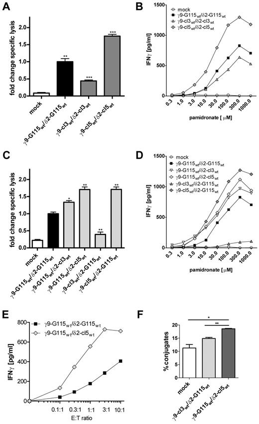
To elucidate differences among γ9δ2TCRs of tumor-reactive clones, sequences of γ9-cl3wt/δ2-cl3wt and γ9-cl5wt /δ2-cl5wt were determined and aligned with γ9δ2TCR G115. All 3 γ9δ2TCRs only differed in their CDR3 domains: 1-3 amino acids between position γ109 and γ111 in γCDR3 and 4-8 amino acids between δ108 and δ112 in δCDR3 (supplemental Table 1, numbering according to the international ImMunoGeneTics information system [IMGT]15 ). To determine whether distinct γ9δ2TCRs mediate differential antitumor reactivity, individual γ9δ2TCR chains were cloned into the retroviral vector pBullet and linked to a selection marker as described previously.31 The wild-type-combinations γ9-cl3wt/δ2-cl3wt, γ9-cl5wt/δ2-cl5wt, and γ9-G115wt/δ2-G115wt were transduced into peripheral blood αβT cells, selected by antibiotics, and further expanded. γ9δ2TCR G115 (γ9-G115wt/δ2-G115wt)9,12 served as a control, as did cells transduced with an empty vector cassette (mock). γ9δ2TCR-transduced T cells showed similar γ9δ2TCR expression (data not shown) and were tested for their lytic activity against the tumor target Daudi in a 51Cr-release assay (Figure 1A). T cells expressing γ9-cl3wt/δ2-cl3wt had a 50% reduced ability to lyse tumor cells (P < .01), whereas T cells with γ9-cl5wt/δ2-cl5wt were nearly twice as potent (P < .01) as the control γ9-G115wt/δ2-G115wt. To determine whether the phenotypes of γ9δ2TCR-transduced cells with decreased or increased functional avidity are also present at the cytokine level, a pamidronate-titration assay was performed. Pamidronate treatment of Daudi cells blocks the mevalonate-pathway downstream to IPP, causing the accumulation of IPP and an enhanced cytokine secretion of responsive T cells. To exclude NK-like activation, CD4+ γ9δ2TCR-transduced T cells, which lack the expression of major NK receptors such as NKG2D, were selected by MACS sorting. Transductants were tested at different concentrations of pamidronate against the tumor target Daudi. Mock-transduced T cells that underwent equivalent stimulation but express an irrelevant αβTCR served as control. IFNγ secretion was measured by ELISA and the half-maximal effective concentration (EC50) was calculated (Figure 1B). Consistent with the changes observed for lytic capacity, T cells transduced with γ9-cl3wt/δ2-cl3wt secreted lower amounts of IFNγ (maximum 600 pg/mL), whereas T cells expressing γ9-cl5wt/δ2-cl5wt produced higher levels of IFNγ (maximum 1300 pg/mL) at all pamidronate concentrations relative to control γ9-G115wt/δ2-G115wt (maximum 800 pg/mL). Despite different plateaus in IFNγ secretion, all selected mutants and the wild-type control had a comparable pamidronate-EC50 (approximately 30 pg/mL). These results indicate that distinct γ9δ2TCR clones mediate different functional avidity, and that the high variability among parental γ9δ2T-cell clones in tumor recognition seems to be substantially regulated by the CDR3 domains of individual γ9δ2TCRs. Therefore, the correlation between CDR3 domains and functional avidity was investigated.
Antitumor reactivity mediated by γ9δ2TCRs. Peripheral blood T cells were virally transduced with indicated wild-type γ9δ2TCRs or CTE-engineered γ9δ2TCRs and tested against Daudi (A,C) in a 51Cr-release assay (E:T, 3:1). Specific lysis is indicated as the fold change 51Cr-release measured in the supernatant after 5 hours compared with reactivity of γ9-G115wt/δ2-G115wt-engineered T cells (B,D) in an IFNγ ELISA in the presence of indicated amounts of pamidronate or (E) different E:T ratios. (F) Percentages of cell-cell conjugates of Daudi and T cells engineered with indicated γ9δ2TCR were determined by flow cytometry. Data represent the means ± SD. *P < .05; **P < .01; and ***P < .001 by 1-way ANOVA.
Antitumor reactivity mediated by γ9δ2TCRs. Peripheral blood T cells were virally transduced with indicated wild-type γ9δ2TCRs or CTE-engineered γ9δ2TCRs and tested against Daudi (A,C) in a 51Cr-release assay (E:T, 3:1). Specific lysis is indicated as the fold change 51Cr-release measured in the supernatant after 5 hours compared with reactivity of γ9-G115wt/δ2-G115wt-engineered T cells (B,D) in an IFNγ ELISA in the presence of indicated amounts of pamidronate or (E) different E:T ratios. (F) Percentages of cell-cell conjugates of Daudi and T cells engineered with indicated γ9δ2TCR were determined by flow cytometry. Data represent the means ± SD. *P < .05; **P < .01; and ***P < .001 by 1-way ANOVA.
CTE as a rapid method to modulate functional avidity of engineered T cells
To make the above determination, we devised a strategy called combinatorial-γδTCR-chain exchange (CTE), which results in the expression of newly combined γ9- and δ2-TCR chains on engineered T cells. During this process, γ9-G115wt was combined with δ2-cl3wt or δ2-cl5wt and δ2-G115wt with γ9-cl3wt or γ9-cl5wt. These combinations were retrovirally transduced into αβT cells. In all transductants, equivalent γδTCR expression was detected, whereas the endogenous αβTCR was clearly down-regulated (supplemental Figure 2A). This resulted not only into a nearly abolished alloreactivity of αβT cells expressing γ9-G115wt/δ2-G115wt, as reported previously,9 but also of selected CTE-engineered αβT cells compared with mock-transduced cells (supplemental Figure 2B). Therefore, reactivity of CTE-engineered T cells primarily depends on expressed γδTCRs and not on residual endogenous αβTCRs. Transductants were functionally tested against the tumor target Daudi in a 51Cr-release assay (Figure 1C). The exchange of γ9- or δ2-chains indeed caused notable differences. Compared with the original TCR γ9-G115wt/δ2-G115wt, the combination of γ9-G115wt/δ2-cl3wt, γ9-G115wt/δ2-cl5wt, or γ9-cl5wt/δ2-G115wt mediated 40%-70% increased specific lysis of tumor cells (all P < .05). The same magnitude of recognition was observed when IFNγ production of CD4+ γδTCR-transduced T cells was tested in a pamidronate titration assay (Figure 1D). Only the combination γ9-cl3wt/δ2-G115wt led to decreased IFNγ production of transduced cells at all pamidronate concentrations (maximum 100 pg/mL), whereas all other CTE-γ9δ2TCRs mediated an increased IFNγ-secretion (maximum ≥ 1000 pg/mL) compared with control TCR γ9-G115wt/δ2-G115wt (maximum 800 pg/mL). Equal pamidronate EC50s of approximately 30 pg/mL were calculated for all responsive γ9δ2TCR-transduced cells.
To determine whether cell-cell interaction influences the response kinetics differently than pamidronate stimulation, CTE-γ9δ2TCR γ9-G115wt/δ2-cl5wt, which mediates improved functional avidity, and control TCR γ9-G115wt/δ2-G115wt were tested in an E:T titration assay (Figure 1E), and an E:T50 was calculated. Interestingly, T cells with γ9-G115wt/δ2-cl5wt responded differently, with an E:T50 of 0.3:1, compared with an E:T50 of 1:1 calculated for control cells expressing γ9-G115wt/δ2-G115wt. To determine whether the interaction between different TCRs and ligands—and thus the affinity—is indeed increased, cell-cell conjugates between Daudi and T cells expressing either potentially high-affinity (γ9-G115wt/δ2-cl5wt) or low-affinity (γ9-cl3wt/δ2-G115wt) TCRs were measured by flow cytometry. Significantly more cell-cell interactions were observed when γ9-G115wt/δ2-cl5wt was expressed compared with γ9-cl3wt/δ2-G115wt and mock-transduced T cells (Figure 1F). This effect did not depend on the presence of pamidronate (data not shown) and G115wt/δ2-cl5wt is thus a high-affinity γ9δ2TCR. Therefore, CTE appears to be an efficient method to rapidly engineer γ9δ2TCRs with increased affinity, mediating improved functional avidity in transduced T cells.
Residues in δCDR3 and Jδ1 are involved in γ9δ2TCR stability and in mediating functional avidity of engineered αβT cells
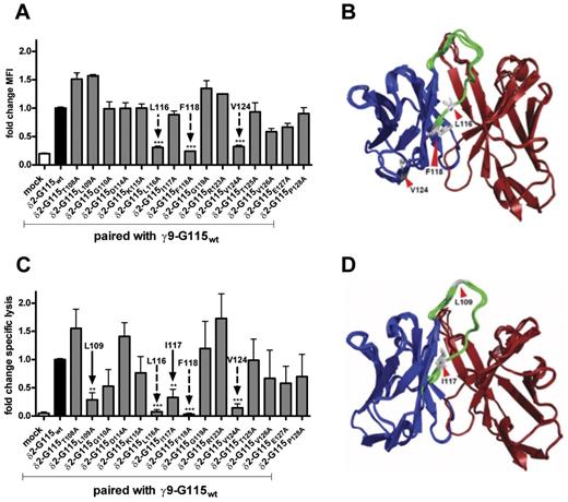
To elucidate the molecular requirements of δCDR3 to mediate optimal functional avidity, alanine mutagenesis of a model δCDR3 (clone G115) was performed; the entire Jδ1 segment was included because important residues have also been reported within Jγ1.17 During an initial screening, 5 sequence areas were found to either influence TCR expression or functional avidity of γ9δ2TCR-transduced T cells (data not shown). To clarify the degree to which single residues are responsible for impaired γ9δ2TCR expression and lower TCR-mediated functional avidity, single alanine mutations were generated. The mutated and wild-type δ2-G115 chains were expressed in combination with γ9-G115wt in αβT cells and tested for γ9δ2TCR expression using a δ2-chain–specific Ab (Figure 2A). Three single alanine mutations caused a 70% lower TCR expression compared with the unmutated δ2-G115wt, namely δ2-G115L116A, δ2-G115F118A, and δ2-G115V124A (supplemental Table 2). Comparable results were observed using Abs directed against the γ9-chain or the constant domain of the γδTCR (data not shown), indicating the importance of δ2-G115L116, δ2-G115F118, and δ2-G115V124 for stable TCR expression. The crystal structure of γ9δ2TCR G115 supports our findings: δ2-G115L116, δ2-G115F118, and δ2-G115V124 are located in hydrophobic cores (Figure 2B) and could thus be crucial for the structural stability of the γ9δ2TCR G115.
γ9δ2TCR expression and functional avidity of transduced T cells expressing single alanine mutated δ2 chain of clone G115. (A) Peripheral blood T cells were virally transduced with indicated γ9 and δ2 TCR chains and analyzed by flow cytometry using a δ2-chain specific Ab. Shown is the fold change in mean fluorescent intensity (MFI) with wild-type controls expressing δ2-G115wt. (C) Lytic activity of transductants was tested in a 51Cr-release assay against the tumor target Daudi (E:T, 10:1). Specific lysis is indicated as the fold change in 51Cr-release measured in the supernatant after 5 hours compared with reactivity of unmutated wild-type (δ2-G115wt). Arrows indicate mutations in δ2-G115 that impaired receptor expression (dashed arrows) or functional avidity (solid arrows). (B,D) Crystal structure of γ9δ2TCR G115 indicating relevant amino acids (red arrows), δ-chain (in blue), δCDR3 (in green), and γ-chain (in brown).
γ9δ2TCR expression and functional avidity of transduced T cells expressing single alanine mutated δ2 chain of clone G115. (A) Peripheral blood T cells were virally transduced with indicated γ9 and δ2 TCR chains and analyzed by flow cytometry using a δ2-chain specific Ab. Shown is the fold change in mean fluorescent intensity (MFI) with wild-type controls expressing δ2-G115wt. (C) Lytic activity of transductants was tested in a 51Cr-release assay against the tumor target Daudi (E:T, 10:1). Specific lysis is indicated as the fold change in 51Cr-release measured in the supernatant after 5 hours compared with reactivity of unmutated wild-type (δ2-G115wt). Arrows indicate mutations in δ2-G115 that impaired receptor expression (dashed arrows) or functional avidity (solid arrows). (B,D) Crystal structure of γ9δ2TCR G115 indicating relevant amino acids (red arrows), δ-chain (in blue), δCDR3 (in green), and γ-chain (in brown).
To address the impact of single alanine mutations on functional avidity, a 51Cr-release assay was performed (Figure 2C). As expected, transductants with low TCR expression (δ2-G115L116A, δ2-G115F118A, and δ2-G115V124A) could not lyse tumor cells effectively, because they demonstrated an 80% lower lytic capacity compared with cells transduced with δ2-G115wt. However, T cells with mutation δ2-G115L109A and δ2-G115I117A (supplemental Table 2) properly expressed the TCR but showed a 70% reduced lytic activity compared with δ2-G115wt–expressing cells. Similar results were obtained when TCR mutants were transduced into CD4+ Jurkat cells and IL-2 production was measured (data not shown). Reduction of lytic activity was also seen when alanine substitutions δ2-G115L109A and δ2-G115I117A were introduced into the δ2-chain of γδTCR clone 3 (data not shown). These results indicate that not only residue δL109,14,16,17 but also δI117 in δCDR3, is generally important for γ9δ2TCRs to mediate functional avidity (Figure 2D). However, sequence alignments between δ2-chains of clones 3, 5, and G115 indicated that δL109 and δI117 are conserved (supplemental Table 1), making it unlikely that these residues mediate different functional avidities of the γ9δ2TCR-transduced cells studied herein.
Influence of CDR3 length on functional avidity of γ9δ2TCR-transduced T cells
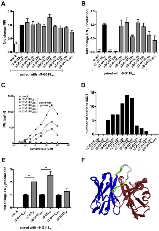
Surprisingly, alanine substitutions during alanine-scanning mutagenesis of γ9δ2TCR G115 could replace large parts of the δCDR3 domain without functional consequences. This raises the possibility that the crucial factor for the differing functional avidities of distinct γ9δ2TCR combinations is not a defined amino acid, but the relative length between the functionally important residues δ2-G115L109 and the structurally important residue δ2-G115L116. Therefore, different δ2-G115 length mutants were generated. Because the triple δ2-G115T113-K115 is also important for stable surface expression (data not shown), 9 length mutants (δ2-G115LM) with 0-12 alanines between δ2-G115L109 and δ2-G115T113 were generated and equally expressed in αβT cells, again in combination with γ9-G115wt (Figure 3A). To determine the functional avidity of δ2-G115LM–transduced T cells, CD4+ TCR-transduced T cells were selected by MACS sorting, and an IFNγ ELISA in response to Daudi was performed in the presence of pamidronate (Figure 3B). Interestingly, engineered T cells expressing δ2-G115LM0 and δ2-G115LM1 were unable to produce IFNγ, and T cells expressing δ-G115LM4 or δ-G115LM12 secreted only approximately half the amount of IFNγ compared with δ2-G115wt–transduced cells. All other mutants (δ2-G115LM2, 3, 5, 6, 9) induced comparable amounts of IFNγ in engineered T cells relative to transductants expressing δ2-G115wt. Mutants with functional impairment (δ2-G115LM0,1,4,12, supplemental Table 2) were further tested against increasing pamidronate concentrations and an EC50 was calculated. Despite different plateaus in maximal IFNγ secretion, all selected δ2-G115LM transduced cells and the wild-type control had a comparable pamidronate-EC50 (approximately 30 pg/mL; Figure 3C). Length mutations were also studied in γCDR3 of γ9δ2TCR G115 by engineering stretches of 1-6 alanines between γ9-G115E108 and γ9-G115E111.1 (γ9-G115LM1-6). However, this did not affect functional avidity (supplemental Figure 3A).
γ9δ2TCR expression and functional avidity of transduced T cells expressing γ9δ2TCR G115 with δ2-CDR3 length mutations. (A) γ9δ2TCR expression of indicated transductants was analyzed by flow cytometry using a γδTCR-pan Ab. Shown is the fold change in mean fluorescent intensity (MFI) compared with wild-type controls expressing δ2-G115wt. (B) IFNγ secretion of δ2-G115LM–transduced T cells against the tumor target Daudi (E:T, 1:1) was measured by ELISA after 24 hours of incubation in the presence of 100μM pamidronate. Shown is the fold change in IFNγ production compared with reactivity of transductants expressing δ2-G115wt. (C) Transductants expressing δ2-G115LM0,1,4,12 were tested in a titration assay against the tumor target Daudi with increasing amounts of pamidronate as indicated. IFNγ production was measured after 24 hours by ELISA. (D) Generated δ2-G115LMs were matched in a BLAST search with γ9δ2TCRs described in the IMGT database. Shown is the number of citations compared with δ2-G115LM of similar δCDR3 length. (E) Transductants with δ2-G115LM2,4,6 were compared side-by-side with transductants expressing individual γ9δ2TCRs of the same δCDR3 length. IFNγ secretion of transduced T cells against the tumor target Daudi (E:T, 1:1) was measured by ELISA after 24 hours in the presence of 100μM pamidronate. Shown is the fold change in IFNγ production compared with reactivity of transductants expressing δ2-G115wt. Data represent the means ± SD. **P < .01; and **P < .001 by 1-way ANOVA. (F) Crystal structure of γ9δ2TCR G115; the region that was used for alanine stretches within δCDR3 is shown in white, the residual δCDR3 in green, the δ chain in blue, and the γ chain in brown.
γ9δ2TCR expression and functional avidity of transduced T cells expressing γ9δ2TCR G115 with δ2-CDR3 length mutations. (A) γ9δ2TCR expression of indicated transductants was analyzed by flow cytometry using a γδTCR-pan Ab. Shown is the fold change in mean fluorescent intensity (MFI) compared with wild-type controls expressing δ2-G115wt. (B) IFNγ secretion of δ2-G115LM–transduced T cells against the tumor target Daudi (E:T, 1:1) was measured by ELISA after 24 hours of incubation in the presence of 100μM pamidronate. Shown is the fold change in IFNγ production compared with reactivity of transductants expressing δ2-G115wt. (C) Transductants expressing δ2-G115LM0,1,4,12 were tested in a titration assay against the tumor target Daudi with increasing amounts of pamidronate as indicated. IFNγ production was measured after 24 hours by ELISA. (D) Generated δ2-G115LMs were matched in a BLAST search with γ9δ2TCRs described in the IMGT database. Shown is the number of citations compared with δ2-G115LM of similar δCDR3 length. (E) Transductants with δ2-G115LM2,4,6 were compared side-by-side with transductants expressing individual γ9δ2TCRs of the same δCDR3 length. IFNγ secretion of transduced T cells against the tumor target Daudi (E:T, 1:1) was measured by ELISA after 24 hours in the presence of 100μM pamidronate. Shown is the fold change in IFNγ production compared with reactivity of transductants expressing δ2-G115wt. Data represent the means ± SD. **P < .01; and **P < .001 by 1-way ANOVA. (F) Crystal structure of γ9δ2TCR G115; the region that was used for alanine stretches within δCDR3 is shown in white, the residual δCDR3 in green, the δ chain in blue, and the γ chain in brown.
These results indicate that considerable alanine stretches within γ9 and δ2CDR3 domains can be tolerated, likely because CDR3 regions are relatively exposed parts of the TCR (Figure 3F). However, too short and very long alanine stretches between δ2-G115L109 and δ2-G115T113 in particular, as well as stretches with 4 alanines, are associated with reduced or absent function of a γ9δ2TCR (Figure 3B-C). Loss of binding in mutants with short alanine stretches is most likely because the middle segment of δCDR3 is crucial for binding to the ligand. That suggests the existence of an optimal δCDR3 length for γ9δ2TCRs. Therefore, the CDR3 length within the γ9δ2TCR repertoire was studied.
Consequences for the physiologic γ9δ2T-cell repertoire
The IMGT database15 was searched for reported stretches between γ9-G115E109 and γ9-G115E111.1, as well as δ2-G115L109 and δ2-G115T113. A preferential length for reported γ9-chains was found for CDR3 regions corresponding to γ9-G115LM2 and γ9-G115LM3, but shorter stretches were also reported (supplemental Figure 3B). In contrast, δ2-chains with short δCDR3 domains, such as δ2-G115LM1 or δ2-G115LM0, were not reported (Figure 3D), consistent with our observation that such chains are not functional. The majority of listed γ9δ2TCRs contain δCDR3 lengths which correspond to δ2-G115LM5,6,7. These findings support the hypothesis that positive selection favors γ9δ2TCRs with an optimal δCDR3 length of 5-7 residues between δ2-G115L109 and δ2-G115T113. However, the individual sequence might still play a role in γ9δ2TCR-mediated functional avidity.
Influence of the CDR3 sequence on γ9δ2TCR-mediated functional avidity
To test the hypothesis that both the length and sequence of δCDR3 can be important to mediate optimal functional avidity, γ9δ2TCR length mutants δ2-G115LM2, δ2-G115LM4, and δ2-G115LM6 were transduced into αβT cells in combination with γ9-G115wt. IFNγ secretion of transductants in response to Daudi was compared with cells transduced with wild-type sequences from δ2-cl3wt (corresponds in length to δ2-G115LM2), δ2-cl5wt (corresponds in length to δ2-G115LM4), and δ2-G115wt (corresponds in length to δ2-G115LM6; supplemental Table 3). Although T cells transduced with δ2-G115LM6 and δ2-G115wt did not differ in the amount of cytokine secretion, all other combinations of wild-type chains showed a more than 2-fold increase in IFNγ compared with the length mutant that selectively contained alanines (Figure 3E). These results were confirmed when the lytic capacity of transduced cells was tested (data not shown). The sequence in δCDR3 is therefore also a significant factor for the optimal functioning of a γ9δ2TCR. We also investigated the sequential importance of γCDR3.
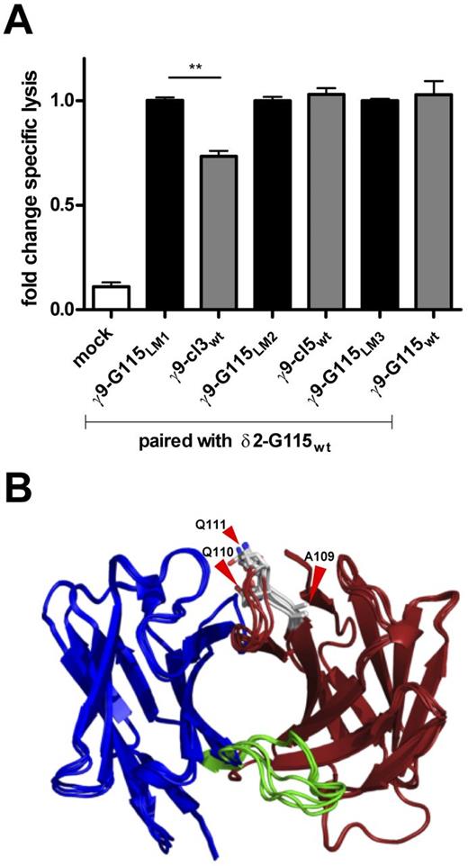
γ9-G115LM1-3 were transduced into T cells in combination with δ2-G115wt. IFNγ secretion of transductants in response to Daudi was compared with cells transduced with γ9-cl3wt (corresponds in length to γ9-G115LM1), γ9-cl5wt (corresponds in length to γ9-G115LM2), and γ9-G115wt (corresponds in length to γ9-G115LM3; supplemental Table 3). T cells expressing γ9-cl3wt/δ2-G115wt selectively produced lower amounts of IFNγ compared with their equivalent γ9-G115LM1 (Figure 4A). Previously, the same γ9δ2TCR combination was also found to mediate reduced functional avidity (Figure 1C-D). Interestingly, loss of activity could be restored to normal levels (referred to γ9δ2TCR G115wt) by mutating γCDR3E109 in γ9-cl3wt to γCDR3A109, which demonstrates that a single change in the variable sequence of γ9CDR3 is sufficient to regulate the functional avidity of the γ9δ2TCR-transduced T cells investigated herein.
Functional avidity of transduced T cells expressing γ9δ2TCR G115 with γ9-CDR3 length mutations. (A) Peripheral blood T cells were virally transduced with indicated γ9 and δ2 TCR chains. Lytic activity of transductants was compared side-by-side with T cells expressing individual γ9δ2TCRs of the same γ9CDR3 length. Specific lysis is indicated as the fold change 51Cr-release measured in the supernatant after 5 hours. Data represent the means ± SD. **P < .01 by 1-way ANOVA. (B) Crystal structure of γ9δ2TCR G115 indicating γ9CDR3 in gray including amino acids γ9-G115A109, γ9-G115Q110, andγ9-G115Q111 (red arrows). δCDR3 is shown in green; the δ chain in blue; and the γ chain in brown.
Functional avidity of transduced T cells expressing γ9δ2TCR G115 with γ9-CDR3 length mutations. (A) Peripheral blood T cells were virally transduced with indicated γ9 and δ2 TCR chains. Lytic activity of transductants was compared side-by-side with T cells expressing individual γ9δ2TCRs of the same γ9CDR3 length. Specific lysis is indicated as the fold change 51Cr-release measured in the supernatant after 5 hours. Data represent the means ± SD. **P < .01 by 1-way ANOVA. (B) Crystal structure of γ9δ2TCR G115 indicating γ9CDR3 in gray including amino acids γ9-G115A109, γ9-G115Q110, andγ9-G115Q111 (red arrows). δCDR3 is shown in green; the δ chain in blue; and the γ chain in brown.
In summary, the length and sequence of the δ2CDR3 domain between L109 and T113 (supplemental Table 1) play a crucial role in γ9δ2TCR-mediated functional avidity. In addition, the individual sequence between E108 and E111.1in γ9CDR3 can hamper the activity of a γ9δ2TCR, and in G115 γCDR3A109 is most likely crucial for ligand interaction (Figure 4B and supplemental Table 1). This provides not only the rationale for CTE-engineered γ9δ2TCRs, but also for random mutagenesis within both the γ9 and δ2CDR3 regions.
CTE-engineered T cells as a tool for cancer immunotherapy
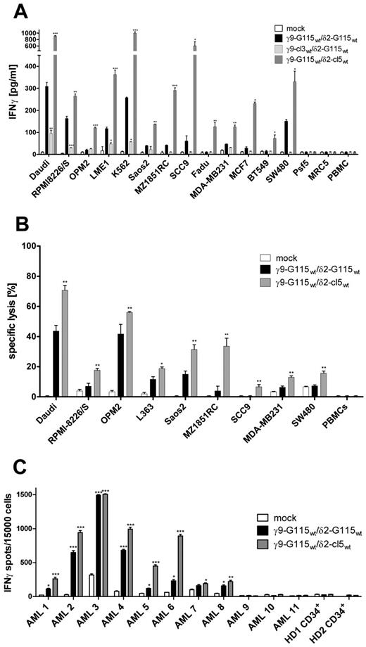
CTE-engineered γ9δ2TCRs with increased activity against tumor cells are interesting candidates for TCR-gene therapeutic strategies. This leads to the question of whether changes in functional avidity mediated by CTE-γ9δ2TCRs constitute a unique phenomenon of a defined γ9δ2TCR pair in response to the B-lymphoblastic cell line Daudi or if this is a general response to most tumor targets. Therefore, CTE-γ9δ2TCRs that mediated increased (γ9-G115wt/δ2-cl5wt) or reduced (γ9-cl3wt/δ2-G115wt) activity were tested against various tumors in an IFNγ ELISA in the presence of pharmacologic concentrations of pamidronate (10μM; Figure 5A).9 Tumor reactivity was significantly increased against a whole range of different tumor entities, including other hematologic cancers such as RPMI8226/S, OPM2, LME1 (all multiple myeloma), and K562 (myelogenous leukemia) and solid cancer cell lines such as Saos2 (osteosarcoma), MZ1851RC (renal cell carcinoma), SCC9, Fadu (head and neck cancer), MDA-MB231, MCF7, BT549 (all breast cancer), and SW480 (colon carcinoma) when taking advantage of γ9-G115wt/δ2-cl5wt, compared with γ9-G115wt/δ2-G115wt and was significantly reduced or even absent for all other targets using γ9-cl3wt/δ2-G115wt. Moreover, CTE-engineered T cells with increased activity against tumor cells still did not show any reactivity toward healthy tissue such as PBMCs and fibroblasts. Superior lytic activity of T cells engineered with γ9-G115wt/δ2-cl5wt was also observed for hematologic cancer cells such as RPMI8226/S, OPM2, and L363 and the solid cancer cell lines Saos2, MZ1851RC, SCC9, MDA-MB231, and SW480 compared with control T cells expressing γ9-G115wt/δ2-G115wt (Figure 5B). Therefore, CTE-engineered γ9δ2TCRs can provide higher tumor reactivity against a broad panel of tumor cells while not affecting normal tissue and thus have the potential to increase efficacy of TCR-engineered T cells.
Antitumor reactivity of T cells transduced with CTE-engineered γ9δ2TCRs in vitro. Peripheral blood T cells were virally transduced with indicated γ9δ2TCRs and tested against indicated tumor cell lines and healthy control tissue. (A) Transductants were incubated with target cells (E:T, 1:1) in the presence of 10μM pamidronate. IFNγ production was measured after 24 hours by ELISA. Data represent the means ± SD. *P < .05; **P < .01; and ***P < .001 by 1-way ANOVA. (B) Transductants were incubated with indicated tumor targets loaded with 51Cr (E:T, 10:1). Percentage of specific lysis was determined by 51Cr-release measured in the supernatant after 5 hours. (C) CTE-engineered T cells were tested against primary AML blasts and healthy progenitor cells in an IFNγ ELISpot assay (E:T, 3:1) in the presence of 10μM pamidronate. Data represent the means ± SD. *P < .05; **P < .01; and ***P < .001 by 1-way ANOVA.
Antitumor reactivity of T cells transduced with CTE-engineered γ9δ2TCRs in vitro. Peripheral blood T cells were virally transduced with indicated γ9δ2TCRs and tested against indicated tumor cell lines and healthy control tissue. (A) Transductants were incubated with target cells (E:T, 1:1) in the presence of 10μM pamidronate. IFNγ production was measured after 24 hours by ELISA. Data represent the means ± SD. *P < .05; **P < .01; and ***P < .001 by 1-way ANOVA. (B) Transductants were incubated with indicated tumor targets loaded with 51Cr (E:T, 10:1). Percentage of specific lysis was determined by 51Cr-release measured in the supernatant after 5 hours. (C) CTE-engineered T cells were tested against primary AML blasts and healthy progenitor cells in an IFNγ ELISpot assay (E:T, 3:1) in the presence of 10μM pamidronate. Data represent the means ± SD. *P < .05; **P < .01; and ***P < .001 by 1-way ANOVA.
To assess the potential clinical impact of CTE-engineered γ9δ2TCRs, we investigated whether an increased efficacy of CTE-γ9δ2TCRs is also present when primary blasts of AML patients are chosen as targets. CTE-γ9δ2TCR–transduced T cells were tested against 11 primary AML blasts and healthy CD34+ progenitor cells in an IFNγ-ELISpot assay (Figure 5C). Transductants expressing γ9-G115wt/δ2-cl5wt recognized 8 of 11 primary AML samples equally or superiorly compared with control γ9-G115wt/δ2-G115wt. Furthermore, CD34+ progenitor cells were not recognized by T cells expressing either γ9-G115wt/δ2-cl5wt orγ9-G115wt/δ2-G115wt. In light of these findings, CTE-engineered TCR γ9-G115wt/δ2-cl5wt appears to be a promising candidate for clinical application.
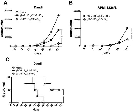
Finally, to demonstrate that CTE-γ9δ2TCRs are safe and function with increased efficacy compared with the original constructs in vivo, adoptive transfer of T cells engineered with CTE-TCRs was studied in a humanized mouse model: protection against outgrowth of Daudi or RPMI8226/S in Rag2−/−γc−/− double-knockout mice. Peripheral blood αβT cells were transduced with CTE-TCR γ9-G115wt/δ2-cl5wt or control TCR γ9-G115wt/δ2-G115wt. CTE-TCR–transduced T cells showed similar expression of homing markers including L-selectin and CCR7 (data not shown). Irradiated Rag2−/−γc−/− mice received luciferase-transduced Daudi (0.5 × 10⁶) or RPMI8226/S cells (5 × 10⁶) and 107 CTE-engineered T cells by IV injection. The frequency of T-cell infusion was reduced to 1 IV injection relative to our previously reported model, in which 2 infusions were given to test the superiority of CTE-TCR–transduced T cells under suboptimal conditions.9 This resulted in loss of protection with TCR G115wt-engineered T cells when tumor growth was measured by bioluminescence imaging (Figure 6A-B). However, CTE-engineered T cells expressing γ9-G115wt/δ2-cl5wt clearly reduced tumor outgrowth for Daudi (20 000 counts/min at day 42, n = 4) and RPMI8226/S (80 000 counts/min at day 35, n = 7) compared with TCR G115wt-engineered T cells (Daudi: 180 000 counts/min at day 42; RPMI8226/S: 210 000 counts/min at day 35). T cells could be found in the periphery until 1-2 weeks after infusion in mice (data not shown), but the frequency of T cells was not correlated with tumor regression. Finally, in the rapidly lethal Daudi model, only mice treated with CTE-engineered T cells had a significant increased overall survival of approximately 2 months relative to mice treated with T cells expressing γ9-G115wt/δ2-G115wt (Figure 6C). These results indicate that CTE-engineered γ9δ2TCRs efficiently mediate antitumor reactivity in vivo, which suggests that CTE is a potential tool with which to optimize γ9δ2TCRs for clinical application.
Antitumor reactivity of T cells transduced with CTE-engineered γ9δ2TCRs in vivo. The functional avidity of T cells expressing CTE-γ9δ2TCR γ9-G115wt/δ2-cl5wt or control γ9δ2TCR (γ9-G115wt/δ2-G115wt) was studied in Rag2−/−γc−/− double-knockout mice (4-7 mice per group). After total body irradiation (2 Gy) on day 0, mice were IV injected with 0.5 × 10⁶ Daudi-luciferase or 5 × 10⁶ RPMI8226/S-luciferase cells and 107 CTE-γ9δ2TCR–transduced T cells at day 1. In addition, 6 × 10⁵ IU of IL-2 in IFA and pamidronate (10 mg/kg body weight) were injected at day 1 and every 3 weeks until the end of the experiment. (A-B) Tumor outgrowth was assessed in vivo by bioluminescence imaging measuring the entire area of mice on both sides. Data represent the means of all animals measured (Daudi, n = 4; RPMI8226/S, n = 7). *P < .05; and **P < .01 by 1-way ANOVA (Daudi at day 42 and RPMI8226/S at day 35). (C) Overall survival of treated Daudi mice was monitored for 72 days. *P < .05; and **P < .01 by log-rank (Mantel-Cox) test.
Antitumor reactivity of T cells transduced with CTE-engineered γ9δ2TCRs in vivo. The functional avidity of T cells expressing CTE-γ9δ2TCR γ9-G115wt/δ2-cl5wt or control γ9δ2TCR (γ9-G115wt/δ2-G115wt) was studied in Rag2−/−γc−/− double-knockout mice (4-7 mice per group). After total body irradiation (2 Gy) on day 0, mice were IV injected with 0.5 × 10⁶ Daudi-luciferase or 5 × 10⁶ RPMI8226/S-luciferase cells and 107 CTE-γ9δ2TCR–transduced T cells at day 1. In addition, 6 × 10⁵ IU of IL-2 in IFA and pamidronate (10 mg/kg body weight) were injected at day 1 and every 3 weeks until the end of the experiment. (A-B) Tumor outgrowth was assessed in vivo by bioluminescence imaging measuring the entire area of mice on both sides. Data represent the means of all animals measured (Daudi, n = 4; RPMI8226/S, n = 7). *P < .05; and **P < .01 by 1-way ANOVA (Daudi at day 42 and RPMI8226/S at day 35). (C) Overall survival of treated Daudi mice was monitored for 72 days. *P < .05; and **P < .01 by log-rank (Mantel-Cox) test.
Discussion
γ9δ2T cells are innate lymphocytes that provide strong antitumor reactivity against solid and hematologic cancers.32,33 However, despite positive preclinical data, adoptive transfer of γ9δ2T cells in clinical studies has provided only limited tumor control.8 In the present study, we found one factor preventing the successful translation of this strategy into humans: the strong tumor-reactive potential of γ9δ2T cells is not a universal feature among all γ9δ2T cells. Individual γ9δ2T-cell clones differ in their antitumor reactivity in specificity and functional avidity. The latter is substantially regulated by CDR3 domains of individual γ9δ2TCRs. Most likely, single amino acid substitutions in CDR3 affect the affinity of a γ9δ2TCR to its ligand. We have also provided a means to engineer immune cells that harbor γ9δ2TCRs with increased antitumor reactivity in vitro and in vivo, and these are promising candidates for clinical applications.
Limited evidence has been provided for an important role of the variable domains of a γ9δ2TCRs in mediating function. The requirement for germline-encoded residues has been reported only within γCDR3 and a hydrophobic residue at position δ109 within δCDR3.14,16 In the present study, Jδ1 residue I117 was found to be important for tumor recognition, which shows that another germline-encoded residue is mandatory in the δ2TCR chain. Mutation of residue δ109 or δI117 partially abrogates γ9δ2TCR-mediated activation, indicating that such residues may be crucial for a first binding of a γ9δ2TCR to its target. However, we have shown that single amino acids in the highly variable part of CDR3, such as γCDR3A109, can clearly affect functional avidity. In the γ9δ2TCR G115, γCDR3A109 points back toward the γ-chain, whereas γCDR3Q110 and γCDR3Q111 point toward the back of the TCR but without contacting any δ-chain residues. In combination with our functional data, this modeling suggests that γCDR3A109 in G115 is important for directly mediating ligand interactions. However, the A/E and Q/E substitutions are fairly nonconservative and could also indirectly alter the structure of the γCDR3 loop and the TCR-binding site through global conformational changes. In addition, our data indicate that diverse amino acid compositions in δ2CDR3 can affect functional avidity; the combination of a defined γ9 and δ2 chain is particularly important. We hypothesize that the highly variable parts of γ9 and δ2CDR3 complement each other to form a structure or allow a conformational change that is favorable or unfavorable for target recognition. Therefore, in contrast to previous results,34 we have shown herein that δCDR3 alone does not correctly reflect the full interaction of a γ9δ2TCR with its target. Receptor flexibility is apparently necessary, for example, to adjust to a variable cell surface of an antigen that might be presented in different ways35 or to respond with different affinities, as was recently demonstrated for T22-reactive γδT cells with variable δCDR3-domains in mice.36 This is also consistent with a 2-step binding model reported for αβTCRs, which requires a preliminary interaction and then adjustment.37 However, these hypotheses are limited by the fact that no direct interaction of a γ9δ2TCR with a ligand has been reported, which prevents further visualization of the proposed receptor-ligand interactions.
Our present data suggest that certain limitations in the γ9δ2TCR repertoire are mediated by the δCDR3 length. If δCDR3 is too short, γ9δ2TCRs are not functional, and such receptors have not been reported within the human δ2TCR repertoire. Although the IMGT database for human γ9δ2TCR repertoire used herein is certainly not complete, it is plausible that alterations in δCDR3 in particular can limit the positive selection of a γ9δ2T cell. The functionally tolerated variability in CDR3 sequences and lengths reported in the present study support the observation that γ9δ2T-cell responses to phosphoantigen stimulation do not further select for defined CDR3 sequences.38,39 Consistent with this, we observed identical dose-response kinetics in pamidronate titration experiments, although the magnitude of response differed significantly. The observation that equal pamidronate EC50s were calculated for all responsive γ9δ2TC-transduced cells that only differ in their CDR3 domains indicates that γ9 and δ2CDR3 binding does not involve substrates directly regulated by pamidronate. Because IPP is enhanced by pamidronate in the mevalonate pathway, it seems unlikely that IPP contacts γ9 or δ2CDR3 directly and is in turn able to regulate the functional avidity of γ9δ2TCR-transduced cells, as was suggested recently.40 However, these theories support the hypothesis that a secondary signal is present41 and therefore a rather multimolecular signature is required for recognition, as has been reported for other γδTCRs.42
Interestingly, T cells engineered to express defined γ9δ2TCRs did reflect the functional avidity of the original γ9δ2T-cell clone tested herein. Therefore, differences in CDR3 domains of γ9δ2TCRs can be responsible for differential functional avidities observed between individual γ9δ2T-cell clones. However, it is still likely that the functionality of distinct γ9δ2T cells is orchestrated by different stimulatory molecules such as NKG2D43 and costimulatory signals derived from molecules that are also expressed on γ9δ2T cells.41 The plethora of specificities and functional avidities of distinct γ9δ2T-cell clones must therefore be taken into account when such cells are ex vivo expanded and adoptively transferred.1
To generate tumor-reactive T cells that mimic the reactivity of a γ9δ2T cell, we proposed to engineer αβT cells to express a defined γ9δ2TCR.9 This allows rapid engineering of tumor-reactive T cells that are not limited by HLA restrictions and are readily available for nearly any patient with any cancer. Moreover, it allows a γδT-cell repertoire to be repleted. The functionality of this repertoire is usually heavily impaired in cancer patients.44 To further improve functional avidity mediated by a TCR, laborious display strategies have been used for αβTCRs.45 Based on our observation that it is mainly the γ9 and δ2CDR3 domains that are involved in mediating functional avidity, we have proposed the concept of CTE as an efficient method to design γ9δ2TCRs that mediate broad and strong antitumor responses. The CTE-γ9δ2TCR–transduced T cells tested remained tumor specific and did not respond to healthy tissues. This indicates that pairing distinct γ9 or δ2 chains only strengthens the response toward malignant cells instead of altering specificity, which reduces the likelihood of unwanted specificities. We conclude that CTE engineering provides an elegant strategy to redirect T cells more effectively against a broad range of tumor cells.
There is an Inside Blood commentary on this article in this issue.
The online version of this article contains a data supplement.
The publication costs of this article were defrayed in part by page charge payment. Therefore, and solely to indicate this fact, this article is hereby marked “advertisement” in accordance with 18 USC section 1734.
Acknowledgments
The authors thank Charles Frink, Jeanette Leusen, Bas Nagelkerken, and Tuna Mutis for helpful discussions.
This work was supported by grant 43400003 from The Netherlands Organization for Health Research and Development; grant 917.11.337 from VIDI-ZonMW, grant 0902 from Landsteiner Foundation for Blood Transfusion Research; grant 10-736 from The Association for International Cancer Research; and grant UU-2010-4669 from The Dutch Cancer Society.
Authorship
Contribution: C.G., S.v.D., S.H., E.D., T.S., S.H., K.S., W.S., Z.S., and A.M. designed and performed the experiments; R.S. produced the modeling data; J.K. supervised all experiments; and C.G. and J.K. wrote the manuscript.
Conflict-of-interest disclosure: The authors declare no competing financial interests.
Correspondence: Jürgen Kuball, UMC Utrecht, Department of Hematology and Department of Immunology, Huispostnr KC02.085.2, Lundlaan 6, 3584 EA Utrecht, The Netherlands; e-mail: j.h.e.kuball@umcutrecht.nl.

This feature is available to Subscribers Only
Sign In or Create an Account Close Modal