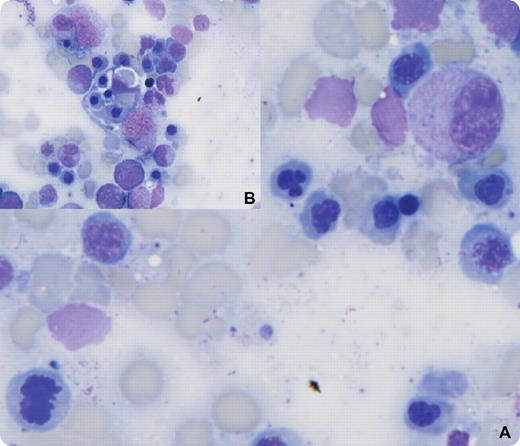
A 34-year-old male with a history of T cell–rich B-cell lymphoma relapsed 1 year after high-dose therapy with recurrent fevers, abdominal pain, and jaundice. He was cachetic with hepatosplenomegaly and ascites. Laboratory findings showed a white blood cell count of 2.7 × 10⁹/L, hemoglobin 8.7 g/dL, platelets of 21 × 109/L, hyperbilirubinemia, and no evidence of hemolysis including a negative Coombs test. Bone marrow examination revealed marked hypercellularity and affirmed recurrent lymphoma (occupying 30%-40% of marrow). In addition, there was dysplasia of red cell precursors (panel A) and phagocytosis of bone marrow elements by lymphohistiocytes (panel B). A diagnosis of hemophagocytic lymphohistiocytosis (HLH) was made. Laboratory data consistent with HLH included albumin of 1.7 gm/dL, fibrinogen 65 mg/dL, ferritin > 15 000 ng/mL, and soluble IL-2 receptor > 6500 units/mL. The relapsed lymphoma was successfully treated with salvage therapy. A repeat bone marrow examination showed no lymphoma, normal hematopoiesis, an absence of dysplasia, and no phagocytosis. Several months later the patient relapsed and died.
HLH can be familial or associated with viral infections, autoimmune disorders, and some malignancies. Treatment of the underlying disorder may reverse HLH, as was true in this case. The appearance of HLH with lymphoma is rare and often associated with a poor outcome.
For additional images, visit the ASH IMAGE BANK, a reference and teaching tool that is continually updated with new atlas and case study images. For more information visit http://imagebank.hematology.org.


This feature is available to Subscribers Only
Sign In or Create an Account Close Modal