Abstract
Monoclonal or oligoclonal T cell receptor positive populations (TCR+) can be observed in elderly individuals with viral infections, patients (pts) with pancytopenia, bone marrow failure syndromes, and in large granular lymphocytic leukemia. Expansion of these T cell clones may indicate that an antigen driven myelosuppressive mechanism is active in pts with MDS. However the clinical significance of clonal T cell population/TCR+ in MDS is not clear. In the present study we have reported on the clinical characteristics and prognostic relevance of monoclonal or oligoclonal TCR+ cells in pts with MDS.
A retrospective chart review was performed for all pts with a diagnosis of MDS in whom TCR clonal assessment was done at our institution between 2005 and 2013. Using labeled V-primers, TCR (beta and gamma) were assessed in the bone marrow (BM) samples by semi quantitative polymerase chain reaction (PCR) assay with a sensitivity of 1:100 to 1:10,000 lymphocytes.
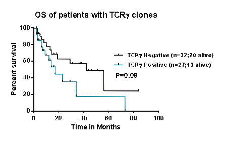
The clinical characteristics and outcomes of 50 pts, with a median age of 67 yrs (range 35-85) were analyzed. 17 (34 %) pts had TCRβ+ clones, 22 pts (44 %) had TCRγ+ clones. Pts with any TCR+ clones (n=27; 54 %) were compared with those pts who were negative for TCR clonality (n=23; 46 %). The median follow-up for the pts with or without TCR+ clones was 17 months vs. 19 months, respectively. Clinical characteristics of the pts with and without TCR+ clones are outlined in Table 1. Overall, the groups were similar with regards to blood counts, blast percentages, bone marrow cellularity, and karyotype. There were trends towards higher percentages of males, lower neutrophil count, higher IPSS category, and more pts with >10% BM blasts in the TCR+ group, but these did not reach statistical significance. Three pts (6%) transformed to acute myeloid leukemia and two of these pts had TCR+ clones in the BM. The majority of these pts were previously untreated 34/50 (68%). We then looked at response to therapy with hypomethylating agents in those pts who were previously untreated and/or who had previously received one type of therapy (N=42). In 20 pts with TCR+ clones, 8 (40%) received hypomethylating agents and 2 went into complete remission (25%), 2 had stable disease and 4 (20%) were non responders. In 22 pts without TCR+ clones 4 (18%) received hypomethylating agents and there were no (0%) responders. The estimated 3 year overall survival was 21% vs 47% between pts with andwithout TCR+ clones, respectively (P=0.67). In a subset analysis restricted to pts with available TCRγ assessment (n=67), the estimated 3 year OS for pts with TCRγ+ vs. those without TCRγ clones was 17% vs 57%, respectively (P=0.08) (Figure 1). No difference in OS was observed when pts with or without TCRβ clones were compared.
We report our experience of the clinical impact of clonal TCR+ populations in pts with MDS. TCR+ clonal T-cells were present in about half of pts with MDS. Although the study is limited with number of pts, those with MDS and clonal TCR+ population tend to have higher proportions of poor risk features and exhibit a trend of inferior outcome as compared to those pts without TCR+ clones. Larger prospective studies are needed to better define the clinical and mechanistic relevance of clonal TCR+ populations in pts with MDS.
Patient Characteristics (N= 50)
| Characteristic | TCR-Positive | TCR-Negative | P-value |
| N | 27 | 23 | |
| Median Age, yrs [Range] | 68 [45-81] | 67 [35-85] | NS |
| Age ≥ 60, N (%) | 20 (74) | 19 (83) | 0.47 |
| Male sex, N (%) | 21 (78) | 13 (57) | 0.11 |
| Median Hgb [Range] | 9.8 [7.1 - 14.6] | 9.2 [4.4-13.5] | 0.33 |
| Median Platelet [Range] | 51 [18-331] | 78 [7-359] | 0.99 |
| Median ANC [Range] | 1.4 [0-3.8] | 1.9 [0-5.6] | 0.14 |
| Median ALC [Range] | 0.9 [0-2.4] | 1.3 [0-3.6] | 0.52 |
| Median BM Blast [Range] | 4 [1-15] | 2 [0-18] | 0.18 |
| Cytogenetic Group | 0.49 | ||
| Dip/-Y | 13 | 14 | |
| -5/-7 | 3 | 4 | |
| Other | 10 | 4 | |
| Insufficient | 1 | 1 | |
| IPSS Group | 0.11 | ||
| High | 4 (15) | 0 (0) | |
| Int-2 | 9 (33) | 6 (26) | |
| Int-1 | 7 (26) | 12 (52) | |
| Low | 7 (26) | 5 (22) | |
| BM Cellularity | 0.73 | ||
| Hypocellular | 5 (18) | 6 (26) | |
| Normocellular | 14 (52) | 12 (52) | |
| Hypercellular | 8 (30) | 5 (22) | |
| BM Blasts > 10%, N (%) | 8 (30) | 2 (9) | 0.18 |
| RAEB | 13 (48) | 7 (30) | 0.44 |
| 3-yr OS (%) | 21 | 47 | 0.67 |
No relevant conflicts of interest to declare.
Author notes
Asterisk with author names denotes non-ASH members.


This feature is available to Subscribers Only
Sign In or Create an Account Close Modal