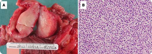
A 70-year-old Hispanic woman with history of coronary arterial disease presented with dull abdominal pain and weight loss for ∼2 weeks. A computed tomography scan of the abdomen revealed a large mass occupying the proximal jejunum. The patient had no history of celiac disease or malabsorption. She underwent surgical resection. Grossly, the tumor measured 10 cm in length and had a multinodular appearance with surface ulceration and a soft “fish flesh,” yellow-white, homogenous cut surface (panel A). Microscopically, the tumor consisted of monomorphic small lymphocytes (panel B) positive for cytoplasmic CD3, CD8, CD56, and T-cell receptor γ (TCR-γ), and negative for CD4, CD20, CD30, and TCR-β.
Enteropathy-associated T-cell lymphoma (EATL) is a lymphoma of intestinal intraepithelial T lymphocytes. The World Health Organization divided EATL into 2 variants: a classical form (type I), which comprises 80% to 90% of cases, and a type II or monomorphic variant, which comprises 10% to 20% of EATL cases. The classical form consists of large pleomorphic lymphoma cells which are usually CD3+, CD5−, CD7+, CD8−/+, CD4−, CD56−, TCRβ+/−. Type II is composed of small round monomorphic cells. The tumor cells in type II are CD3+, CD4−, CD8+, CD56+, and TCRβ+. Type I is mostly associated with celiac disease. The monomorphic variant may also be preceded by celiac disease but it has not been well characterized.
For additional images, visit the ASH IMAGE BANK, a reference and teaching tool that is continually updated with new atlas and case study images. For more information visit http://imagebank.hematology.org.


This feature is available to Subscribers Only
Sign In or Create an Account Close Modal