Key Points
Gα12 interaction with α-SNAP regulates basal EC vWF secretion.
PAR-1 activation-dependent signaling via Gα12/RhoA and Gαq/11 enhances vWF secretion.
Abstract
von Willebrand factor (vWF) secretion by endothelial cells (ECs) is essential for hemostasis and thrombosis; however, the molecular mechanisms are poorly understood. Interestingly, we observed increased bleeding in EC-Gα13−/−;Gα12−/− mice that could be normalized by infusion of human vWF. Blood from Gα12−/− mice exhibited significantly reduced vWF levels but normal vWF multimers and impaired laser-induced thrombus formation, indicating that Gα12 plays a prominent role in EC vWF secretion required for hemostasis and thrombosis. In isolated buffer-perfused mouse lungs, basal vWF levels were significantly reduced in Gα12−/−, whereas thrombin-induced vWF secretion was defective in both EC-Gαq−/−;Gα11−/− and Gα12−/− mice. Using siRNA in cultured human umbilical vein ECs and human pulmonary artery ECs, depletion of Gα12 and soluble N-ethylmaleimide-sensitive-fusion factor attachment protein α (α-SNAP), but not Gα13, inhibited both basal and thrombin-induced vWF secretion, whereas overexpression of activated Gα12 promoted vWF secretion. In Gαq, p115 RhoGEF, and RhoA-depleted human umbilical vein ECs, thrombin-induced vWF secretion was reduced by 40%, whereas basal secretion was unchanged. Finally, in vitro binding assays revealed that Gα12 N-terminal residues 10-15 mediated the binding of Gα12 to α-SNAP, and an engineered α-SNAP binding-domain minigene peptide blocked basal and evoked vWF secretion. Discovery of obligatory and complementary roles of Gα12 and Gαq/11 in basal vs evoked EC vWF secretion may provide promising new therapeutic strategies for treatment of thrombotic disease.
Introduction
von Willebrand factor (vWF) is important for platelet adhesion and subsequent thrombus formation1 ; therefore, a qualitative or quantitative change in plasma vWF is a predictive factor for cardiovascular disease or bleeding diathesis.2 Whereas an elevated plasma level of vWF is associated with increased risk for venous thrombosis, coronary artery disease, and stroke,3-6 inherited or acquired vWF deficiency syndrome (von Willebrand disease [vWD]) results in frequent hemorrhagic disorders.7 Measurement of vWF continues to be the most important laboratory test for vWD diagnosis.7 Type 3 vWD, the most severe form as a result of a complete absence of vWF in all vascular compartments including plasma, endothelial cells (ECs), and platelets,8 is a life-threatening condition because of major defects in coagulation and primary hemostasis.
vWF is stored in Weibel-Palade bodies (WPBs),9 the unique secretory granules in ECs10 that also contain P-selectin11 and other procoagulation and proinflammatory factors.12 vWF is also stored in and released from platelet α-granules during platelet activation.13 Release of vWF from ECs is thought to be mediated by both constitutive (basal) and agonist-induced signaling pathways, whereas vWF secretion from platelet α-granules is entirely stimulus-induced.14 Circulating vWF levels are likely maintained by basal secretion of vWF from ECs, and thus a change in this process would dramatically affect plasma vWF levels.15 Delivery of EC-specific vWF to patients and mice with vWD is sufficient to correct defects in hemostasis,7,13 whereas platelet-derived vWF only partially restores defects in thrombosis.13
Exocytosis, mediated by the fusion of secretory vesicles with the plasma membrane,16 requires the ATP-ase N-ethylmaleimide-sensitive-fusion factor (NSF), soluble NSF attachment protein α (α-SNAP), and SNAP receptors (SNAREs).14,17 α-SNAP binding to the cis-SNARE complex in the fused membrane mediates recruitment of NSF that together promotes exocytosis and SNARE disassembly.14 Although this core secretory machinery is widely expressed, there is significant heterogeneity in the signaling mechanisms that control endothelial secretory granule exocytosis.18
Activation of G protein-coupled receptors and G protein α subunits (Gs, Gi, and Gq/11) can stimulate secretory granule release.19 Thrombin (enzyme IIa) produced during vascular injury activates the coagulation cascade and also potentiates vWF secretion via stimulation of protease-activated receptor 1 (PAR-1)-dependent signaling.20,21 The general mechanism by which G protein α subunits regulate exocytosis is not known, although it has been suggested that G proteins mediate cell-type and compartment-specific functions and early transport events.19 Direct G protein–dependent regulation of the SNARE protein fusion machinery required for secretory granule exocytosis has been previously reported.22 Distinct from their traditional plasma membrane localization,23 heterotrimeric G proteins have been shown to shuttle rapidly between plasma membrane and intracellular membranes, where they have distinct roles on specific organelles, including those in exocytotic pathways.24 A yeast 2-hybrid screen using activated Gα12 as bait identified α-SNAP, an intrinsic component of the endothelial exocytotic machinery,14 as a potential binding partner for Gα12.25 Thus, we reasoned that Gα12 may play an important role in promoting membrane trafficking and exocytosis in ECs and assessed the role of Gα12/α-SNAP interactions as well as Gα12/13 and Gαq/11 signaling in basal and thrombin-induced vWF secretion, primary hemostasis, and thrombosis. Collectively, our results describe a molecular mechanism of vWF release that is dependent on Gα12 and Gαq/11 signaling via RhoA- and Ca2+/α-SNAP-dependent mobilization and fusion of WPBs, with additional evidence implicating that direct interaction of Gα12 with α-SNAP serves as a catalyst for basal vWF secretion.
Materials and methods
Mouse monoclonal antibodies against guanine nucleotide G protein α subunits Gα12, Gα13, and Gαq/11 were generated as previously described.26 Human plasma–derived vWF was purified as described.1 Tamoxifen-inducible tie2-CreERT2;Gα13flox/flox;Gα12−/− (EC-specific Gα13 knockout [EC-Gα13−/−] in a global Gα12−/− background) and tamoxifen-inducible tie2-CreERT2;Gαq flox/flox ;Gα11− /− (EC-Gαq−/− in a global Gα11−/− background) were as described.27,28 vWF secretion assay was as previously described.29 Tail-snip blood loss was determined as described.30,31 Real-time intravital microscopy of cremaster muscle arteriole thrombus formation was performed as previously described.32 Myc-tagged constitutively activated Gα12Q229L mutant and Gα12WT constructs bearing a N-terminal Gα12QL substitution mutants with sequence Asn-Ala-Ala-Ile-Arg-Ser (NAAIRS) cassette were as described.33 Data are presented as mean ± standard error of the mean. Statistical significance was assessed by 1-way analysis of variance with Bonferoni post hoc testing, and P < .05 was considered significant. All procedures on mice were approved by the University of Illinois Animal Care and Use Committee in accordance with the Guide for the Care and Use of Laboratory Animals. Human blood was drawn under steady conditions from nonmedicated healthy donors, according to a standard institutional review board-approved protocol, on informed consent in accordance with the Declaration of Helsinki.
Results
Blood levels of vWF in Gα12/13- and Gαq/11-deficient mice
To assess the physiologic role of G protein signaling in the production of circulating vWF, we measured plasma levels of vWF in Cre-Gα13flox/flox; Gα12−/− and Cre-Gαqflox/flox;Gα11−/− mice,27 with or without tamoxifen (TX) treatment, to induce endothelial Cre activation and resultant Gαq gene deletion in global Gα11−/− or Gα13 deletion in global Gα12−/− mice. To exclude the effects of TX per se on plasma levels of vWF, we administered TX (1 mg/d for 5 days) to WT mice (n = 2) and collected blood from the tail vein on days 1 (just before the first TX injection), 3, 7, 10, and 14. On day 3, a small transient increase in plasma vWF level was observed (supplemental Figure 1A, available on the Blood Web site), and then plasma vWF level normalized on days 7, 10, and 14 (the day of the experimental procedure).
Plasma from Gα12−/− and EC-Gα13−/−;Gα12−/− mice exhibited significantly reduced plasma levels of vWF compared with WT, Gα11−/−, and EC-Gαq−/−;Gα11−/− mice (Figure 1A). However, there was no significant difference in the percentage of low–, intermediate–, or high–molecular weight vWF multimers in Gα12−/− plasma (Figure 1B), indicating that vWF secretion rather than vWF packaging or processing was defective in the absence of Gα12. These data further suggest that Gα12 may play an important role in maintaining basal blood levels of vWF.
The role of Gα12 and Gαq in vWF secretion and primary hemostasis. (A) vWF levels in plasma from WT vs Gα12−/−, Gα11−/−, EC-Gαq−/−;Gα11−/−, and EC-Gα13−/−;Gα12−/− mice. *P < .05 vs WT; n = 6/group. (B) Representative vWF multimer gel from WT, Gα12−/−, and Gα11−/− mouse plasma. Quantitative multimer analysis shows no difference between Gα12−/− (n = 3) and WT or Gα11−/− mouse vWF. (C) Constitutive and PAR-1 agonist peptide-induced vWF release from WT, Gα12−/−, and EC-Gαq/;Gα11−/− isolated perfused mouse lungs. Time-course of basal and PAR-1-specific peptide TFLLR-evoked vWF release into mouse lung perfusate. (D) vWF in WT, Gα12−/− and EC-Gαq−/−;Gα11−/− mouse lung extracts from untreated lungs or after PAR-1 peptide stimulation. *P < .05 vs WT + TFLLR; n = 6/group. See also supplemental Figure 1B.
The role of Gα12 and Gαq in vWF secretion and primary hemostasis. (A) vWF levels in plasma from WT vs Gα12−/−, Gα11−/−, EC-Gαq−/−;Gα11−/−, and EC-Gα13−/−;Gα12−/− mice. *P < .05 vs WT; n = 6/group. (B) Representative vWF multimer gel from WT, Gα12−/−, and Gα11−/− mouse plasma. Quantitative multimer analysis shows no difference between Gα12−/− (n = 3) and WT or Gα11−/− mouse vWF. (C) Constitutive and PAR-1 agonist peptide-induced vWF release from WT, Gα12−/−, and EC-Gαq/;Gα11−/− isolated perfused mouse lungs. Time-course of basal and PAR-1-specific peptide TFLLR-evoked vWF release into mouse lung perfusate. (D) vWF in WT, Gα12−/− and EC-Gαq−/−;Gα11−/− mouse lung extracts from untreated lungs or after PAR-1 peptide stimulation. *P < .05 vs WT + TFLLR; n = 6/group. See also supplemental Figure 1B.
Role of Gα12 and Gαq/11 in constitutive vs evoked endothelial vWF secretion
To address the role of G protein α subunits in constitutive (basal, spontaneous) vs PAR-1-mediated vWF secretion from ECs, we measured vWF in the ex vivo isolated buffer-perfused mouse lung. Lung preparations were used to exclude platelets as a source of vWF and hepatic stellate cells as contributors of proteases that cleave ultralarge vWF strings.5 Ventilated WT, Gα12−/−, and EC-Gαq−/−; Gα11−/− mouse lungs were cannulated and perfused with warmed recirculating RPMI media via pulmonary artery and left atrial catheters in situ. To assess basal vWF secretion, precleared blood-free lungs were perfused for 15 minute with 5 mL 37°C recirculating RPMI media (approximately the same volume as the normal mouse blood volume). Samples of the recirculating buffer were collected at 5-minute intervals over a period of 30 minutes, and vWF (mU/mL perfusate) was measured by enzyme-linked immunosorbent assay (ELISA).
vWF was detectable in the lung perfusate immediately after initiating perfusion with fresh media (116.2 ± 7.7 mU/mL at the 5-minute point in WT mice, and 106.6 ± 13.6 mU/mL in EC-Gαq−/−;11−/− mice, but only 6.2 ± 0.8 mU/mL in Gα12−/− mice; Figure 1C). The accumulation of vWF in recirculating lung perfusate over time described a straight line in WT mice (secretion rate, 2.5 mU/mL per minute). Intriguingly, baseline vWF values were significantly lower in Gα12−/− mouse lungs (Figure 1C) (secretion rate, 0.6 ± 0.03 mU/mL per minute).
To assess the mechanism of thrombin-induced vWF release from ECs via PAR-1 activation, we used the PAR-1-specific synthetic peptide TFLLR to stimulate lung ECs. The addition of 30 µM TFLLR to the recirculating lung perfusate sharply increased vWF levels in WT mice, which peaked after 10-15 minutes and remained constant for up to 1 h. The initial slope of the vWF secretion curve was used to estimate the rate of vWF release after the addition of TFLLR, which was approximately 25 mU/mL per minute in WT mouse lungs. In Gα12−/− mouse lungs, TFLLR-induced vWF secretion was significantly reduced (3 mU/mL per minute; Figure 1C). Under these same conditions, basal vWF secretion in the pulmonary circulation of EC-Gαq−/−;Gα11−/− lungs was not different from that observed in WT mice, although TFLLR-induced vWF release was reduced by approximately 40% (Figure 1C). Consistent with these findings, we observed equivalent vWF immunoreactivity in blood-free homogenates of nonstimulated WT, Gα12−/−, and EC-Gαq−/−; Gα11−/− mouse lungs (Figure 1D, open bars). However, significantly greater vWF remained in Gα12−/− and EC-Gαq−/−; Gα11−/− lungs after TFLLR stimulation (Figure 1D, solid bars), suggesting vWF is being made, although thrombin-evoked secretion from lung vascular endothelium is significantly reduced in the absence of Gα12 or Gαq/11. As summarized in supplemental Figure 1B, the majority of vWF in Gα12−/− mouse lungs was retained in the lung (not secreted), whereas in EC-Gαq−/−;Gα11−/− mouse lungs, basal secretion was normal but TFLLR-evoked EC-derived vWF secretion was significantly reduced. The presence of EC WPB vWF in en face descending aorta and paraffin-embedded lung sections was verified by immunostaining and confocal microscopy. WT, Gα12−/−, and Gα11−/− tissues stained positive for vWF (supplemental Figure 2A-B), consistent with there being equivalent vWF synthesis and storage in WPBs in WT, Gα12−/−, and Gα11−/− mice.
Regulation of hemostasis and thrombosis by Gα12
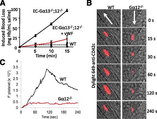
In a previously published study performed using megakaryocyte-restricted-Gα12/13 double knockout mice, it was observed that Gα13-dependent signaling in platelets is important for hemostasis and thrombosis.28 However, the molecular mechanisms and functional significance of EC Gα12 and Gα13 in primary hemostasis were not assessed. As a direct measurement of primary hemostasis, we determined blood loss after tail-snip in anesthetized EC-Gα13−/−;Gα12−/− vs WT mice (n = 3 male mice/group, age 8 weeks) as previously described.31 Hb accumulation from the cut tail immersed in saline solution was measured every 5 minutes for 15 minutes. The induced hemorrhagic event was significantly prolonged in EC-Gα13−/−;Gα12−/− mice compared with WT mice (Figure 2A). In addition, EC-Gα13−/−;Gα12−/− mice were prone to spontaneous hemorrhagic incidents in the abdominal cavity and recurrence of bleeding after stoppage. Consistent with the primary hemostasis defect being a result of low plasma level of vWF, hemostasis was corrected by i.v. administration of purified human plasma–derived vWF (2 μg/g BW) (Figure 2A).
Effect of Gα12 on platelet accumulation at the site of vascular injury in live mice. (A) Induced tail-snip blood loss in EC-Gα13−/−;Gα12−/− vs WT mice, measured as Hb concentration accumulating in a saline-filled microfuge tube over the course of 15 minutes. Infusion of purified human vWF (vehicle in WT mice) into the jugular vein of EC-Gα13−/−;Gα12−/− mice induced partial recovery of the hemorrhagic event (n = 3 mice/group). (B) WT and Gα12−/− mice (n = 3) were infused with Dylight 649–conjugated anti-mouse CD42c antibody (0.05 μg/g body weight) through a cannulus placed in a jugular vein. After laser injury, platelet thrombus formation was monitored via real-time intravital microscopy. Representative images of fluorescence associated with platelets (red) over the course of 240 seconds after vascular injury within the context of bright-field arteriolar histology. (C) Median integrated fluorescence intensity of anti-CD42c antibodies (Fplatelets) in 25 thrombi in 3 WT and 3 Gα12−/− mice.
Effect of Gα12 on platelet accumulation at the site of vascular injury in live mice. (A) Induced tail-snip blood loss in EC-Gα13−/−;Gα12−/− vs WT mice, measured as Hb concentration accumulating in a saline-filled microfuge tube over the course of 15 minutes. Infusion of purified human vWF (vehicle in WT mice) into the jugular vein of EC-Gα13−/−;Gα12−/− mice induced partial recovery of the hemorrhagic event (n = 3 mice/group). (B) WT and Gα12−/− mice (n = 3) were infused with Dylight 649–conjugated anti-mouse CD42c antibody (0.05 μg/g body weight) through a cannulus placed in a jugular vein. After laser injury, platelet thrombus formation was monitored via real-time intravital microscopy. Representative images of fluorescence associated with platelets (red) over the course of 240 seconds after vascular injury within the context of bright-field arteriolar histology. (C) Median integrated fluorescence intensity of anti-CD42c antibodies (Fplatelets) in 25 thrombi in 3 WT and 3 Gα12−/− mice.
Mice with Gα12 deficiency exhibited low vWF levels in blood-free lung vascular perfusates and elevated vWF level in blood-free lung homogenates both before as well as after PAR-1 stimulation. Thus, Gα12 may be important for maintaining basal levels of circulating vWF required for platelet thrombus formation. To investigate the role of Gα12 in platelet thrombus formation in vivo, we performed real-time fluorescence intravital microscopy in anesthetized mice. After arteriolar wall injury induced by micropoint laser ablation, platelets were monitored by Dylight 649–conjugated anti-mouse CD42c antibody. In both WT and Gα12−/− mice, platelet thrombus formation was initiated at the site of arteriolar injury (Figure 2B). However, the platelet thrombus did not grow at the injury site in Gα12−/− mice compared with WT mice (Figure 2B-C). From these results, we concluded that Gα12-dependent EC vWF secretion is required for hemostasis and thrombosis and focused on this mechanism in comparison with EC Gαq signaling in subsequent studies.
Potentiation of Gα12-dependent basal EC-vWF release by thrombin-stimulated Gαq/Gα11- and Gα12-regulated EC-vWF secretion in vitro
We next investigated the role of G protein signaling in basal vs PAR-1-stimulated vWF secretion from confluent, low passage, and serum-deprived human umbilical vein ECs (HUVECs) and human pulmonary artery ECs (HPAECs). Using a siRNA approach, G protein α subunits Gαq, Gα12, and Gα13 were depleted from HUVECs (Figure 3A; supplemental Figure 3A-B) and HPAECs (supplemental Figure 3C). To quantify vWF release in response to thrombin stimulation, we established 2 complementary protocols, secretion/ELISA for quantification of vWF release from EC monolayers (Figure 3B) and fluorescence microscopy for visualization/quantification of WPBs in individual cells (Figures 4-5), which provide a quantitative measure of vWF exocytosis/secretion kinetics from ECs both basally and on PAR-1 activation.
Effect of siRNA-mediated depletion of Gα12, Gα13, and Gαq on constitutive vs thrombin-induced vWF release from HUVECs. (A) Western blot analysis shows specific and complete downregulation of Gα12, Gα13, and Gαq. (B) In Gα12-deficient HUVECs, both constitutive and thrombin-induced vWF release was inhibited. Gα13 depletion had no effect on basal or thrombin-induced vWF release. In Gαq-deficient HUVECs, thrombin-induced vWF secretion was reduced by 40%, whereas basal vWF secretion was not affected. (C) Western blots show siRNA-mediated downregulation of α-SNAP, p115 RhoGEF, and RhoA. (D) α-SNAP siRNA completely blocked constitutive and thrombin-induced vWF release by HUVECs. Depletion of p115 RhoGEF and RhoA had no effect on constitutive vWF secretion, although both reduced thrombin-induced vWF secretion. Data are mean ± SEM; n = 3. *P < .05 vs respective (basal or thrombin-treated) control siRNA values.
Effect of siRNA-mediated depletion of Gα12, Gα13, and Gαq on constitutive vs thrombin-induced vWF release from HUVECs. (A) Western blot analysis shows specific and complete downregulation of Gα12, Gα13, and Gαq. (B) In Gα12-deficient HUVECs, both constitutive and thrombin-induced vWF release was inhibited. Gα13 depletion had no effect on basal or thrombin-induced vWF release. In Gαq-deficient HUVECs, thrombin-induced vWF secretion was reduced by 40%, whereas basal vWF secretion was not affected. (C) Western blots show siRNA-mediated downregulation of α-SNAP, p115 RhoGEF, and RhoA. (D) α-SNAP siRNA completely blocked constitutive and thrombin-induced vWF release by HUVECs. Depletion of p115 RhoGEF and RhoA had no effect on constitutive vWF secretion, although both reduced thrombin-induced vWF secretion. Data are mean ± SEM; n = 3. *P < .05 vs respective (basal or thrombin-treated) control siRNA values.
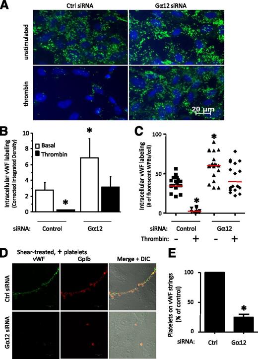
Impaired basal and thrombin-induced vWF secretion from WPBs in Gα12 depleted HUVECs. HUVECs were treated with a pool of 4 different oligonucleotides directed against Gα12 or a control siRNA. (A) Epifluorescent images of vWF immunostaining (green) in control siRNA- or Gα12 siRNA–treated HUVECs after incubation for 15 minutes with 25 nM thrombin or serum-free medium alone (unstimulated). Scale bars = 20 µm. (B) Intracellular vWF staining (corrected integrated density) in individual, randomly selected control siRNA and Gα12 siRNA–treated cells (mean ± SEM; 10-30 cells/group; *P < .05 vs basal signal in control cells). (C) Number of fluorescent WPBs per randomly selected control and Gα12 siRNA–treated cell (*P < .05 vs unstimulated control; n = 10-30 cells/group; red horizontal bars depict median values). (D) Confluent HUVECs were incubated with normal human platelets and exposed to uniform and constant shear for 5 minutes. After washing, platelets bound to HUVEC-anchored vWF strings were detected by anti-GpIb monoclonal antibody staining (red) and rabbit anti-human vWF polyclonal antibody staining (green). (E) Graph shows fluorescence intensity of individual platelets per vWF string per field (percentage of control siRNA treatment; *P < .05 vs control; n = 6).
Impaired basal and thrombin-induced vWF secretion from WPBs in Gα12 depleted HUVECs. HUVECs were treated with a pool of 4 different oligonucleotides directed against Gα12 or a control siRNA. (A) Epifluorescent images of vWF immunostaining (green) in control siRNA- or Gα12 siRNA–treated HUVECs after incubation for 15 minutes with 25 nM thrombin or serum-free medium alone (unstimulated). Scale bars = 20 µm. (B) Intracellular vWF staining (corrected integrated density) in individual, randomly selected control siRNA and Gα12 siRNA–treated cells (mean ± SEM; 10-30 cells/group; *P < .05 vs basal signal in control cells). (C) Number of fluorescent WPBs per randomly selected control and Gα12 siRNA–treated cell (*P < .05 vs unstimulated control; n = 10-30 cells/group; red horizontal bars depict median values). (D) Confluent HUVECs were incubated with normal human platelets and exposed to uniform and constant shear for 5 minutes. After washing, platelets bound to HUVEC-anchored vWF strings were detected by anti-GpIb monoclonal antibody staining (red) and rabbit anti-human vWF polyclonal antibody staining (green). (E) Graph shows fluorescence intensity of individual platelets per vWF string per field (percentage of control siRNA treatment; *P < .05 vs control; n = 6).
Confocal microscopy of Gα12 and vWF. Mutationally-activated Gα12 stimulates WPB exocytosis. (A) HUVECs were transfected with GFP alone (negative control) or GFP-tagged Gα12WT or Gα12Q229L constructs. After 4 h of serum starvation, GFP-expressing HUVECs were incubated for 15 minutes with vehicle (unstimulated) or 25 nM thrombin. WPBs were visualized by vWF immunofluorescent staining (red). Single confocal optical sections (pinhole set to achieve 1 Airy unit) are shown; bars correspond to 20 µm. (B) Fluorescent WPBs per cell were quantified as described in Materials and methods. Red horizontal bars represent median values. *P < .05 by analysis of variance vs unstimulated GFP-alone expressing cells. *,#P < .05 vs all other groups.
Confocal microscopy of Gα12 and vWF. Mutationally-activated Gα12 stimulates WPB exocytosis. (A) HUVECs were transfected with GFP alone (negative control) or GFP-tagged Gα12WT or Gα12Q229L constructs. After 4 h of serum starvation, GFP-expressing HUVECs were incubated for 15 minutes with vehicle (unstimulated) or 25 nM thrombin. WPBs were visualized by vWF immunofluorescent staining (red). Single confocal optical sections (pinhole set to achieve 1 Airy unit) are shown; bars correspond to 20 µm. (B) Fluorescent WPBs per cell were quantified as described in Materials and methods. Red horizontal bars represent median values. *P < .05 by analysis of variance vs unstimulated GFP-alone expressing cells. *,#P < .05 vs all other groups.
To demonstrate that Gα12 signaling in ECs regulates vWF secretion, HUVECs and HPAECs were transfected with control or Gα12 siRNA, and 48 hours later, cells were serum starved for a minimum of 4 hours and then stimulated with 25 nM α-thrombin for up to 1 hour. After Gα12 siRNA treatment, the level of Gα12 expression decreased to a level not detected by western blotting but had no effect on Gαq or Gα13 expression (Figure 3A). Consistent with the knockout mouse studies, Gα12 siRNA significantly reduced basal as well as thrombin-induced vWF secretion from HUVECs as well as HPAECs (Figure 3B; supplemental Figure 3). After the transfection of Gα13 siRNA, which depleted Gα13 but not Gα12 or Gαq protein (Figure 3A), however, we observed no significant effect on basal or thrombin-induced vWF secretion (Figure 3B). These data suggest that Gα12, but not Gα13, mediates EC-specific basal as well as thrombin-induced vWF release from cultured HUVEC and HPAEC.
We next investigated the involvement of Gαq in the regulation of the vWF secretory pathway. Gαq siRNA resulted in nearly complete Gαq depletion, whereas the Gα12 protein level remained unchanged (Figure 3A). After Gαq depletion, the amount of vWF released on thrombin stimulation was reduced by 40% in HUVECs (Figure 3B; supplemental Figure 3B) and 60% in HPAECs, whereas basal vWF secretion was not affected (Figure 3B; supplemental Figure 3C). These data suggest that Gαq is important in thrombin-evoked EC vWF secretion.
Role of NSF/αSNAP, p115RhoGEF, and RhoA in basal vs thrombin-induced vWF release
N-Ethylmaleimide is a well-known inhibitor of exocytosis in ECs.34 Consistent with the notion that WPB exocytosis requires NSF/α-SNAP,17 the addition of 1 mM N-ethylmaleimide to the recirculating lung perfusion system for 1 minute abolished vWF secretion (supplemental Figure 4A). To explore the role of α-SNAP in constitutive and thrombin- or calcium ionophore–induced vWF release from human ECs, siRNA was used to deplete α-SNAP from HUVEC, and then vWF was measured by ELISA as described earlier. Cell lysates were also saved for western blot analysis (Figure 3C). We observed that α-SNAP depletion completely blocked basal and thrombin- as well as calcium ionophore–induced vWF secretion (Figure 3D; supplemental Figure 4B). These data demonstrate that α-SNAP is required for all forms of vWF secretion.
In p115 RhoGEF and RhoA siRNA–depleted HUVECs (Figure 3C), thrombin-induced vWF secretion was reduced by 20%-40%, although basal vWF secretion was not affected (Figure 3D). Thus, Gα12-mediated basal vWF secretion occurs independent of the RhoA pathway, whereas receptor-mediated Gα12 and/or Gαq/11 signaling enhance vWF secretion via p115 RhoGEF and RhoA signaling.
siRNA-mediated Gα12 depletion blocks thrombin-induced WPB exocytosis in vitro
To distinguish whether the inhibitory effect of Gα12 depletion on vWF secretion is a result of the inhibition of vWF secretion as opposed to impaired vWF production or storage, we performed vWF immunostaining and confocal microscopy of control siRNA and Gα12 siRNA-treated HUVECs in the presence or absence of thrombin stimulation. As shown in Figure 4A, unstimulated HUVEC transfected with control or Gα12 siRNA both exhibited vWF-staining patterns consistent with localization in WPBs, and siRNA-downregulation of Gα12 did not impair synthesis or packaging of vWF. Quantification of vWF staining and the number of WPBs per cell revealed that more than 80% of resting cells contained multiple WPBs/cell. Interestingly, image analysis revealed greater intracellular vWF staining in Gα12 siRNA–treated HUVECs compared with control siRNA-treated cells in the absence of thrombin stimulation (Figure 4A, upper right panel). Thirty minutes after thrombin treatment of control cells, we observed a more than 80% decrease in intracellular vWF staining, which was uniform throughout the EC monolayer (Figure 4A, lower left panel), suggesting thrombin-induced WPB exocytosis of vWF. However, total intracellular vWF staining, as well as the number of vWF-positive fluorescent structures (ie, WPBs) per cell observed after thrombin stimulation, were significantly greater in Gα12-depleted HUVECs compared with control siRNA-treated cells (Figure 4A-C), indicating that thrombin-induced vWF secretion via WPB exocytosis was prevented in absence of Gα12.
Depletion of EC Gα12 impairs vWF-dependent platelet adhesion in vitro
Platelet adhesion is one of the earliest responses to vascular injury and is directly influenced by blood flow.35 To elucidate the role of Gα12 signaling in vWF secretion and thrombosis evoked by shear stress, we used a functional flow-based assay and imaged platelet adhesion along secreted strands of vWF.36 A shear rate of 600 reciprocal seconds (∼6 dynes/cm2) induced by a cone-plate rheometer was used to mimic physiologic arterial or arteriolar wall shear forces37 and promote the formation of vWF strings emanating from cultured ECs (supplemental Figure 5). Control siRNA or Gα12 siRNA–treated HUVECs were exposed to uniform and constant shear in presence or absence of washed platelets. Platelet adhesion/alignment on vWF “strings” was assessed 5 minutes after shear initiation by fixation with 2% paraformaldehyde and immunostaining with anti-vWF and anti-Gp1b antibodies. Representative confocal images (Figure 4D) and summarized data (Figure 4E) indicate that depletion of Gα12 impaired shear-induced platelet adhesion along vWF strings. Therefore, EC vWF secretion and subsequent formation of vWF filaments to which platelets adhere requires EC Gα12.
Activated Gα12 stimulates WPB exocytosis from HUVECs
To elucidate the role of Gα12 in WPBs exocytosis, HUVECs were transfected with a constitutively active green fluorescent protein (GFP)-tagged Gα12Q229L mutant, GFP-tagged Gα12WT, or GFP alone, and then confocal images of vWF immunostaining were quantified. Transfection with GFP alone did not affect the distribution or number of WPBs per cell (Figure 5A). As expected, on 25 nM thrombin stimulation for 15 minutes, the majority (80%) of GFP-positive control cells showed reduced vWF-positive WPBs (Figure 5A-B). In nonstimulated cells expressing Gα12WT or constitutively active Gα12Q229L mutant (10-30 cells per experiment; n = 4), we observed less intracellular vWF staining (P < .05 vs GFP alone), suggesting the WPBs had been depleted of vWF as a result of Gα12-dependent exocytosis (Figure 5A-B).
Gα12 modulation of α-SNAP regulates vWF exocytosis in vitro
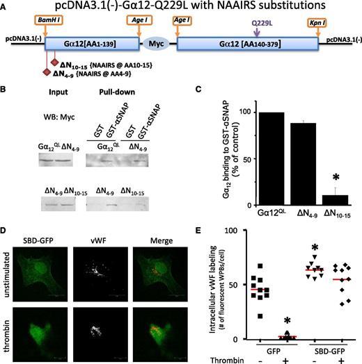
We observed that α-SNAP siRNA blocks basal, thrombin-evoked, and Ca2+ ionophore-induced secretion of vWF (Figure 3D; supplemental Figure 4B), indicating that α-SNAP-mediated fusion of WPBs is required for vWF secretion. We next explored the role of Gα12/αSNAP interactions in the regulation of vWF secretion, using a 2-step experimental approach. First, we used a library of Gα12-substitution mutants33 (Figure 6A) in which the primary sequence of Gα12 was replaced with the NAAIRS cassette to assess domain-specific in vitro binding of Myc-tagged Gα12Q229L with or without NAAIRS substitution of N-terminal amino acids 4-9 (ΔN4-9) or 10-15 (ΔN10-15) (Figure 6A) to GST-αSNAP. As shown in Figure 6B-C, 6 residues in the primary sequence of the Gα12 (R10CLLPA15 ) were required for Gα12 binding to α-SNAP, whereas ΔN4-9 mutant binding to α-SNAP was not significantly different from Gα12QL. Second, to assess the role of Gα12 interaction with α-SNAP in vWF secretion, we constructed a GFP-tagged α-SNAP binding domain (aa 10-15) minigene (GFP-SBD) based on the identified Gα12⋅GST-αSNAP interaction. In unstimulated cells, vWF staining in GFP-SBD-expressing HUVECs was similar that in to control GFP-expressing cells (Figure 6D). However, intracellular vWF staining was significantly reduced after thrombin stimulation of control cells (expressing empty GFP vector), whereas vWF staining remained unchanged in GFP-SBD-expressing cells (Figure 6D-E). These results suggest that the GFP-SBD minigene inhibited thrombin-induced WPB exocytosis in support of our thesis that Gα12 promotes vWF secretion from WPBs by directly interacting with α-SNAP.
Gα12 interaction with α-SNAP. (A) Design of N-terminal Gα12QL substitution mutants with sequence Asn-Ala-Ala-Ile-Arg-Ser (NAAIRS). The ΔN10-15 Gα12QL construct was generated by replacing 6 consecutive residues (aa 10-15) with NAAIRS; the ΔN4-9 Gα12QL construct was generated by replacing aa 4-9 with NAAIRS. (B) Coimmunoprecipitation of Myc-tagged constitutively active Gα12Q229L mutant expressed in HEK 293A cells with endogenous α-SNAP. Myc tagged Gα12QL mutants33 were expressed in HEK 293A cells, and their interaction with GST-αSNAP was analyzed by pull-down assay. Constitutively active Gα12-ΔN4-9 was observed to bind αSNAP similar to full-length Gα12QL, whereas Myc-ΔN10-15 mutant bound much less to α-SNAP. (C) Densitometry values (mean ± SEM; n = 3 independent experiments). *P < .05 vs full length Gα12QL. (D) Confocal images of functional dominant-negative effect of GFP-tagged-SBD (aaN10-15, the putative αSNAP binding domain) on vWF secretion in HUVECs. Twenty-four hours after transfection, cells were serum-starved for 4 hours, washed, incubated with 25 nM thrombin for 15 minutes, fixed, and stained for vWF (red). (E) Each symbol represents the number of WPBs per individual cell. GFP-tagged SBD functioned as a competitive inhibitor of basal and thrombin-induced vWF exocytosis from WPBs. *P < .05 vs unstimulated empty GFP vector transfected cells; n = 7-15 cells/group; red line represents median value.
Gα12 interaction with α-SNAP. (A) Design of N-terminal Gα12QL substitution mutants with sequence Asn-Ala-Ala-Ile-Arg-Ser (NAAIRS). The ΔN10-15 Gα12QL construct was generated by replacing 6 consecutive residues (aa 10-15) with NAAIRS; the ΔN4-9 Gα12QL construct was generated by replacing aa 4-9 with NAAIRS. (B) Coimmunoprecipitation of Myc-tagged constitutively active Gα12Q229L mutant expressed in HEK 293A cells with endogenous α-SNAP. Myc tagged Gα12QL mutants33 were expressed in HEK 293A cells, and their interaction with GST-αSNAP was analyzed by pull-down assay. Constitutively active Gα12-ΔN4-9 was observed to bind αSNAP similar to full-length Gα12QL, whereas Myc-ΔN10-15 mutant bound much less to α-SNAP. (C) Densitometry values (mean ± SEM; n = 3 independent experiments). *P < .05 vs full length Gα12QL. (D) Confocal images of functional dominant-negative effect of GFP-tagged-SBD (aaN10-15, the putative αSNAP binding domain) on vWF secretion in HUVECs. Twenty-four hours after transfection, cells were serum-starved for 4 hours, washed, incubated with 25 nM thrombin for 15 minutes, fixed, and stained for vWF (red). (E) Each symbol represents the number of WPBs per individual cell. GFP-tagged SBD functioned as a competitive inhibitor of basal and thrombin-induced vWF exocytosis from WPBs. *P < .05 vs unstimulated empty GFP vector transfected cells; n = 7-15 cells/group; red line represents median value.
Discussion
Using a combination of several independent model and assay systems, including G protein–deficient mice, blood loss measurement after tail-snip, cremaster arteriolar laser-injury thrombus formation, isolated buffer-perfused mouse lungs, and specific siRNA-depleted cultured human ECs derived from umbilical vein and pulmonary artery (HUVEC and HPAEC), we investigated the molecular mechanism of EC-specific vWF secretion.
Previous studies using megakariocyte-restricted-Gα12 and Gα13-deficient mice demonstrated that Gα13-dependent signaling regulates platelet aggregation and shape change, as well as U46619- and PAR-4-induced P-selectin degranulation.28 In contrast, the present study in ECs indicates that Gα13 knock-down has no effect on basal or evoked vWF release. Rather, using a loss-of-function approach as well as exogenous expression of mutationally activated Gα12, we observed that Gα12 mediates both basal (or constitutive-like, which maintains the circulating concentration of vWF) and thrombin-evoked (or regulated) vWF secretion (which results in the local release of vWF at the site of vascular injury).
We identified 2 independent mechanisms that may explain how Gα12 modulates both basal and evoked secretion of vWF from ECs. First, Gα12 interacts directly with α-SNAP and functions to facilitate WPB exocytosis at the plasma membrane. Second, Gα12 appears to be involved in the delivery of WPBs containing vWF to the plasma membrane. In eukaryotic cells, Gα12 in its GTP-bound form is known to activate p115 RhoGEF and its downstream effector RhoA, leading to increased actin polymerization and actinomyosin cross-bridge cycling.33 As recently described by Nightingale et al,38 dynamic assembly and disassembly of actin rings at the base of WPBs promotes the translocation of WPBs to the plasma membrane and exocytosis of vWF once fused. In support of this assertion, our studies suggest that classical Gα12-dependent signaling via p115RhoGEF and RhoA activation may promote actin-dependent delivery of WPBs to the membrane independent of Gα12-mediated activation of the exocyst complex.
In cultured HUVECs and HPAECs, we observed impaired thrombin-evoked vWF secretion resulting from siRNA-mediated Gα12 and Gαq depletion, whereas only Gα12 deficiency abolished basal secretion. Thus, Gα12 and Gαq/11 signaling appears to be required for basal and/or evoked vWF secretion from mouse and human ECs. HUVECs depleted of α-SNAP by specific siRNA downregulation were also unable to release vWF basally or in response to thrombin or calcium ionophore A23187. Therefore, Gα12 and α-SNAP are critical components of the vWF release mechanism, whereas Gαq/11 activation downstream of PAR-1 appears to be involved in potentiating vWF secretion.
Consistent with in vitro findings, experiments employing the isolated perfused mouse lung model demonstrated decreased basal EC-specific vWF release in Gα12−/− mice compared with WT, Gα11−/−, and EC-Gαq−/−;Gα11−/− mice. After stimulation of pulmonary vascular ECs with the PAR-1-activating peptide TFLLR, we observed significantly less vWF release from Gα12−/− and EC-Gαq−/−;Gα11−/− mouse lung ECs compared with WT mice. Mice lacking Gα12 displayed low plasma levels of vWF, which correlated well with defective exocytosis of vWF from WPBs in the absence of Gα12. Lack of EC Gα12/13 was also associated with increased blood loss after tail-snip in anesthetized mice, which could be rescued by infusion of purified human vWF. Furthermore, mice with Gα12 deficiency alone exhibited reduced platelet thrombus formation after laser injury to the isolated cremaster artery, as well as reduced flow-dependent adhesion of platelets on vWF strings in vitro. These functional defects were shown to be a result of impaired secretion and low plasma vWF level in absence of Gα12, rather than abnormal vWF multimerization.
vWF secretion has been described to occur via 3 pathways: constitutive, constitutive-like, and regulated, in which only regulated secretion requires agonist stimulation.20,39,40 Resting ECs reportedly secrete a small amount of pro-vWF via a canonical constitutive secretory pathway that is susceptible to protein synthesis inhibition.39 Constitutive-like (basal or spontaneous) secretion of vWF is thought to occur from storage granules in the absence of agonist stimulation.39,40 Thus, our studies support the contention that the majority of the vWF secreted from unstimulated ECs does not occur via the classical constitutive secretory pathway but, rather, via basal (constitutive-like) release from a post-Golgi storage compartment,40 presumably WPBs.39 On the basis of the results of the present study, we speculate that the low level, but otherwise normal-appearing, low–, intermediate–, and high–molecular weight plasma vWF multimers observed in Gα12−/− mice is a result of the lack of WPB priming and vWF secretion via Gα12-αSNAP-dependent activation of exocytosis.
As basal vWF secretion was reportedly calcium-independent,41 Gα12-mediated basal vWF secretion is also likely to occur independent of an increase in intracellular [Ca2+]. In contrast, endothelial Gαq/11 signaling may be important for potentiating vWF release in response to thrombin via Ca2+-dependent activation of αSNAP.42 With regard to the molecular mechanism by which α-SNAP is involved in fusion and in exocytosis, it is known that the binding of α-SNAP and stimulation of NSF ATP-ase activity is Ca2+-dependent,42 that α-SNAP can be activated by posttranslational modification,43 and that S-nitrosylation of NSF inhibits WPB exocytosis.17 On the basis of the observed reduction in basal vWF secretion in Gα12-depleted ECs, we speculate that Gα12 facilitates the final step of secretion by potentiating the binding of NSF to α-SNAP, thereby increasing NSF ATP-ase activity and fusion of WPBs with the plasma membrane. Gα12 activation by PAR-1 at the plasma membrane appears to also play a role by promoting delivery of WPBs to the plasma membrane, whereas Ca2+ signaling liberated by the Gαq/11-dependent pathway may further activate α-SNAP activity (Figure 7). In summary, absence of Gα12 in ECs renders them unable to maintain receptor-independent basal vWF secretion or integrate on-demand signals coming from the coagulation cascade (ie, thrombin stimulation), whereas Gαq/11 deficiency resulted in moderate but significant attenuation of thrombin-mediated vWF secretion.
Working model of G protein signaling-dependent EC vWF secretion. vWF secretion occurs via Gα12-αSNAP interaction resulting in constitutive (basal) exocytosis (left). On thrombin stimulation (right), the rate of vWF secretion is potentiated by Gαq-dependent and Ca2+-induced activation of α-SNAP and by Gα12-RhoA-actin-dependent delivery of WPBs to the plasma membrane.
Working model of G protein signaling-dependent EC vWF secretion. vWF secretion occurs via Gα12-αSNAP interaction resulting in constitutive (basal) exocytosis (left). On thrombin stimulation (right), the rate of vWF secretion is potentiated by Gαq-dependent and Ca2+-induced activation of α-SNAP and by Gα12-RhoA-actin-dependent delivery of WPBs to the plasma membrane.
The online version of this article contains a data supplement.
There is an Inside Blood commentary on this article in this issue.
The publication costs of this article were defrayed in part by page charge payment. Therefore, and solely to indicate this fact, this article is hereby marked “advertisement” in accordance with 18 USC section 1734.
Acknowledgments
The authors thank Maricela Castellon, Oleg Chaga, Tiffany Sharma (University of Illinois at Chicago), and Paula Jacobi (Blood Center of Wisconsin) for excellent technical assistance; Barry Kreutz (University of Illinois at Chicago) for critical reading of the manuscript; Thomas Meigs (University of North Carolina, Ashville) for G12 constructs; and Stefan Offermanns (Max-Planck Institute) for providing G protein–deficient mice.
This work was supported by National Institutes of Health Heart, Lung and Blood Institute Research Program Project grants P01 HL60678 and R01 HL71626 (to R.D.M.), HL062350 and HL080264 (to X.D.), P30 HL101302 and HL109439 (to J.C.), HL081588 (to S.L.H.), and T32 HL072742 (Training Program in Cardiovascular Sciences, S.C. Dudley). Funding was also provided by the NFAT Project of the New Energy and Industrial Technology Development Organization and the FIRST program from the Japan Society for the Promotion of Science.
L.R. is a PhD candidate at the University of Illinois at Chicago. This article is based on a thesis submitted in partial fulfillment of the requirements for a doctoral degree.
Authorship
Contribution: L.R. designed and performed experiments, analyzed data, made the figures, and wrote the paper; A.A. designed research and analyzed data; S.L.H., G.L., O.C., D.J.V., F.R.B., and S.M.V. performed experiments and analyzed data; H.I., O.K.-A., T.H., N.S., T.K., and L.R. were involved in G protein monoclonal antibody production and screening; A.S.-T. and X.D. provided crucial reagents and equipment and analyzed data; K.K. and J.C. conducted experiments and analyzed data; and R.D.M. designed the research, analyzed the results, and wrote the paper.
Conflict-of-interest disclosure: The authors declare no competing financial interests.
Correspondence: Richard D. Minshall, Department of Anesthesiology and Department of Pharmacology, University of Illinois at Chicago, 835 S Wolcott Ave (m/c 868), Chicago, IL 60612; e-mail: rminsh@uic.edu.


