Abstract
Acute myeloid leukemia (AML) is a heterogeneous disease with a great variety of somatic driver mutations. Each AML carries specific (combinations of) acquired mutations, which enables minimal residual disease (MRD) detection in virtually every patient with next-generation sequencing (NGS). In recent years there has been an increasing interest to conduct longitudinal molecular MRD detection to better estimate prognosis, monitor treatment efficacy, as well as prediction of an impending relapse. The development of molecular monitoring on archived samples has been slow because only limited numbers of samples collected during the course of the disease were biobanked. We have systematically investigated whether archived May-Giemsa Grünwald (MGG)-stained bone marrow slides could be an alternative source of material to conduct NGS-based molecular MRD detection in AML. Since MGG-stained bone marrow evaluations are performed at high frequency to evaluate the response on therapy and repopulation of the bone marrow, we focused on these bone marrow slides taken at regular intervals during treatment.
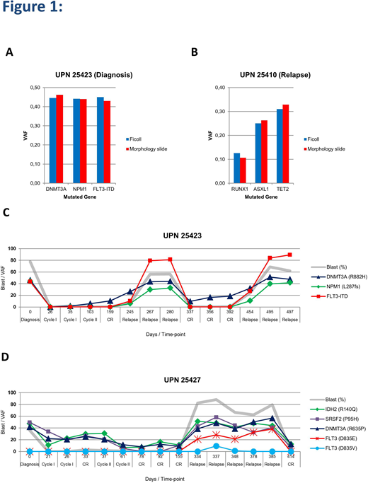
We initial tested DNA isolation protocol on MGG-stained and unstained morphology slides, which were archived over the years. The DNA could be isolated from the MGG-stained (n=16) and unstained bone marrow slides (n=16). However, we were unable to isolate genomic DNA from all of the unstained slides (11 out of 16). Furthermore, the DNA concentration from the unstained slides was much lower than the MGG-stained slides. We next isolated DNA from MGG-stained bone marrow slides of 43 AML samples at diagnosis and relapse of which the mutation status of genes frequently mutated in myeloid diseases was known. DNA isolated from Ficoll-purified mononuclear cells of the same pairs was previously sequenced by NGS using the Illumina Trusight Myeloid panel. NGS-based mutation screening with the same panel on DNA obtained from morphology slides revealed that detection of the driver mutations was feasible. In fact, all known mutations were detectable at diagnosis and relapse. Interestingly, in 91% (39 out of 43) of cases, the variant allele frequency (VAF) of the mutations were virtually identical to those detected using the DNA from MNCs. Representative examples are shown in Figure 1 (A and B). In a minority of samples, the VAFs differed between MNCs and bone marrow slides (3 cases VAF higher in MNCs and 1 case VAF lower in MNCs). These findings indicate that DNA isolated from bone marrow slides are a rich source for NGS-based mutation detection in AML at diagnosis and relapse.
We next sought to evaluate the use of MGG-stained bone marrow slides to study the mutations kinetics in time from AML diagnosis to relapse. To perform this analysis 30 paired-AML (diagnosis - relapse) with variable time to relapse (range: 200 to 4004 days) were selected. We collected the archived bone marrow slides at different time points between diagnosis and relapse, isolated DNA and performed NGS-based mutation detection. The NGS analysis of the 30 paired-AML cases showed different patterns of mutation dynamics and clonal evolution. Representative examples are shown in Figure 1 (C and D). Interestingly, by this systematic analyses of somatic mutations at various time points clear distinction could be made between leukemia-specific mutations and mutations associated with clonal hematopoiesis at the time of complete remission in the individual AML cases.
Our study uncovers an unutilized source of material for mutation detection at diagnosis and relapse in AML. These materials are widely available and well archived. Furthermore, our study reveals the potential use of MGG stained bone marrow morphology slides to track mutations and (sub)clones in AML, which is applicable to virtually every AML patient and possibly valuable for other hematological malignancies as well.
No relevant conflicts of interest to declare.
Author notes
Asterisk with author names denotes non-ASH members.


This feature is available to Subscribers Only
Sign In or Create an Account Close Modal