Current iron overload therapeutics have inherent drawbacks including perpetuated low hepcidin. Here, we unveiled that lactate, a potent hepcidin agonist, effectively reduced serum and hepatic iron levels in mouse models of iron overload with an improved erythropoiesis in β-thalassemic mice.
TO THE EDITOR:
Iron overload disorders are defined by the storage of excess iron in the body. The liver acts both as an iron reservoir and a central iron regulator to govern iron distribution and iron storage. Hepcidin, a 25 amino acid peptide hormone secreted from the liver, plays a pivotal role in iron homeostasis.1 Ferroportin (FPN) on the membranes of cells transports iron into blood plasma. Once bound by hepcidin, FPN undergoes occlusion, ubiquitination, and proteolysis.2 Therefore, the hepcidin-FPN axis determines systemic iron balance and iron deployment.3 Patients with iron overload require phlebotomy or iron chelators to prevent or manage the toxicity of high systemic iron levels. However, chelation therapy causes side effects that frequently limit compliance and therapeutic efficacy. Based on preclinical and very early clinical data, restoration of hepcidin levels could be an effective alternative strategy to prevent iron accumulation and ameliorate iron overload diseases.4,5 Recently, our study uncovered that lactate (LA) induces hepcidin via cyclic adenosine monophosphate (cAMP)-protein kinase A (PKA)-Smad pathway, which couples energy metabolism with iron metabolism.6 Considering that LA, the metabolic intermediate, exhibited low toxicity, we hypothesized that LA treatment could improve iron overload diseases.
Consistent with our recent findings,6 LA treatment increased the phosphorylation of Smad1/5/8 and hepcidin expression in AML-12, Hepa1-6, and HCCLM3 hepatocyte cell lines, dependent on soluble adenylyl cyclases–mediated cAMP production (supplemental Figure 1, available on the Blood website). To explore the effect of LA on serum hepcidin levels, 129S wild-type mice received a single intravenous injection of LA at a dose of 150 mg/kg body weight. As shown in supplemental Figure 2A, the serum LA level increased quickly after injection of LA, reached its peak concentration at 5 minutes with 1.6-fold increase relative to the baseline, and dropped rapidly afterward. In contrast, the LA level in the liver tissue exhibited longer retention than the serum level, with 2.3-fold increase at 20 minutes, reaching the peak level with 3.9-time elevation at 30 minutes, relative to the baseline, followed by a gradual decline and a return to baseline after 12 hours (supplemental Figure 2B). In response to the LA elevation, the serum hepcidin was significantly induced at 6 hours with 2.2-fold increase, and the induction reached the peak at 12 hours with 3.1-fold increase, followed by a decline and a turn to the baseline at 48 hours (supplemental Figure 2C). Therefore, these results showed that LA administration greatly induced serum hepcidin. Similar to our previous findings in vitro that LA acts as a hepcidin activator through the cAMP-PKA-Smad pathway, LA administration markedly enhanced the phosphorylation of CREB1 and Smad1/5/8 in vivo (supplemental Figure 2D).
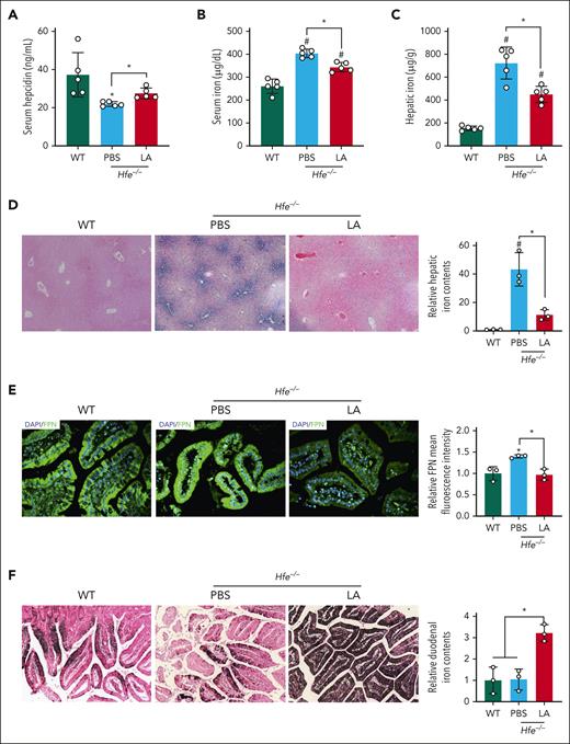
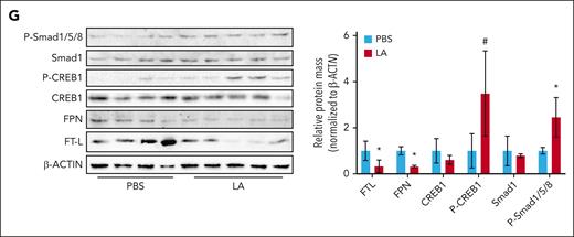
We next tested the stimulatory effect of LA on hepcidin expression in 2 hepcidin-deficient models, namely hemochromatosis (Hfe−/−) and β-thalassemia (Hbbth3/+) mice, and explored the ability of LA to modify iron overload and distribution. After treatment, no toxicity was observed in Hfe−/− mice, as evidenced by minimal changes in organ histology and markers of inflammation and tissue injury (supplemental Figure 3). Of note, no pH change was observed in sera. Intriguingly, LA administration increased serum hepcidin levels by 25.3% relative to phosphate-buffered saline (PBS)-treated mice (Figure 1A). As a consequence, serum iron and hepatic tissue iron levels were respectively reduced by 14.8% and 37.8% in LA-challenged Hfe−/− mice relative to control mice (Figure 1B-C). Perls staining corroborated the reduction of hepatic iron accumulation in LA-treated mice in comparison with untreated control (Figure 1D). In response to elevated hepcidin levels in LA-treated Hfe−/− mice, FPN was greatly diminished in the duodenum sections relative to untreated controls (Figure 1E). In agreement, the iron transport into plasma was inhibited in the duodenum, as characterized by substantial iron accumulation in duodenal sections (Figure 1F). The phosphorylation of Smad1/5/8 and CREB1 was induced, and the FPN and ferritin light chain (FTL) were decreased in the liver of mice with LA treatment (Figure 1G). Encouragingly, consistent results were found in Hfe−/− mice with LA treatment for 6 weeks (supplemental Figure 4A-C). Consistently, our ex vivo data revealed that LA treatment increased hepcidin expression of isolated primary Hfe−/− hepatocytes with 2.5-fold induction, compared with untreated control (supplemental Figure 4D). In analogy, LA induced intracellular cAMP levels and enhanced the phosphorylation of Smad1/5/8 and CREB1 (supplemental Figure 4E-F). Additionally, LA treatment ameliorated the hepatic oxidative stress, as evidenced by the reduced hepatic malondialdehyde contents, compared with untreated control (supplemental Figure 4G).
LA reduces iron burden in Hfe−/− mice. Four-week-old Hfe−/− mice were treated with intravenous LA (150 mg/kg body weight) or PBS for 4 weeks (n = 5 mice per group). (A) Changes in serum hepcidin levels of Hfe−/− mice receiving LA treatment. (B-C) Changes in serum (B) and hepatic tissue (C) iron contents in Hfe−/− mice with LA treatment. (D) Perls Prussian blue staining of liver sections (×100) from Hfe−/− mice after LA treatment, with quantitative analysis of iron content in the right panels. (E) Immunofluorescent staining of FPN (in green), and 4′,6-diamidino-2-phenylindole (DAPI, blue) in the duodenum sections (×400) from Hfe−/− mice upon LA treatment with quantitative analysis in the right panel. (F) Enhanced 3,3'-diaminobenzidine (DAB) iron staining of duodenum sections (×200) from Hfe−/− mice after LA treatment, with quantitative analysis of iron content in the right panels. (G) Western blotting of Smad1, P-Smad1/5/8, CREB1, P-CREB1, FPN, and FTL in liver specimens from LA-treated mice relative to untreated control with quantitative analysis in the right panel. ∗P < .05 and #P < .001, relative to untreated WT mice or as indicated. Data are represented as mean ± standard deviation. WT, wild type.
LA reduces iron burden in Hfe−/− mice. Four-week-old Hfe−/− mice were treated with intravenous LA (150 mg/kg body weight) or PBS for 4 weeks (n = 5 mice per group). (A) Changes in serum hepcidin levels of Hfe−/− mice receiving LA treatment. (B-C) Changes in serum (B) and hepatic tissue (C) iron contents in Hfe−/− mice with LA treatment. (D) Perls Prussian blue staining of liver sections (×100) from Hfe−/− mice after LA treatment, with quantitative analysis of iron content in the right panels. (E) Immunofluorescent staining of FPN (in green), and 4′,6-diamidino-2-phenylindole (DAPI, blue) in the duodenum sections (×400) from Hfe−/− mice upon LA treatment with quantitative analysis in the right panel. (F) Enhanced 3,3'-diaminobenzidine (DAB) iron staining of duodenum sections (×200) from Hfe−/− mice after LA treatment, with quantitative analysis of iron content in the right panels. (G) Western blotting of Smad1, P-Smad1/5/8, CREB1, P-CREB1, FPN, and FTL in liver specimens from LA-treated mice relative to untreated control with quantitative analysis in the right panel. ∗P < .05 and #P < .001, relative to untreated WT mice or as indicated. Data are represented as mean ± standard deviation. WT, wild type.
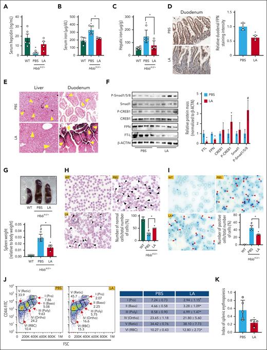
Following the same experimental plan as that with Hfe−/− mice, no overt signs of toxicity were observed in Hbbth3/+ mice (supplemental Figure 5A). LA treatment induced remarkable hepatic hepcidin messenger RNA and protein levels and reduced serum and hepatic iron by 31.9% and 47.9% in Hbbth3/+ mice, compared with untreated control, respectively (Figure 2A-C and supplemental Figure 5B), nearly to the levels seen in wild-type mice, indicative of a robust therapeutic effect. In agreement, decreased FPN staining and increased iron accumulation were observed in duodenum sections from LA-treated mice compared with untreated control (Figure 2D-E). Similarly, enhanced phosphorylation of Smad1/5/8 and CREB1 and decreased FPN and FTL protein levels were detected in the liver of mice following LA administration (Figure 2F). Additionally, LA treatment reduced hepatic malondialdehyde levels by 25.1% and 4-hydroxynonenal mass by 66.0% in liver specimens from Hbbth3/+ mice, compared with untreated control (supplemental Figure 5C-D). Together, these findings suggested that LA induces hepcidin expression to decrease the FPN protein and consequently prevent iron absorption in the duodenum, thus lowering serum and tissue iron load and oxidative stress in Hfe−/− and Hbbth3/+ mice.
LA ameliorates iron burden in Hbbth3/+ mice. Four-week-old Hbbth3/+ mice were treated with intravenous LA (150 mg/g body weight) or vehicle for 4 weeks (n = 4-5 mice per group). (A-C) Serum hepcidin levels (A) and serum (B) and hepatic tissue (C) iron contents of wild-type (WT) and Hbbth3/+ mice in response to LA administration. (D) Immunohistochemical staining of FPN on duodenum sections (×100) from Hbbth3/+ mice after LA administration, with quantitative analysis in the right panel. (E) Enhanced DAB iron staining of liver (×200) and duodenum (×200) sections from Hbbth3/+ mice with LA treatment. Brown represents iron deposition (denoted by yellow arrows). (F) Western blotting of Smad1, P-Smad1/5/8, CREB1, P-CREB1, FPN, and FTL in liver specimens from LA-treated mice relative to untreated control, with quantitative analysis in the right panel. (G) Spleen morphology (the upper panel) and spleen weight per body weight (the lower panel) of WT and Hbbth3/+ mice treated with LA or solvent. (H) Blood smears stained with Wright-Giemsa stain, with black arrows indicating damaged or deformed erythrocytes (×1000) and quantification of normal cells. (I) Reticulocyte counts were performed using new methylene blue staining with red arrows indicating positive cell (×1000) and quantification of positive cells. (J) Representative erythropoiesis profiles of bone marrow from Hbbth3/+ mice treated with LA or PBS. (K) Index of splenic erythropoiesis (as calculated by spleen weight × % spleen erythroid cells determined in flow cytometry analysis). ∗P < .05 and #P < .001, relative to untreated WT mice or as indicated. Data are represented as mean ± standard deviation. Baso, basophilic erythroblasts; Ortho, orthochromatic erythroblasts; Poly, polychromatic erythroblasts; Pro, proerythroblasts; Retic, reticulocytes.
LA ameliorates iron burden in Hbbth3/+ mice. Four-week-old Hbbth3/+ mice were treated with intravenous LA (150 mg/g body weight) or vehicle for 4 weeks (n = 4-5 mice per group). (A-C) Serum hepcidin levels (A) and serum (B) and hepatic tissue (C) iron contents of wild-type (WT) and Hbbth3/+ mice in response to LA administration. (D) Immunohistochemical staining of FPN on duodenum sections (×100) from Hbbth3/+ mice after LA administration, with quantitative analysis in the right panel. (E) Enhanced DAB iron staining of liver (×200) and duodenum (×200) sections from Hbbth3/+ mice with LA treatment. Brown represents iron deposition (denoted by yellow arrows). (F) Western blotting of Smad1, P-Smad1/5/8, CREB1, P-CREB1, FPN, and FTL in liver specimens from LA-treated mice relative to untreated control, with quantitative analysis in the right panel. (G) Spleen morphology (the upper panel) and spleen weight per body weight (the lower panel) of WT and Hbbth3/+ mice treated with LA or solvent. (H) Blood smears stained with Wright-Giemsa stain, with black arrows indicating damaged or deformed erythrocytes (×1000) and quantification of normal cells. (I) Reticulocyte counts were performed using new methylene blue staining with red arrows indicating positive cell (×1000) and quantification of positive cells. (J) Representative erythropoiesis profiles of bone marrow from Hbbth3/+ mice treated with LA or PBS. (K) Index of splenic erythropoiesis (as calculated by spleen weight × % spleen erythroid cells determined in flow cytometry analysis). ∗P < .05 and #P < .001, relative to untreated WT mice or as indicated. Data are represented as mean ± standard deviation. Baso, basophilic erythroblasts; Ortho, orthochromatic erythroblasts; Poly, polychromatic erythroblasts; Pro, proerythroblasts; Retic, reticulocytes.
Next, we explored the role of LA in improving ineffective hematopoiesis in Hbbth3/+ mice. As shown in Figure 2G, mice with LA treatment exhibited significant reduction in the spleen-to-body weight ratio by 52.2% compared with mice treated with PBS. And the spleen histological examination also underlined modestly reduced splenic erythropoiesis (supplemental Figure 5A). The red cell morphology in blood smears was characterized by widely varying sizes and shapes and cell fragmentation in PBS-treated Hbbth3/+ mice. Strikingly, the appearance of erythrocytes in blood smears significantly improved with increased proportion of normal cells in peripheral blood (Figure 2H) and declined reticulocyte counts after LA treatment (Figure 2I). Of note, the serum erythropoietin and erythroferrone levels were significantly reduced by 27.1% and 62.1%, respectively, in Hbbth3/+ mice in comparison with untreated control (supplemental Figure 5E-F). Thereby, our results suggested that LA administration indeed improved erythropoiesis.
Hematological data showed that LA treatment did not fully rescue the red blood cell (RBC) indexes of Hbbth3/+ mice, as comparable values were observed for RBC, hemoglobin, mean corpuscular volume, and hematocrit with or without treatment. Nonetheless, the red cell distribution width was significantly reduced by 17.3% in LA-treated Hbbth3/+ mice compared with untreated control (supplemental Figure 5G), which echoes the results of blood smear. In mice treated with LA, the proerythroblasts, basophilic erythroblasts, polychromatic erythroblasts, and orthochromatic erythroblasts in the bone marrow were modestly decreased, and the proportions of reticulocytes and RBCs were slightly elevated, indicating modestly improved erythropoietic function of the bone marrow (Figure 2J).
Next, we closely looked at the erythropoiesis in the spleen. As shown in supplemental Figure 5H and Figure 2K, the proportion of total erythroid cells (Ter119+ cells) was reduced by 17.8% and the index of splenic erythropoiesis was decreased by 57.4% in the LA-treated mice relative to untreated control. Moreover, the proportions of polychromatic erythroblasts and orthochromatic erythroblasts were modestly elevated and the proportion of mature RBCs was reduced in the spleen (supplemental Figure 5I), suggesting somewhat alleviated splenic erythropoiesis in mice receiving LA treatment. Collectively, these results suggested that LA treatment reduced extramedullary erythropoiesis in Hbbth3/+ mice, showing improvement of ineffective erythropoiesis (albeit to a mild extent).
Here, we found that LA administration markedly increased hepcidin expression and decreased the serum and hepatic tissue iron concentrations as well as oxidative stress in Hfe−/− and Hbbth3/+ mice. Moreover, LA had slight effect on ineffective erythropoiesis in Hbbth3/+ mice, although more investigations are warranted in the future. These findings may have implications for pathophysiology and therapeutics of iron overload disorders.
Acknowledgements
The authors thank all laboratory members for their assistance with experiments and reagents.
This research was supported by the National Natural Science Foundation of China (grants 22021003, 21920102007 and 22150006) and National Key Research and Development Program of China (grant 2021YFA0719302).
Authorship
Contribution: S.L. and S.Z. supervised and designed the study; W.L., Y.W., and H.W. performed experiments and analyzed data; S.L., S.Z., and W.L. wrote the manuscript; W.F. and Q.Y. participated in experiments, data analysis, and the manuscript preparation; S.L., T.G., S.Z., and J.M. advised on experimental design and helped with experiments; T.G. participated in study design and revised the manuscript; and all authors discussed the results, revised the manuscript, and contributed to the manuscript.
Conflict-of-interest disclosure: The authors declare no competing financial interests.
Correspondence: Sijin Liu, Research Center for Eco-Environmental Sciences, Chinese Academy of Sciences, 18 Shuangqing, Haidian District, Beijing, China 100085; email: sjliu@rcees.ac.cn; and Shuping Zhang, Medical Science and Technology Innovation Center, Shandong First Medical University and Shandong Academy of Medical Sciences, 6699 Qingdao, Huaiyin District, Jinan, China 250117; email: spzhang@sdfmu.edu.cn.
References
Author notes
W.L., Y.W., and H.W. contributed equally to this work.
Data are available on request from the corresponding authors, Sijin Liu (sjliu@rcees.ac.cn) and Shuping Zhang (spzhang@sdfmu.edu.cn).
The online version of this article contains a data supplement.


This feature is available to Subscribers Only
Sign In or Create an Account Close Modal