Abstract
Bone marrow cells (BMCs) from CXB-12/HiaJ (CXB-12) mice had 14 times the total long-term repopulating ability found in the best of 11 other CXB recombinant inbred (RI) lines. BMCs from each RI line donor were mixed with genetically marked standard competitor BMCs from the BALB/cBy×C57BL/6 F1 (CByB6F1) hybrid, the mice used to produce the RI lines, and the mixtures repopulated lethally irradiated CByB6F1 recipients. Percentages of donor-type erythrocytes and lymphocytes measured the actual long-term repopulating functions of the donor RI lines relative to the standard competitor. CXB-12 BMCs repopulated better after 3 or 6 months than after 1 month, suggesting that the most primitive precursors were involved. Compared to CByB6F1 standard competitor cells, CXB-12 cells repopulated 3 to 12 times as well, with their advantage increasing when higher doses of cells were transplanted, probably because of hybrid resistance of the recipient against low doses. This was far better than expected, because F1 cells normally function 2 to 3 times as well as cells from an inbred strain. In competitive dilution, the advantage resulted from 2 factors: more precursor cells and more function per precursor. In the model that best fit the data, CXB-12 donors had 2.4 times the concentration of hematopoietic stem cells (HSCs) as the CByB6F1 standard, and each HSC repopulated 1.4 times as well. CXB-12 mice did not have elevated erythrocyte and lymphocyte numbers in blood and marrow and did not have unusually elevated concentrations of colony-forming unit spleen, cobblestone colonies, and long-term colony-initiating cells in marrow.
Introduction
Hematopoietic stem cells (HSCs) function long term and proliferate to self-renew and differentiate to produce progenitors of all hematopoietic lineages.1-5 Cells obtained from bone marrow, cord blood, or mobilized peripheral blood of healthy donors are clinically useful as sources of transplantable HSCs. After transplantation, donor HSCs compete for engraftment with residual host HSCs.6 High levels of function from donor HSCs are extremely valuable for competing effectively with host HSCs in many clinical conditions.7-10 Unfortunately, donor HSCs are often damaged by clinical manipulations so that they repopulate less well than residual host HSCs.11-13 Analysis of the genetically superior HSCs reported here may suggest how functional abilities of donor HSCs can be improved.14-17
A broad range of genetic backgrounds and defined genetic combinations are available in mouse models,18,19 including recombinant inbred (RI) lines.20-22 The set of CXB RI lines used in the current study were derived from inbred BALB/cBy (BALB) and C57BL/6By (B6) strains by intercrossing individuals from the F2 generation and then inbreeding for at least 20 generations of brother–sister mating. All mice of a particular RI line are genetically identical and homozygous at all loci, with approximately half the loci derived from B6 and half from BALB.20
The competitive repopulation assay directly measures the ability of a population of donor HSCs to engraft recipients relative to the repopulating ability of a population of standard competitor HSCs.23-25 When genetically distinguishable donor and standard competitor cells are mixed in a specific ratio and injected into lethally irradiated recipients, the fraction of donor-type blood cells is proportional to the fraction of donor cells injected if neither type of HSC has a repopulating advantage. If a mixture of such donor–competitor cells were engrafted in a 2:1 ratio, two thirds of the blood cells would be donor type. If the observed proportion of donor-type blood cells is significantly different from the proportion of donor cells transplanted, a repopulating advantage or disadvantage in the donor HSCs is detected. The percentage of donor type blood cells in the recipient circulation is used to calculate total repopulating units (RUs) in a donor cell population relative to the standard competitor,25 where 1 donor RU has the same repopulating ability as 105 standard competitor bone marrow cells (BMCs).
We compared the relative long-term repopulating abilities of 12 CXB RI lines in vivo in the current study, using the CByB6F1 hybrid of the BALB and B6 parent strains as recipients and standard competitors because they carry all B6 and BALB alleles. Therefore, they provide the best system available in which long-term functional ability of HSCs from multiple CXB RI lines can be studied in a constant environment, though F1 hybrid resistance in the recipients must be taken into consideration. The advantage of this technique is the direct measurement of long-term function in vivo relative to a CByB6F1 standard. BMCs from CXB-12 donors had the highest long-term repopulating advantage ever reported. The new competitive dilution assay shows that CXB-12 BMCs have both an increased concentration of HSCs and an increased functional ability per HSC.
Materials and methods
Mice
Normal inbred B6 (Gpi1b/Gpi1b), BALB (Gpi1a/Gpi1a), congenic B6, and BALB strains differing at Gpi1, hybrid CByB6F1 (Gpi1a/Gpi1b), and recombinant inbred CXB lines 1-12 were raised at The Jackson Laboratory in a specific pathogen-free facility. CByB6F1 competitors of Gpi1b/Gpi1b genotype were produced from B6 × congenic BALB (Gpi1b/Gpi1b) matings, whereas CByB6F1 competitors of Gpi1a/Gpi1a genotype were produced from BALB×congenic B6 (Gpi1a/Gpi1a) matings. All mice in the current study were used between 2 and 4 months of age, except for old CXB-12 donors that were 24 months old. Mice were fed NIH-31 (4% fat) diet ad libitum with standard animal husbandry management as published.18
Bone marrow transplantation and competitive repopulation
BMCs were flushed from 2 femurs and 2 tibias of each donor or competitor mouse through a 23-gauge needle with 2.0 mL Iscove modified Dulbecco medium and filtered through a 100-μm mesh nylon cloth to remove debris. Nucleated cells were counted using a model ZBI Coulter Counter (Coulter Electronics, Hialeah, FL). Donor BMCs were mixed with aliquots of competitor BMCs in specific ratios (see figure legends) and injected intravenously into recipients. Except where noted differently, CByB6F1 recipients were sublethally irradiated with 5 Gy (500 rads) 14 days before transplantation and intraperitoneally injected with 100 μg/mouse of a natural killer (NK) cell antibody (anti-NK1.1, clone PK 136) 3 days before transplantation to deplete NK cells (NK cell–depleted recipients). This alleviates hybrid resistance against B6, BALB, and CXB RI donor cells.26 27 In all cases, recipients were given 11 Gy (1100 rads) lethal irradiation 4 to 6 hours before marrow injection. Irradiation was from a cesium Cs 137 gamma source (Shepard Mark 1; Shepard & Associates, Glendale, CA) at 1.6 Gy (160 rads)/minute.
CXB RI lines 2, 4, 5, 6, 10, and 12 are Gpi1a/Gpi1a type, whereas CXB RI lines 1, 3, 7, 8, 9, and 11 are Gpi1b/Gpi1b type; each type of donor BMCs was mixed with BMCs from CByB6F1 competitors specially bred to have a different Gpi1 allele. Because recipients were normal CByB6F1 mice, their cells would have been detected easily by their heterozygous Gpi1a/Gpi1belectrophoretic bands; however, this was not found. From blood collected 1, 3, and 6 months after reconstitution by the mixtures of marrow, percentages of donor-derived erythrocytes and lymphocytes were measured after separating Gpi1 types by cellulose acetate gel electrophoresis, as reported earlier.25
Repopulating units
Relative repopulating abilities are most accurately compared when cells from each of the different donors are mixed with cells from the same standard competitor pool in the same experiment. Each RU is the relative repopulating ability of 105 fresh marrow cells from the same standard competitor pool. So that independent experiments could be compared, fresh marrow from standard, young congenic CByB6F1 hybrid competitors was used with a variety of donors in each experiment. Numbers of RUs from each donor were calculated from the observed percentage donor cells, where the number of fresh competitor marrow cells used per 105 equalled C. Then by definition, the percentage is 100 (RU/RU + C). By algebraic rearrangement, this gives RU = % (C)/(100 − %). Thus, if a donor population contains 20 RUs, it repopulates as well as 20 × 105 standard competitor marrow cells, and mixtures of 20 donor RUs plus 20 × 105 competitor cells produce an average of 50% donor-type erythrocytes and lymphocytes in the recipients. Total RUs per donor were estimated, assuming that the BMCs in both femurs and tibias were 25% of the total marrow cells in an adult mouse.
Blood and bone marrow composition and colony-forming unit spleen
Blood samples were taken from young male and female B6, BALB, CByB6F1, and CXB-12 mice through the orbital sinus. Concentrations of white blood cells (WBCs) and red blood cells (RBCs) were counted using the ZBI Coulter Counter described earlier. Portions of peripheral blood samples were incubated in Gey solution to lyse RBCs and then were stained with B220, CD4, and CD8 antibodies and analyzed through fluorescence activated cell staining (FACS) using a FACScan II (Becton Dickinson, Mountain View, CA) flow cytometer to determine percentages of B220, CD4, and CD8 lymphocytes. BMCs from young B6, BALB, CByB6F1, and CXB-12 mice were also tested for lymphocyte composition through FACS analysis.
To measure colony-forming unit spleen (CFU-S), 105 BMCs were injected into each of 4 strain-matched, lethally irradiated recipients. All recipients were killed at day 12. The spleen of each recipient was removed, fixed in Bouin fixative, and examined for the numbers of macroscopic colonies. Short-term production of CFU-S was tested as above, except in lethally irradiated CByB6F1 recipients, using 4 × 10⁵ BMCs from lethally irradiated CByB6F1 carriers given 106 BMCs 7 days earlier or using 2 × 10⁵ BMCs from lethally irradiated CByB6F1 carriers given 106 BMCs 14 days earlier.
Competitive dilution using Poisson modeling
Portions containing 5 × 10⁴ CXB-12 donor BMCs (Gpi1a/Gpi1a) and 5 × 10⁵ CByB6F1 standard competitor BMCs (Gpi1b/Gpi1b) were injected into each of 38 NK cell–depleted CByB6F1 recipients. The Poisson function, Pi = e−N × (Ni/i!), gives the probability (Pi) that a recipient gets a certain number (i) of HSCs, where N is the average number of HSCs injected into each recipient.27 28 The proportion of recipients with 0% CXB-12–type cells 6 months after reconstitution was used to compute the number of HSCs (N) in CXB-12 BMCs using the i = 0 term of the Poisson function: P0 = e−N or n = −lnP0. This gave n = 1.2 for 5 × 10⁴ CXB-12 donor BMCs. Then the probabilities (Pi) of having 0, 1, 2, 3, or 4 donor HSCs in a recipient were, according to the Poisson function: 0.3012, 0.3614, 0.2169, 0.0867, or 0.0260, respectively. Because 5 × 10⁵competitor cells were used, n = 5.0 for the competitor, giving probabilities of having 0, 1, 2, 3, 4, 5, 6, 7, 8, or 9 competitor HSCs in a recipient as: 0.0067, 0.0337, 0.0842, 0.1404, 0.1755, 0.1755, 0.1462, 0.0653, 0.0363, or 0.0181, respectively. In practice, we calculated 2 arrays of probabilities, one for the donor, and one for the competitor, with i = 0 to 15 for 16 probabilities in each case. These form a 16 × 16 matrix of 256 combined probabilities, and each is associated with a donor:competitor HSC ratio that can be used to predict a value representing donor contribution. In the current study we used the highest 37 probability values because there were 37 recipients available 6 months after transplantation.
We used the 37 corresponding donor:competitor ratios in the Poisson modeling to estimate repopulating ability per cell for CXB-12 HSCs by comparing 3 hypothesized levels of repopulating abilities per cell for CXB-12 HSCs: F = 1, F = 1.4, and F = 2, each relative to a repopulating ability per CByB6F1 standard HSC of F = 1. From the value of F, a predicted percentage donor value was calculated for each of the 37 donor:competitor ratios associated with the 37 highest probabilities. This produced 3 sets of predicted donor percentage values, each ranked from low to high and compared to the 37 ranked observed data in a paired t test. The hypothesized F values that generated predictions significantly different from observed data were rejected. The F value whose predictions were not significantly different from the observed data were accepted to represent repopulating ability per cell for CXB-12 donor HSCs relative to those of the CByB6F1 standard.
Long-term colony-initiating cells
The long-term colony-initiating cell (LTC-IC) assay was performed as described by Müller-Sieburg and Riblet.14 Briefly, bone marrow cells were seeded onto confluent layers of the stromal cell line S17 in 96-well plates. At least 48 wells were seeded per cell dilution, and the dilations covered the range of 1 × 10⁴ to 625 cells/well. Wells that contained colonies of small granulocytic cells were enumerated at the indicated time points, and LTC-IC frequencies were calculated from maximum likelihood statistics. The B6-Ly5 congenic mouse was used because, as described previously, it has the same frequency of LTC-IC as the parental C57BL/6 mouse.
Cobblestone area–forming cell assay
Procedures for cobblestone area–forming cell assay (CAFC) were adopted as described by Ploemacher et al,29 with a feeder layer from an S17 stroma cell line in 96-well microplates.14,27,30 At confluence, the S17 feeder cells were irradiated with 50 Gy (5000 rads) and overlaid with BMCs from individual donors. Each well contained 0.2 mL cobblestone media (10% fetal calf serum, 10% horse serum, 80% α-MEM, 50 IU/mL penicillin, 50 μg/mL streptomycin, 50 μmol/L β-mercaptoethanol, 10 μmol/L hydrocortisone sodium succinate, and 2 mmol/L L-glutamine). On each plate, 20 wells were used for each of 4 cell doses (1644, 3780, 8695, or 20 000 donor BMCs), and 16 wells with no cells were controls.31 All plates were cultured at 33°C with 5% CO2 and fed weekly by changing half the media in each well. At 3 and 5 weeks of incubation, wells were scored positive if they had one or more colonies of at least 6 cells within the stromal layer with a flat cobblestone shape under a phase-contrast microscope. CAFC frequencies (N) in the cell dose were calculated from the fractions of negative wells (P0) where n = −lnP0.
Statistical analyses
During Poisson modeling, differences between ranked predicted values and ranked observed data were analyzed through pairedt test. Strain differences were tested through variance analyses using the JMP statistical discovery software (SAS Institute, Cary, NC) on “Fit Y by X” and “Fit Model” platforms, respectively.32
Results
CXB-12 BMCs repopulate far better than BMCs from 11 other CXB lines
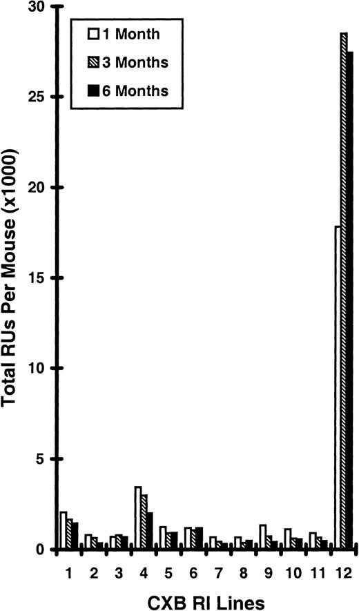
HSC function was directly compared in 12 different CXB RI lines (Figure 1) by mixing BMCs from 2 donors of each line with a portion from 1 of 2 pools of standard competitor CByB6F1 marrow. Recipients were NK cell–depleted and lethally irradiated, and each was given 4 × 10⁶ donor BMCs mixed with 2 × 10⁶ CByB6F1 competitor BMCs of the opposite Gpi1 type. After 1, 3, and 6 months, numbers of RUs were calculated as detailed in “Materials and methods.” The Gpi1 marker used to distinguish donor and competitor standard cells in competitive repopulation does not affect repopulating abilities25,28 33and thus could not cause the CXB-12 advantage.
Relative HSC repopulating abilities of CXB RI lines 1 to 12.
Four million donor cells (CXB RI lines) plus 2 million standard competitor cells (CByB6F1) were given to each NK cell–depleted, lethally irradiated CByB6F1 recipient, all of the same gender. Donor RU concentrations were calculated as described in “Materials and methods” and were multiplied by total BMCs per donor, assuming that 2 tibias and 2 femurs contain 25% of the total BMCs, to compute total RUs per mouse. Because CXB-12 percentages were 98% and 96%, so high that an error of a few percentage points greatly alters RU numbers, the data from the 3 × 10⁶ CXB-12:107 F1 ratio in Table 1 were considered more accurate and used in this Figure. Data are presented as means, with standard error bars from 2 donors tested separately for each CXB RI line.
Figure 1 shows an enormous (P < .0001) strain effect as CXB-12 donors repopulated far better than donors of the other 11 CXB RI lines. CXB-12 donors had 14 times more RUs at 6 months, representing the most primitive HSCs, than did CXB-4 donors, the next highest, and 36 times more than the average (764 RUs per donor) for the other 11 RI lines. The repopulating advantage of CXB-12 BMCs increased greatly between 1 month and 3 months. This increase with time did not occur with the other RI lines. The major histocompatibility locus (H2) does not explain why CXB-12 mice have such a high repopulating ability because the CXB-1, 4, 9, and 12 lines are all H2d/H2d, but CXB-1 and CXB-4 repopulate only slightly better than the other CXB RI lines (all H2b/H2b homozygotes), whereas CXB-9 does not repopulate any better than the others.
Repopulating advantage of CXB-12 BMCs increases with time and with increasing numbers of BMCs
Repopulating abilities of CXB-12 BMCs were tested in a separate study, using 10 × 105 or 30 × 105 CXB-12 BMCs mixed with 100 × 105 standard competitor BMCs from CByB6F1. This study is displayed with 2 independent studies in Table1. Although some CXB-12 repopulating advantage was present in the short-term HSCs responsible for repopulation after 1 month, by far the strongest advantage occurred in the more primitive HSCs, which repopulate after 3 and 6 months (Table1). In all cases, the repopulating advantage of CXB-12 BMCs increased greatly between 1 month and 3-6 months after transplantation.
Dose-dependent hematopoietic stem cell repopulating advantage in CXB-12 mice
| Months . | CXB-12:CByB6F1 BMC ratio . | |||
|---|---|---|---|---|
| 5 × 10⁴:5 × 10⁵ | 106:107 | 3 × 10⁶:107 | 4 × 10⁶:2 × 10⁶ | |
| % CXB-12 cells* | ||||
| 1 | 10.2 ± 1.3 | 29.0 ± 4.0 | 66.0 ± 13.3 | 90.4 ± 2.8 |
| 3 | 19.5 ± 2.5 | 39.8 ± 2.7 | 82.6 ± 8.9 | 98.4 ± 1.3 |
| 6 | 21.9 ± 2.9 | 39.0 ± 2.4 | 78.5 ± 9.3 | 95.8 ± 2.5 |
| Expected CXB-12:CByB6F1 RUs (% CXB-12) without CXB-12 repopulating advantage† | ||||
| 0.5:5 (9.1) | 10:100 (9.1) | 30:100 (23.1) | 40:20 (66.7) | |
| Observed CXB-12:CByB6F1 RUs‡ | ||||
| 1 | 0.6:5 (1.2) | 41:100 (4.1) | 194:100 (6.4) | 188:20 (4.7) |
| 3 | 1.2:5 (2.4) | 66:100 (6.6) | 474:100 (16) | 1232:20 (31) |
| 6 | 1.4:5 (2.8) | 64:100 (6.4) | 366:100 (12) | 456:20 (11) |
Recipients were natural killer cell–depleted, lethally irradiated, young CByB6F1 hybrids. Cells descended from CXB-12 donor and CByB6F1 competitors were distinguished by their Gpi1 marker. Mean ± SE are given for 37 recipients per donor for the 5 × 10⁴:5 × 10⁵ donor:competitor ratio from the experiment detailed in Figure 3; for 4 recipients per donor in a second experiment for the 106:107 and 3 × 10⁶:107 ratios; and for 8 recipients per donor for the 4 × 10⁶:2 × 10⁶ ratio from the experiment in Figure 1.
RU indicates repopulating unit.
Average of erythrocytes and lymphocytes.
The RU is defined as 1 per 105 BMCs for CByB6F1 competitors.
Fold CXB-12 advantage.
Percentages of CXB-12 type erythrocytes and lymphocytes in recipients after 3 and 6 months were far higher than expected if CXB-12 donor and CByB6 F1 competitor cells repopulated equally (Table 1). However, the advantage of CXB-12 BMCs increased with total numbers of donor cells. It was only 2.4- to 2.8-fold using the lowest cell numbers, 5 × 10⁴ donor cells plus 5 × 10⁵ standard F1 cells. When cell numbers were increased, using 106 donor cells plus 107 standard F1 cells, the CXB-12 advantage increased to 6.4- to 6.6-fold. It further increased to a 12- to 16-fold advantage using 3 × 10⁶ donor plus 107standard F1 cells (Table 1), probably because some hybrid resistance against CXB-12 cells remained in the recipients and was saturated when higher numbers of CXB-12 BMCs were used. The 11- to 31-fold advantage with 4 × 10⁶ donor plus 2 × 10⁶ standard F1 cells showed the same effect; the wide range resulted from inaccuracies when donor percentages were greater than 90%, so that a difference of a few percentage points greatly affected RU calculations.
CXB-12 mice have normal myeloid, lymphoid, and 12-day CFU-S values
CXB-12 mice have similar peripheral blood WBC and RBC concentrations in comparison to B6, BALB, and CByB6F1 mice (Table2). Although there were significant strain differences in peripheral blood B220 and CD4 lymphocyte percentages, total BMC numbers, and marrow percentages of B220 or CD8 cells, in all cases values for CXB-12 mice were similar to those of at least one of the controls (Table 2). Furthermore, day 12 CFU-S from fresh marrow or pre–CFU-S from carriers after 7 or 14 days was not increased in CXB-12 mice (Table 2). Despite high primitive HSC function, regulatory mechanisms in CXB-12 mice maintain blood, marrow, and short-term precursor cell concentrations at normal levels.
CXB-12 mice have normal numbers of peripheral blood cells, bone marrow cells, and colony-forming unit spleen
| Parameter . | B6 . | BALB . | CByB6F1 . | CXB-12 . |
|---|---|---|---|---|
| In peripheral blood | ||||
| WBC (109/L) | 15.0 ± 0.9 | 10.7 ± 1.9 | 11.9 ± 2.4 | 10.2 ± 2.3 |
| RBC (109/mL) | 1.12 ± 0.31 | 1.38 ± 0.33 | 1.51 ± 0.41 | 1.43 ± 0.28 |
| B220 (%) | 54.6 ± 4.5 | 20.5 ± 1.9 | 30.3 ± 2.1 | 32.5 ± 3.7 |
| CD4 (%) | 11.5 ± 0.1 | 29.4 ± 3.5 | 32.0 ± 1.0 | 20.1 ± 1.3 |
| CD8 (%) | 12.0 ± 0.3 | 14.8 ± 0.9 | 16.0 ± 0.1 | 14.3 ± 1.3 |
| In bone marrow | ||||
| Total BMC (105) | 3040 ± 288 | 2163 ± 32 | 2528 ± 87 | 3043 ± 149 |
| B220 (%) | 27.7 ± 0.1 | 28.1 ± 1.1 | 23.1 ± 1.3 | 24.3 ± 0.2 |
| CD4 (%) | 0.84 ± 0.07 | 0.78 ± 0.06 | 0.72 ± 0.07 | 0.74 ± 0.08 |
| CD8 (%) | 1.53 ± 0.05 | 0.66 ± 0.05 | 0.82 ± 0.10 | 0.53 ± 0.11 |
| CFU-S, day 12 | 8.8 ± 0.8 | 9.5 ± 1.0 | 9.0 ± 1.5 | 10.0 ± 0.6 |
| Pre–CFU-S, day 7 | 2.5 ± 1.2 | 3.7 ± 1.2 | 2.4 ± 1.0 | 1.0 ± 0.3 |
| Pre–CFU-S, day 14 | 3.0 ± 0.6 | 3.0 ± 0.5 | 1.0 ± 0.5 | 1.6 ± 0.4 |
WBC indicates white blood cell; RBC, red blood cell; BMC, bone marrow cell; CFU-S, colony-forming unit spleen.
Data given as mean ± SEM after variance analysis; N = 3–8. Pre–CFU-S were retransplanted to give numbers of 12 day CFU-S in CByB6F1 recipients of 4 × 10⁵ BMCs from CByB6F1 carriers given 106 BMCs 7 days earlier or 2 × 10⁵ BMCs 14 days earlier.
Both HSC concentration and repopulating potential per HSC are increased in CXB-12 mice
We used competitive dilution to test whether the CXB-12 advantage is caused by increased HSC numbers, functional ability per HSC, or both. Each of 38 CByB6F1 recipients was given 5 × 10⁴CXB-12 donor BMCs mixed with 5 × 10⁵ CByB6F1 competitor BMCs. Six months after reconstitution, 11 of 37 recipients (1 died after 2 months) had 0% CXB-12 type (Gpi1a/Gpi1a) blood cells, giving a Poisson (n = 1.2) for 5 × 10⁴ CXB-12 donor BMCs or 2.4 HSCs in 105 CXB-12 BMCs (Table3).
Hematopoietic stem cell concentration and repopulating ability per hematopoietic stem cell using competitive dilution
| CXB-12 BMC injected in each recipient | 5 × 10⁴ |
| CByB6F1 BMC injected in each recipient | 5 × 10⁵ |
| No. recipients injected | 38 |
| No. recipients available at 6 months | 37 |
| No. recipients with 0% donor type blood cell at 6 months | 11 |
| No. HSCs in 5 × 10⁴ CXB-12 BMCs | 1.2 |
| P0 = 11/37 = 0.2973; N = −lnP0 = −ln 0.2973 = 1.2 | |
| No. HSCs in 105 CXB-12 BMCs | 2.4 |
| No. HSCs in 5 × 10⁵CByB6F1 BMCs (assumed)3-150 | 5 |
| Repopulating potential per CByB6F1 HSC (defined) | 1 |
| Repopulating potential per CXB-12 HSC relative to CByB6F1 standard | 1.4 |
BMC indicates bone marrow cell; HSC, hematopoietic stem cell.
Recipients are 2- to 4-month-old, natural killer cell–depleted, lethally irradiated, male CByB6F1 mice. BMCs were pooled from 3 young (2- to 3-month-old) CXB-12 males (Gpi1a/Gpi1a) and competed with BMCs pooled from 3 young CByB6F1 males (Gpi1b/Gpi1b).
For CByB6F1 competitors, HSC concentration is taken as 1 per 105 BMCs; repopulating ability per HSC is defined as 1.
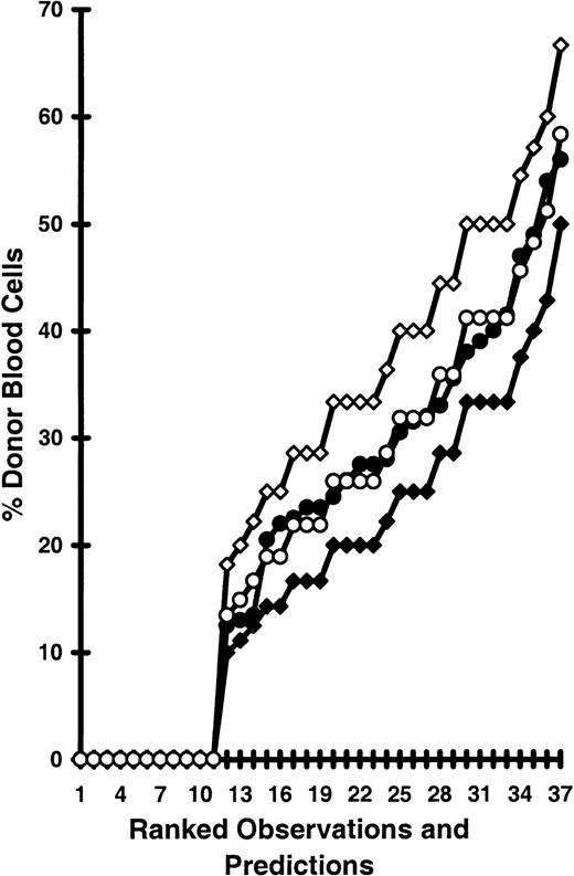
In Figure 2, F values (repopulating function relative to competitor HSCs) of 1.0, 1.4, and 2.0 in donor HSCs were compared. Of the 3 sets of predictions, the F = 1.4 model best fit observed data, whereas models using F = 1 or F = 2 generated predictions significantly below or above the observed data. Thus, each CXB-12 HSC repopulated 1.4 times as well as each CByB6F1 HSC (Table 3). Because the dose of CXB-12 BMCs used in this study was very low, the hybrid resistance that remained in the CByB6F1 recipients probably reduced the CXB-12 HSC functional advantage. Nevertheless, the concentration of HSCs was 2.4 times that of the F1 hybrid standard, and the functional ability was 1.4 times the standard.
CXB-12 cell numbers and repopulating abilities per cell.
The observed donor cell percentages after 6 months for the 37 recipients in the competitive dilution assay described in “Materials and methods” are ranked from low to high and shown as OBS (●). We hypothesized 3 levels of repopulating potential—“F” for each CXB-12 donor HSC relative to F = 1 for each CByB6F1 competitor HSC; F = 1 (♦), F = 2 (⋄), and F = 1.4 (○)—and generated 3 sets of predictions based on combined Poisson probabilities as described in “Materials and methods.” The predictions from models F = 1 and F = 2 were significantly different from actual observations; thus, models F = 1 and F = 2 were rejected. Predictions from model F = 1.4 were not significantly different from actual observations. Thus, F = 1.4 best represents the repopulating ability of a CXB-12 HSC relative to that of a CByB6F1 HSC.
Repopulating advantage of CXB-12 cells in recipients with full hybrid resistance
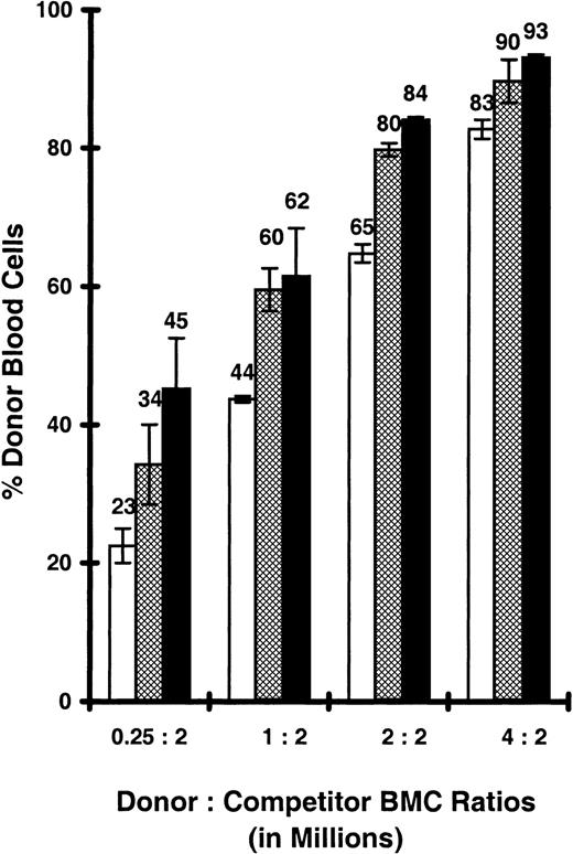
Hybrid resistance reduced but did not remove the CXB-12 advantage in recipients that were not treated to remove NK cells. Figure3 shows percentages, and RU values are given in the legend. Compared to the CByB6F1 standard competitor, CXB-12 BMCs had a 2-fold repopulating advantage after 1 month, despite the presence of recipient NK cells. After 3 and 6 months, the advantage averaged 3.9- and 5.4-fold. Although the increase with time was still obvious, the increased repopulation with donor cell numbers was only proportional to their 16-fold increase in donor BMC numbers, from 0.25 to 4.0 million BMCs per recipient, whereas the amount of standard F1 cells was constant at 2.0 million BMCs. There was no longer an increase in donor cell efficiency that would have been shown by an increase in RU concentration. Because the CByB6F1 recipients were not treated to remove hybrid resistance, its effect may have been constant, not saturated by increasing doses of donor cells, as in Table 1.
CXB-12 HSC engraftment advantage persists despite hybrid resistance.
Bone marrow cells pooled from young CXB-12 donors (Gpi1a) were mixed with 2 × 10⁶BMCs from CByB6F1 competitors (Gpi1b) using 0.25, 1, 2, or 4 × 10⁶ donor cells, respectively, and were injected into lethally irradiated CByB6F1 recipients that were not pretreated to remove NK cells, using 4 recipients per dose. At 1 (■), 3 (▩), and 6 (▪) months after reconstitution, percentage donor erythrocytes and percentage donor lymphocytes were averaged for each recipient and presented as means with standard error bars. If there were no repopulating advantage in CXB-12 HSCs, the expected percentages from CXB-12 donors were 11%, 33%, 50%, and 68% for the 4 donor:competitor ratios, respectively. The differences between observed and expected CXB-12 contributions translate to 2.3-, 1.6-, 1.8-, and 2.4- (average, 2.0)-fold CXB-12 HSC repopulating advantages after 1 month; 4.2-, 2.9-, 4.0-, and 4.4- (average, 3.9)-fold advantages after 3 months; and 6.6-, 3.2-, 5.3-, and 6.7- (average, 5.4)-fold advantages after 6 months.
In separate studies, repopulating abilities of 2 million BALB or B6 donor BMCs in CByB6F1 recipients with intact hybrid resistance were tested with 2 million CByB6F1 competitor standard BMCs. BALB cells contained between 39% and 48% as many RU as found in CByB6F1 cells, approximately 10-fold less than the relative RU concentrations of CXB-12 donors in Figure 3. BMCs from B6 donors contained only 10% to 15% as many RU as found in CByB6F1 cells because of the strong resistance in H-2d/H-2bF1 hybrids against H-2b/H-2b donors.
LTC-IC frequency in CXB-12 bone marrow
In the long-term LTC-IC assay, the LTC-IC is detected by its ability to repopulate a stromal layer in limiting-dilution cultures by forming colonies of myeloid cells. Concentrations of LTC-ICs in vitro from BMCs of B6, BALB, CXB-12, and CXB-4 BMCs were compared at 4, 5, and 6 weeks (Table 4). As previously reported, B6 values were approximately 10% of those found in BALB mice.14,27 30 The CXB-12 strain had values similar to those of BALB, and the CXB-4 values were intermediate. However, in competitive repopulation, BALB cells repopulate approximately 10 times less well than CXB-12 cells, as noted above. Thus, the CXB-12 advantage is not detected by the LTC-IC assay.
Long-term colony-initiating cell concentrations per 105 bone marrow cells
| Time in culture . | B6 . | BALB . | CXB-12 . | CXB-4 . |
|---|---|---|---|---|
| Experiment 1 | ||||
| 4 weeks | 2.9 (1.9–4.4) | 13.9 (11.1–17.5) | 14.0 (11.2–17.6) | 5.4 (3.9–7.4) |
| 5 weeks | 1.8 (1.0–3.2) | 7.4 (5.6–9.8) | 8.6 (6.6–11.1) | 2.8 (1.8–4.2) |
| Experiment 2 | ||||
| 4 weeks | 1.7 (0.9–2.8) | 15.2 (12.1–18.9) | 16.1 (12.3–21) | 7.7 (5.9–10.1) |
| 5 weeks | 1.0 (0.5–2.1) | 4.8 (3.5–6.6) | 7.0 (5.6–11.3) | 3.3 (2.3–4.9) |
| 6 weeks | 0.7 (0.3–1.7) | 4.1 (2.9–5.7)* | 5.2 (3.4–8.1) | 2.6 (1.7–4.0) |
Data given as mean (95% confidence limits) after limiting-dilution analysis.
CAFC frequency in CXB-12 bone marrow
The ability of B6, BALB, CXB-12, and CByB6F1 BMCs to form long-term CAFC colonies in vitro were compared at 5 weeks using S17 feeder cells in limiting-dilution analysis. Results, given as mean ± SD per 105 BMCs for the following strains, were: B6 = 1.6 ± 0.7; BALB = 1.8 ± 0.8; CXB-12 = 5.3 ± 1.8, and CByB6F1 = 3.6 ± 1.2. CXB-12 donors had significantly higher CAFC frequencies than B6 and BALB donors showing nonoverlapping 95% confidence limits. However, though CXB-12 BMCs had higher CFAC concentrations than CByB6F1 BMCs, the differences were not significant. Thus, the CXB-12 advantage was not detected by the CAFC assay.
CXB-12 HSC repopulating abilities in old donors
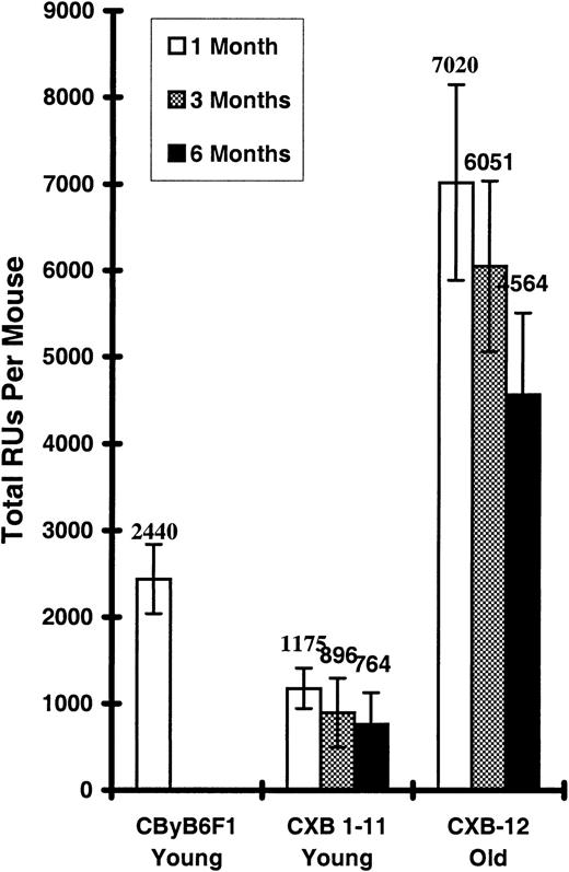
There was a substantial decline with age in HSC function in CXB-12 mice, with old/young RU ratios of 0.21 after 3 months and 0.17 after 6 months (comparing Figures 1 and 4). Still, old CXB-12 BMCs had uniquely high functional ability. Figure 4shows 24-month-old CXB-12 donors tested in the same experiment presented in Figure 1. When comparing levels of cell production by old CXB-12 donors with average values for young donors in the other 11 CXB RI lines, the old CXB-12 donors still repopulate at least 6 times better. Repopulating abilities of young donors from CXB RI lines 1 to 11 declined relative to the CByB6F1 competitor as the time after transplantation increased from 1 to 6 months, probably because of a repopulating advantage in the F1 hybrid competitor cells. This also occurred for old CXB-12 donors. Nevertheless, even after 6 months, old CXB-12 BMCs still contained approximately twice the repopulating ability of the young CByB6F1 competitors (Figure 4).
Relative HSC repopulating abilities in old CXB-12 donors.
Bone marrow cells from 4 male 24-month-old CXB-12 donors (Gpi1a/Gpi1a) were mixed with BMCs from young congenic CByB6F1 standard competitors (Gpi1b) and injected into NK cell–depleted lethally irradiated CByB6F1 recipients using 4 recipients per donor, each given 4 × 10⁶ donor and 2 × 10⁶competitor BMCs. Experimental conditions, analyses, and calculations were the same as in Figure 1. “CXB1-11 young” gives the average total RUs per mouse of the CXB RI lines 1 to 11 shown in Figure 1. Total RUs per CByB6F1 competitor is 1 RU per 105 BMCs multiplied by the average total BMCs ( × 10−5) per competitor mouse, assuming 2 tibias and 2 femurs contain 25% of the total bone marrow.
Discussion
Competitive repopulation directly measures how well donor HSCs engraft recipients relative to standard competitor HSCs.23Among all the assays for HSCs, it best models reconstitution during a therapeutic transplantation in which donor HSCs are delivered into the recipient bloodstream, home to functional sites, and compete with residual host HSCs.6 A population of donor HSCs, with the large repopulating advantage seen in CXB-12 mice (Figure 1), would dramatically enhance the effectiveness of HSC transplantation as a therapeutic procedure.
Hybrid effects
F1 HSCs probably benefit from heterosis, and even after treatments to remove NK cells, recipients probably still have residual hybrid resistance. These factors explain why BMCs from CXB RI lines 1 to 11 all contain less long-term repopulating ability than BMCs from the CByB6F1 competitor (Figure 1). Hybrid resistance explains why the advantage of CXB-12 BMCs increases with higher doses of cells, even though the competitor cell numbers are increased proportionally (Table1). The residual resistance is likely saturated by using donor cell doses of 3 to 4 million.26 When NK cells were not removed, hybrid resistance reduced the CXB-12 advantage to approximately 3-5 fold; there was no effect of cell number because the resistance in untreated recipients was not saturated by the donor cell doses used (Figure 3).
Individual HSCs
The CXB-12 advantage was due to both a 2.4-fold increased HSC concentration and a 1.4-fold proliferative advantage (Figure 2, Table3). Either increased numbers of HSCs in CXB-12 marrow or increased ability to home to sites supporting long-term HSC function, or both, can explain the increase in HSC concentration. The CXB-12 advantage in this competitive dilution experiment was greatly reduced by residual hybrid resistance because the dose of donor cells was only 50 000 BMCs, and the total repopulating advantage at this dose was approximately 5 times less than at higher doses (Table 1). Hybrid resistance in CByB6F1 recipients reduced BALB HSC concentrations from 10 to 4 per million BMCs27; if hybrid resistance had the same type of effect on CXB-12 HSC, their concentration of 24 per million is several times too low.
Donor reactions against competitors
The extremely good fit between the F = 1.4 model predictions and the observed data in Figure 2 suggests that there is no killing of standard competitor CByB6F1 HSCs by CXB-12 marrow. The substantial degree of killing required to produce the CXB-12 repopulating advantage would have reduced the observed concentration of competitor HSCs and increased the number of recipients that had no competitor HSC engraftment. This did not occur. In fact, the HSC concentration of 1 per 105 competitor BMCs gave an excellent fit (Figure 2). Furthermore, marrow cells in adult mice from the other 11 CXB RI lines did not repopulate as well as the F1 hybrid competitor (Figure 1). Each of the 12 CXB RI lines tested in the current study is homozygous for either the b or the d allele at the major histocompatibility locus, so each could recognize the alternate allele carried by the CByB6F1 hybrid as foreign. If the repopulating advantage resulted from killing antigenically disparate competitor cells, all the CXB RI lines would have shown this advantage, not just the CXB-12. Finally, mouse marrow has only weak graft-versus-host activity because of low concentrations of T cells; percentages of CD4- and CD8-bearing marrow cells were low in CXB-12 marrow (Table 2), so it would not react significantly against the F1 hybrid cells.
Other assays for hematopoietic precursor cells
A particularly interesting aspect of CXB-12 marrow is that its long-term repopulating advantage does not cause higher numbers of 12 day CFU-S (Table 2) than found in control inbred or F1 hybrid mice. Apparently, precursor differentiation is regulated to produce normal numbers of CFU-S and normal numbers of myeloid and lymphoid cells (Table 2).
Neither CAFC or LTC-IC (Table 4) frequencies are significantly higher in CXB-12 mice than in controls. LTC-IC populations contain precursors capable of long term repopulation in vivo30; perhaps those in CXB-12 mice have an inferior homing ability in vitro, or a superior homing ability in vivo, relative to those in controls. This is not the first case in which LTC-IC and competitive repopulation assays differ. Both here and in prior work,14 LTC-IC concentrations in BALB BMCs were approximately 10-fold higher than those in B6 BMCs. Yet competitive dilution in congenic recipients with congenic competitors gave similar HSC concentrations of approximately 10 per million BMCs in BALB and B6.34 35
Proliferative exhaustion
Total RUs per mouse were reduced approximately 5-fold with age in CXB-12 donors (Figures 1, 4). One possibility for this reduction is that increasing age alters HSC homing during transplantation, as in B6 mice.28 Another possibility is that accelerated HSC proliferation in young CXB-12 mice leads to early exhaustion. In the latter case, the functional ability of old CXB-12 HSCs should be lower than the average for old donors from the other 11 CXB RI lines. In fact, old CXB-12 BMCs repopulated better than those of young CByB6F1 competitors or those of young donors from CXB RI lines 1 to 11 (Figure4). We have reported previously that 9 of 11 other CXB RI lines had low old/young RU ratios.27 The loss with age in CXB-12 is similar to the reduction with age seen in these strains and in the BALB strain, but not in the other 2 CXB RI lines or in the B6 strain.27,35 The high rate of loss with age segregated in the RI lines with the BALB locus at the D12Nyu17 marker on chromosome 12.27
Genetic implications
Earlier reports, based on LTC-IC and CAFC frequency measurements in vitro, pointed to a few chromosome regions that may contain genes regulating HSC concentration in the mouse.14-16 It will be interesting to test whether the CXB-12 HSC hyperfunction phenotype is related to these chromosome regions. However, it is important to distinguish the phenotypes tested in prior studies from the phenotype in the current report. CXB-12 BMCs show far more effective repopulation and function than young F1 BMCs in F1 recipients for 6 months, approximately a quarter of the murine life span. It is the unprecedented degree of engraftment advantage relative to high doses of healthy competitor BMC that makes the CXB-12 hyper-HSC phenotype novel.
The phenotypes in Figure 1 were tested in a genome-wide QTL mapping analyses using the Map Manager QTX-06 program with the CXB RI mice genotype data, both available online (http://www.jax.org). This analysis did not produce any linkage that is considered significant (lod score, greater than 3.0). Only insignificant linkages to D6Mit15 (lod score, 0.8) and D17Mit22 (lod score, 1.0) were found, probably by random chance, because more than 500 markers were tested. The CXB-12 hyper-HSC phenotype is not controlled by a single Mendelian locus differing between the BALB or the B6 parent strains because the hyper phenotype does not occur in either. It is found in only 1 of the 12 RI lines, so the simplest genetic explanations are either a new mutation or a unique combination of BALB and B6 genes. Testing HSC functional phenotypes in hybrid and backcross mice between CXB-12 and its parental strains will distinguish these 2 possibilities. Based on preliminary data, it could be a codominant mutation or a combination in which a single locus has a significant effect in approximately 40% of the backcross mice, though the effect is reduced from that in the CXB-12. Future studies using B6-H-2d × BALB F1 hybrid recipients and competitors to remove the strong hybrid resistance associated with a difference at the major histocompatibility locus may give clearer results.
If high stem cell function results from a unique combination of 2 alleles, at least 1 must be recessive—CByB6F1 mice have both BALB and B6 alleles at all loci, but they do not express the phenotype. One allele must be required from each parent because neither expresses the phenotype, and the combination must occur in CXB-12 but not in any of the other 11 RI lines. In theory, the chances that this occurs are [2 (¼) (¾)11] = 0.0211, or approximately 1 in 50 combinations of any 2 alleles. We tested this using a list of 253 loci defined in CXB RI lines 1 to 12 (from The Jackson Laboratory Informatics web site:http://www.informatics.jax.org/searches/riset_form.shtml). We compared alleles at pairs of loci for CXB-12 with those of the other 11 lines using an Excel function. In the first 2485 tested, there were 99 pairs unique to CXB-12, approximately 1 in 25. However, half of those pairs were both from the same parental strain, so, in fact, as expected 1 in 50 were pairs of alleles unique to CXB-12, with 1 allele from each strain. There are 253 × 252 = 63 756 possible allele pair permutations of 253 loci. Even considering that this is reduced by linkage, there are too many possibilities for our results using only 12 RI lines to have the statistical power to give significant, or even suggestive, map locations.
Primitive precursors are affected
Regulation of the hyper-HSC phenotype appears to be focused on more primitive HSCs because CXB-12 mice have normal numbers of myeloid cells, lymphoid cells, and less primitive precursors (Tables2, 4). In competitive repopulation, the phenotype is expressed most strongly after 3 and 6 months in the recipients (Table 1; Figures 1,3), which is the time required for primitive HSCs to repopulate. The high number and function of CXB-12 primitive HSCs may be maintained by alterations in a receptor that regulates primitive HSCs or in its ligand. Increases in numbers of HSCs could result from changes in either ligand or receptor; however, the increased proliferative capacity of CXB-12 HSCs after transplantation in normal recipients suggests that the change is intrinsic to the HSCs, perhaps an altered receptor. Understanding the CXB-12 phenotype may eventually lead to improved clinical transplantation procedures to enhance the effectiveness of primitive HSCs.
Acknowledgments
We thank Avis Silva, Bee Stork, and Karen Davis for their excellent technical assistance. We thank Dr Ben Taylor for helpful discussions about RI line analyses and Dean Campagna, an academic year student, and Benjamin H. Schmidt, a summer student, for performing the CAFC assays.
Supported by National Institutes of Health grants HL-58820, HL-58705, and DK-48015. J.C. was an NRSA postdoctoral fellow and was supported by grant AG-05754 from the National Institute on Aging.
The publication costs of this article were defrayed in part by page charge payment. Therefore, and solely to indicate this fact, this article is hereby marked “advertisement” in accordance with 18 U.S.C. section 1734.
References
Author notes
David E. Harrison, The Jackson Laboratory, Campus Box 41, 600 Main St, Bar Harbor, ME 04609; e-mail: deh@jax.org.


This feature is available to Subscribers Only
Sign In or Create an Account Close Modal