Abstract
Serine protease factor Xa plays a critical role in the coagulation cascade. Zymogen factor X is synthesized and modified in the liver. To understand the mechanisms governing the liver-specific expression of factor X, the proximal promoter of human factor X was previously characterized. Two crucial cis elements at −73 and −128 and their cognate binding proteins, HNF-4 and NF-Y, respectively, were identified. In this report, studies are extended to 3 additionalcis elements within the factor X promoter. Using gel mobility shift assays, the liver-enriched protein GATA-4 was identified as the protein binding to the GATA element at −96. GATA-4 transactivates the factor X promoter 28-fold in transient transfection experiments. It was also determined that the Sp family of transcription factors binds 2 DNase I–footprinted sites at −165 and −195. Disruption of Sp protein binding at either site reduces the promoter activity by half. Simultaneous disruption of both sites reduces the promoter activity 8-fold. This is the first report indicating the involvement of GATA-4 in the regulation of clotting factor expression. These observations provide novel insight into mechanisms by which the vitamin K–dependent coagulation factors are regulated.
Introduction
Blood coagulation factor X, in its activated form, plays a central role in the coagulation cascade. Circulating zymogen factor X is activated by the factor VIIa–tissue factor complex (extrinsic tenase) or by factor Ixa–VIIIa (intrinsic tenase). Membrane-bound factor Xa, together with its cofactor factor Va, converts prothrombin to thrombin, which in turn cleaves fibrinogen and results in the formation of the fibrin clot.1 In addition to its role in coagulation, factor Xa has recently been found to elicit intracellular signaling in leukocytes, endothelial cells, and vascular smooth muscle cells through binding to the effector cell protease receptor-1 and other yet to be identified receptors.2-5Activation of cells by factor Xa leads to the release of growth factors and cytokines important in the acute inflammatory response. Thus, factor Xa provides a crucial interface between the coagulation and inflammation processes.
Factor X, like other vitamin K–dependent coagulation factors, is primarily synthesized in the liver, where it undergoes extensive post-translational modifications including the γ-carboxylation of specific glutamic acid residues at the amino terminus.6This unique modification requires vitamin K as a cofactor and is essential for the biologic activity of factor X. Low levels of factor X transcripts are also found in nonhepatic tissues such as the reproductive organs (testis and ovary) and the digestive organs (small intestine and colon).7 The significance of extrahepatic expression of factor X remains to be determined.
To understand the mechanisms governing the expression of factor X, we have carried out functional characterization of the 5′ flanking region of the human factor X gene.7,8 Transcription of factor X initiates at multiple sites, a feature characteristic of TATA-less genes. The proximal 209 base pair (bp) 5′ to the translation start site are sufficient for maximal promoter activity. Using DNase I footprinting analysis with nuclear extracts from HepG2 cells, we identified 4 transcription factor–binding sites in this promoter fragment. Site 1(−73 to −44) binds the liver-enriched transcription factor hepatocyte nuclear factor 4 (HNF-4) and the ubiquitous factor Sp1. Notably, the promoters of 2 other vitamin K–dependent coagulation factors, factor VII and factor IX, also possess HNF-4 binding sites at similar positions with respect to the translation start sites. The recent identification of fatty acyl-CoA thioesters as ligands for HNF-4 further suggests that dietary fatty acid intake could affect the expression of these coagulation factors.9 Site 2 (−128 to −93) binds the ubiquitous transcription factor nuclear factor Y (NF-Y). Mutagenesis experiments demonstrated that both HNF-4 and NF-Y binding sites are critical for the promoter activity of factor X.
In this report, we further characterize the factor X promoter and identify GATA-4, Sp1/Sp3 as important positive regulators. We found that a gut-specific transcription factor GATA-4 binds a canonical GATA site at the 3′ end of site 2. Expression of GATA-4 significantly increases factor X promoter activity. We also identified the proteins binding at site 3 (−165 to −132) and site 4 (−195 to −169). Both Sp1 and Sp3 bind sites 3 and 4. Mutation of either site 3 or site 4 to disrupt Sp1/Sp3 binding only moderately affects the factor X promoter activity. In contrast, simultaneous disruption of both sites results in markedly reduced activity.
Materials and methods
Plasmids
The factor X promoter reporter construct, FXpGH-279, was described previously.8 Mutant constructs were generated from the wild-type FXpGH-279 by overlapping PCR mutagenesis. For the GATA site mutant, GATA at −96 (relative to the translation start site) was changed to CATA (mut GATA). For the Sp1/Sp3 site mutants, the following constructs were generated: m1, GCTGGGGCGT (−68 to −59) was changed to GCTGGTTCGT; m3, CTCCCCTGCC (−149 to −140) was changed to CTAACCTGCC; m4, GGTGGGGCC (−185 to −177) was changed to GGTGCTACC. Sequential mutagenesis was used to create constructs with simultaneous disruption of multiple Sp1/Sp3 binding sites (m1 + 3, m1 + 4, m3 + 4, m1 + 3 + 4). All mutations installed have been tested in gel mobility shift assays to confirm the complete loss of binding to their cognate transcription factors. The factor X promoter constructs contain 108 bp of the promoter fragment and were described previously.8FXpLuc-279 constructs expressing the luciferase reporter were generated by cloning the HindIII (blunted)–BamHI factor X promoter fragments (wild-type and mutants) into theSmaI–BglII sites of pGL3-basic (Promega, Madison, WI). Expression vector for GATA4, pCMV5-GATA-4 was generated by cloning the SacI–SalI fragment containing the coding sequence of GATA-4 (gift from Dr Yamagata) into pCMV5.Drosophila expression vectors for Sp1 and Sp3, pPacSp1 and pPacSp3, were gifts from Dr Horowitz.10
Reporter gene assays
Transient transfections and reporter gene assays in HepG2 and NIH3T3 cells were carried out as described before using FXpGH constructs.7 Secreted growth hormone was measured using radio-immunoassays (Nichols Institute, San Juan Capistrano, CA).Drosophila SL2 cells were maintained in Schneider's medium (Gibco BRL Life Technologies, Rockville, MD) supplemented with 10% heat-inactivated fetal bovine serum and grown at room temperature. SL2 cells were transfected by the calcium phosphate precipitation method as in HepG2 cells except that the precipitates were left on the cells for 48 hours until assay for reporter expression. Because DrosophilaSL2 cells do not express transfected human growth hormone gene, all reporter gene assays in SL2 cells were performed with FXpLuc constructs. Expression of the luciferase reporter was determined as instructed (Promega).
Gel mobility shift assays
Gel mobility shift assays for protein binding at site 3 and site 4 were carried out as described previously.7 Nuclear extracts used in the gel shift assays were prepared as described previously.7 For binding at the GATA site, salmon sperm DNA was omitted, and the concentration of MgCl2 was reduced to 1 mM. For supershift assays, the following antibodies were used: GATA-2 (C-20), GATA-4 (C-20), Sp1 (PEP-2), and Sp3 (D-20) (all from Santa Cruz Biotechnology, Santa Cruz, CA).
Modified aPTT assays using murine plasma
Modified aPTT tests were performed as described in the presence of human factor X–deficient plasma (Organon Teknika, Durham, NC).11 Plasma samples from heterozygous GATA-4 mice and wild-type littermates were gifts from Dr Leiden.
Results
DNase I footprint site 2 of the human factor X promoter contains a canonical GATA site
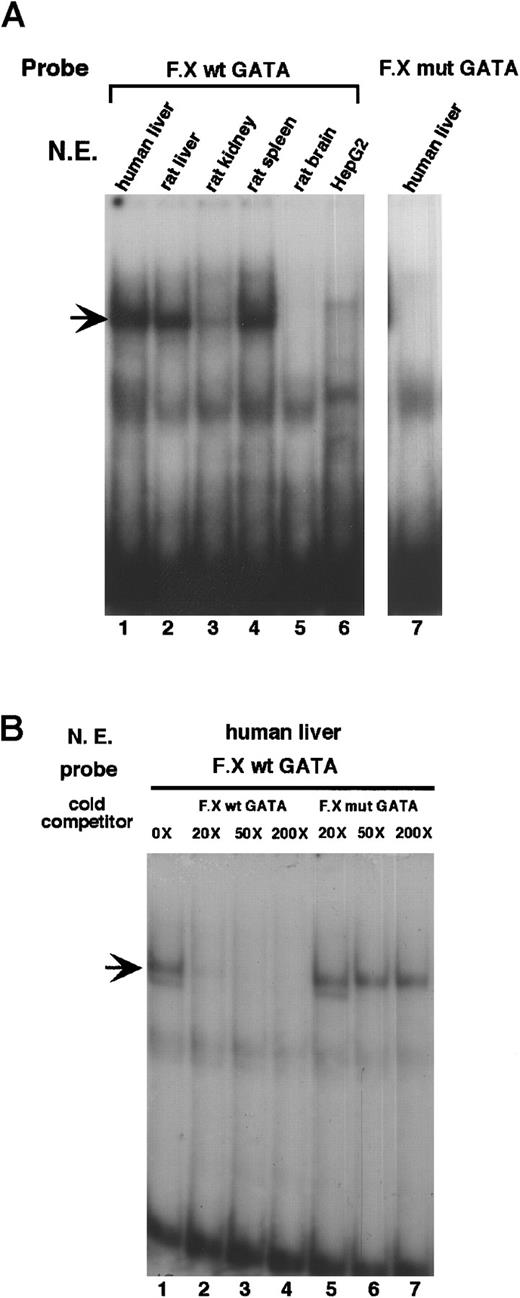
We previously carried out DNase I footprinting assays to determine transcription factor binding sites at the proximal promoter of factor X. Of the 4 protein-protected sites, site 2 (from −128 to −93) contains a CCAAT box and a canonical GATA binding site, TGATAA. We identified NF-Y as the protein that binds the CCAAT box. We next sought to determine whether the GATA site binds one of the GATA transcription factors. An oligonucleotide spanning −109 to −80 of the factor X promoter was radiolabeled and used in gel mobility shift assays. As shown in Figure 1A, the GATA site from the factor X promoter binds a specific protein in nuclear extracts from human liver, rat liver, rat kidney, and rat spleen. Interestingly, there is little binding activity in HepG2 nuclear extracts. This preparation of HepG2 nuclear extracts binds a site 3 oligonucleotide equally well when compared to the human liver extracts (data not shown), indicating that the low binding activity in HepG2 nuclear extracts is not an artifact caused by poor preparation. A single nucleotide substitution changing the GATA sequence toCATA completely abolishes binding of the oligonucleotide to the factor in liver nuclear extracts (Figure 1A, lane 7). To further demonstrate the specificity of the binding, wild-type and mutant oligonucleotides were used as cold competitors in gel mobility shift assays (Figure 1B). The addition of unlabeled wild-type oligonucleotide, even at a low concentration (20×), effectively competes away the specific protein-DNA complex (lanes 1 to 4), whereas the addition of unlabeled mutant oligonucleotide (up to 200×) has no effect on the formation of the complex (lanes 5 to 7).
The factor X promoter contains a canonical GATA site that binds a GATA factor in the liver.
(A) An oligonucleotide containing the GATA site from the factor X promoter, F.X wt GATA (−109 GCCTAAGCCAAGTGATAAGCAGCCAGACAA −80) was radiolabeled and incubated with 10 μg nuclear extracts from various tissues as indicated. F.X mut GATA contains a point mutation that changes GATA to CATA. An arrow indicates the position of the specific DNA-protein complex. (B) F.X wt GATA probe was incubated with liver extracts in the presence of molar excess of unlabeled competitors as indicated. The faster migrating band under the specific complex in lanes 1 and 5 is not always present and is considered nonspecific.
GATA-4 from liver extracts binds the GATA site
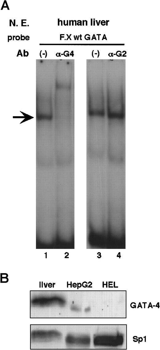
Although several GATA factors—GATA-2, -4, -5, and -6—are expressed in the liver,12,13 only GATA-4 has been demonstrated to bind the GATA site in the enhancer of a liver-specific gene, the mouse albumin gene.14 Therefore, we sought to determine whether GATA-4 is the liver nuclear protein that binds the GATA site in the factor X promoter. As shown in Figure2, the addition of an antibody against GATA-4 completely supershifts the DNA–protein complex to a slower mobility (compare lane 2 to lane 1). Because this antibody does not cross-react with other members of the GATA family, this result indicates that GATA-4 is the sole GATA factor in liver that binds the factor X GATA site. In contrast, an antibody against GATA-2 has no effect on the formation of the DNA–protein complex (lane 4). To confirm the finding in Figure 1A, where HepG2 nuclear extracts are shown to contain little GATA binding activity, we performed Western blot analysis of nuclear extracts from liver and HepG2 with anti–GATA-4. Consistent with the gel shift results, HepG2 cells do not express significant levels of GATA-4 when compared to the liver (Figure 2B).
The factor X GATA site binds GATA-4 from liver extracts.
(A) F.X wt GATA probe was incubated with 10 μg human liver nuclear extracts in the presence of 2 μg antibodies against GATA-4 (αG4) or GATA-2 (αG2). (B) Approximately 100 μg extracts from liver, HepG2, and human erythroleukemia (HEL) cells were analyzed on Western blots using antibodies (Ab) against GATA-4 and Sp1. HepG2 cells contain much less GATA-4 when compared to the liver. Control Western blot indicates that liver and HepG2 cells contain similar amounts of Sp1 in quality and quantity. HEL cell extracts serve as negative control because they do not contain GATA-4.
GATA-4 transactivates the factor X promoter
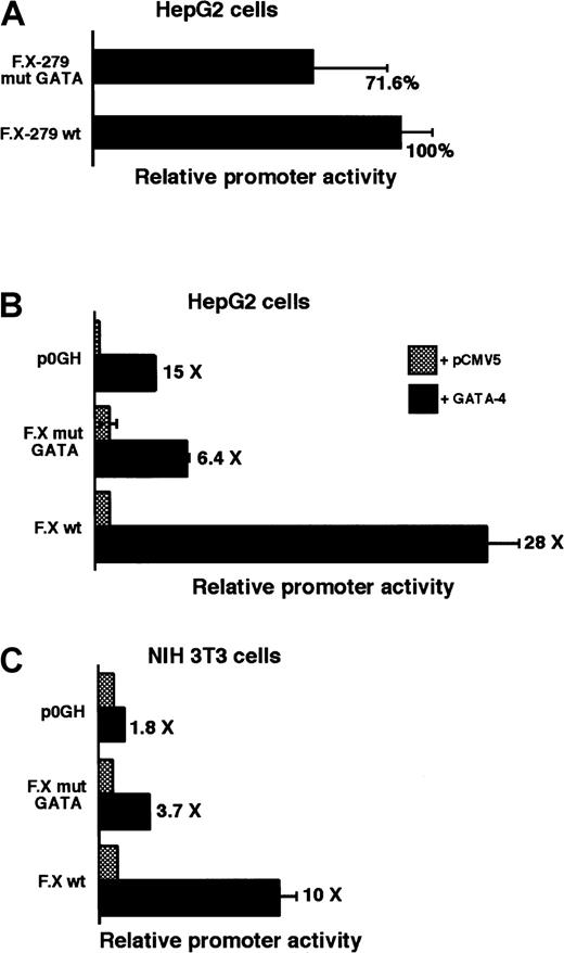
To evaluate the functional significance of the GATA site in the factor X promoter, we introduced a mutant promoter construct defective in GATA-4 binding (mut GATA, where GATA is changed to CATA) into HepG2 cells and performed reporter gene assays. Disruption of the GATA site results in a moderate reduction of the promoter activity (71.6% of wild type; Figure 3A). This is not surprising because HepG2 cells contain little, if any, GATA binding activity (Figure 1A). Therefore, we co-transfected a GATA-4 expression vector with the factor X promoter reporter construct into HepG2 cells (Figure 3B). We used a truncated factor X promoter construct (FX-108GH); the full-length promoter construct has such a high activity that the addition of GATA-4 cannot further increase its level. GATA-4 significantly stimulates the factor X promoter (28-fold). Mutation at the GATA site significantly diminishes the response to GATA-4 (6.4-fold). The residual response is attributed to a possible cryptic GATA site in the growth hormone structural gene because a promoter-less reporter, p0GH, is also stimulated by GATA-4. We also performed transactivation experiments in a nonhepatic environment, NIH 3T3 cells (Figure 3C). GATA-4 activates the factor X promoter to a lesser extent (10-fold) in NIH 3T3 cells. This result suggests that GATA-4 may cooperate with factors present only in the liver.
GATA-4 transactivates the factor X promoter.
(A) An F.X promoter construct containing a mutation at the GATA site (GATA → CATA) in the context of 279 bp full-length promoter was transfected into HepG2 cells and compared to the wild-type construct. (B,C) One microgram of the F.X promoter construct containing 108 bp of the promoter fragment was co-transfected with 0.5 μg of either pCMV5-GATA-4 or pCMV5 vector into HepG2 and NIH 3T3 cells. Results are derived from averages of 4 independent transfections. p0GH, promoter-less reporter construct.
Heterozygous GATA-4 knockout mice express normal levels of factor X
To address the requirement of GATA-4 for the expression of factor X, we assessed plasma factor X levels in a sample from a heterozygous GATA-4 knockout mouse. Murine plasma was added to human factor X–deficient plasma, and the aPTT was determined and compared to a standard curve generated using normal mouse plasma. These experiments showed no difference between the heterozygote and a wild-type littermate (heterozygote, 39.9 seconds; wild type, 39.2 seconds; 1 to 40 dilution). Because homozygous GATA-4 knockout is embryonic lethal at day E.7.5, we were unable to evaluate factor X expression in GATA-4 null mice. It is likely that GATA-4 levels in heterozygotes are adequate for normal factor X expression.
Sp1 and Sp3 bind site 3 and site 4
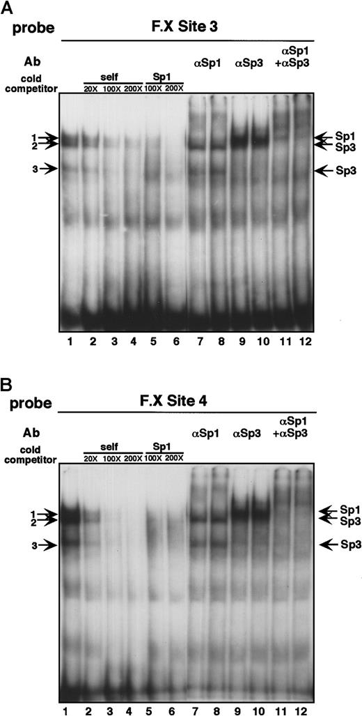
Two protein-protected sites in the factor X promoter, site 3 at −165 to −132 and site 4 at −195 to −169, were delineated in our previous report by DNase footprinting analysis. However, the identities of the proteins binding at these sites were not determined. The promoter fragment lacking site 3 and site 4—that is, a fragment containing −121 to −1 of the factor X promoter—confers 22.8% of the maximal promoter activity in HepG2 cells. This suggests that proteins binding at site 3 and site 4 may be important for the promoter activity of factor X. We performed gel mobility shift assays to determine whether liver extracts bind oligonucleotides derived from site 3 and site 4. To our surprise, site 3 and site 4 oligonucleotides give rise to identical mobility patterns when incubated with liver nuclear extracts, suggesting that the same proteins bind at these 2 sites (Figure 4A-B). The 3 DNA–protein complexes are sequence-specific because the addition of the unlabeled oligonucleotide (lanes 1 to 4, self) abolishes complex formation. Closer inspection of the sequences reveals that the centers of site 3 and site 4 are G rich. This observation raises the possibility that Sp1 and related Sp proteins bind site 3 and site 4. Indeed, the addition of an unlabeled Sp1 consensus site eliminates the complex formation at both site 3 and site 4 (lanes 5 and 6). Because Sp1 and Sp3 are the 2 ubiquitous Sp proteins among family members, we investigated the possibility that Sp1 and Sp3 are the proteins binding at site 3 and site 4. The addition of an antibody against Sp1 supershifts the slowest migrating band (complex 1, lanes 7 and 8). The addition of an antibody against Sp3 supershifts both complex 2 and complex 3 (lanes 9 and 10). This is consistent with the finding that at least 2 forms of Sp3, a full-length and a truncated form, are expressed in cells.15 The addition of both antibodies abolishes the formation of all 3 complexes (lanes 11 and 12). Together, these results show that Sp1 and Sp3 bind at site 3 and site 4 and may regulate the activity of the factor X promoter.
Site 3 and site 4 from the factor X promoter bind Sp1/Sp3 from the liver.
(A) An oligonucleotide containing site 3 from the factor X promoter (−165 GTTCCCAGCGTGGTCACTCCCCTGCCTCGCCAG −132) was radiolabeled and incubated with 10 μg liver nuclear extracts. Lanes 2 to 6 contain molar excess of unlabeled competitor oligonucleotides from either site 3 itself or a consensus Sp1 binding site (5′ ATTCGATCGGGGCGGGGCGAGC 3′). Lanes 7 to 12 contain 2 μg antibodies against Sp1 and Sp3. (B) All lanes contain the same components as Figure 3A except that the probe was derived from site 4 (−191 TGCCTGGGGAAGGTGGGGCCAGAGTGG −169).
Sp1/Sp3 binding to site 3 and site 4, but not site 1, is important for the factor X promoter activity
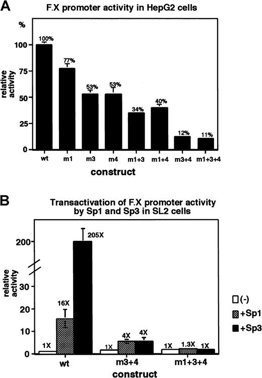
To determine whether Sp1/Sp3 binding at the factor X promoter is functionally significant, mutations that abolish Sp1/Sp3 binding were introduced in the factor X promoter reporter constructs and analyzed in reporter gene assays in HepG2 cells (Figure5A). Mutation at site 3 (m3) moderately reduces the promoter activity to 53% of wild type. Similarly, mutation at site 4 (m4) also reduces the promoter activity to 53%. However, simultaneous mutations at site 3 and site 4 (m3 + 4) reduces the promoter activity to 12% of wild type. We have previously shown that Sp1/Sp3 also bind at site 1. Mutation of the Sp1 sequence in site 1 does not have a significant effect on factor X promoter activity (77% of wild type).7 To determine whether the significance of Sp1 binding at site 1 can only be revealed when sites 3 and 4 are disrupted, we installed a mutation in the Sp1 binding site in site 1 in the context of site 3 mutant (m3) and site 4 mutant (m4). Introduction of the site 1 mutant affected the promoter activity of m3 and m4 only modestly (compare m1 + 3 to m3 and m1 + 4 to m4). Similarly, introduction of the site 1 mutant to m3 + 4 did not significantly reduce the promoter activity (compare m1 + 3 + 4 to m3 + 4). These results are consistent with the previous finding that Sp1 binding at site 1 does not contribute significantly to the promoter activity of factor X. In contrast, Sp1/Sp3 binding at site 3 and site 4 is important for the activity.
Sp1/Sp3 binding to the factor X promoter is required for the maximal promoter activity.
(A) Reporter constructs (2 μg each) containing mutations that disrupt Sp1/Sp3 binding at site 1, site 3, and site 4 (m1, m3, and m4) were transfected into HepG2 cells and compared to the wild-type. m1 + 3, m1 + 4, m3 + 4, and m1 + 3 + 4 contain simultaneous mutations. (B) Reporter constructs (1 μg each) were co-transfected with 1 μg pPac Sp1 or pPacSp3 into Drosophila SL2 cells. Fold increases indicate activities over co-transfection with pPac empty vector (−). Results represent averages of 4 independent transfections.
Both Sp1 and Sp3 are positive regulators of the factor X promoter
Because Sp1 and Sp3 recognize identical sequences, we could not discern which of the Sp factors is a positive regulator of the factor X promoter based on the promoter reporter analysis in Figure 5A. We therefore performed transactivation experiments inDrosophila SL2 cells, which lack endogenous Sp proteins. As shown in Figure 5B, both Sp1 and Sp3 transactivate the factor X promoter significantly (16-fold by Sp1 and 205-fold by Sp3). Consistent with the results in Figure 5A, the mutant construct (m3 + 4) that does not bind Sp proteins demonstrates a markedly reduced response to Sp protein stimulation. We also carried out transactivation experiments by Sp1/Sp3 in HepG2 cells. Transfection with Sp1/Sp3 expression plasmids did not stimulate factor X promoter activity (data not shown). It is likely that endogenous Sp1 and Sp3 are already saturating the binding sites on the factor X promoter reporter, so that there is no further increase in activity on transfection of the Sp1/Sp3 expression vectors.
Discussion
Factor VII, factor IX, and factor X share a high degree of similarity in terms of their gene organization and protein structural domains, suggesting that they derived from a common ancestral gene. We have previously shown that they also share a common transcriptionalcis element, namely, the HNF-4 binding site. However, the steady-state factor X mRNA level in the liver is significantly higher than that of factor VII,7 suggesting that different mechanisms are involved in the regulation of expression of these genes. To extend our understanding of the transcriptional control of factor X, we undertook a complete characterization of its promoter region. We have previously identified HNF-4 and NF-Y as crucial activators of the factor X promoter; in this report, we identified GATA-4, Sp1, and Sp3 as additional positive regulators. Our current understanding of the regulation of the factor X promoter is summarized in Figure6.
Summary of the cis and transelements of the factor X promoter.
The translation start site is designated as +1. Boundaries of the protein-protected sites defined by the DNase I footprint assays are shown. Identities of the transcription factors binding at these sites are noted. Promoter activities in HepG2 cells of constructs containing mutations at these sites (expressed as a percentage of the wild type) are shown below the sites.
We found that GATA-4 binds a canonical GATA site at −96 of the factor X promoter (Figures 1 and 2). Expression of GATA-4 significantly increases factor X promoter activity in both HepG2 and NIH 3T3 cells (Figure 3). Mutation of the GATA site attenuates the response of the factor X promoter to GATA-4. GATA-4 is known to be an important regulator in heart development and the expression of several cardiac-specific genes.16 Its involvement in the expression of clotting factors has not been documented. Figueiredo and Brownlee17 have identified a GATA-like protein-binding site in the human factor VIII promoter region, but, based on the observation that this site is dispensable for factor VIII promoter activity in a hepatoma cell line (PLC/PRF/5 cells), they concluded that the site is not functionally important. Our results suggest that GATA-4 is present only in low abundance in hepatoma cells when compared to liver (Figure1); this could likely explain their finding that the promoter construct defective in GATA protein binding retains activity close to that of the wild type in hepatoma cells. Perhaps like C/EBPα and many other liver-specific transcription factors, GATA-4 is only expressed at high levels in well-differentiated hepatocytes but not in proliferating hepatoma cells.18 Only a handful of liver-specific genes has been shown to be regulated by GATA-4.14,19 In the case of the albumin enhancer, binding of GATA-4, as demonstrated by in vivo footprinting, precedes the expression of the albumin gene during ontogeny.14 The authors suggested that the GATA-4 binding may open the chromatin around the albumin enhancer and potentiate expression of the gene. Similarly, it is possible that GATA-4 could play a role in initiating the expression of factor X during development.
We also found that the ubiquitous factors Sp1 and Sp3 bind at both site 3 and site 4 of the factor X promoter (Figure 4). Disruption of Sp1/Sp3 binding to site 3 and site 4 severely reduces factor X promoter activity, indicating that these sites are important (Figure 5A). Sp3 is a bifunctional protein; in many promoter contexts, it acts primarily as a repressor.15 To determine whether only Sp1 is the positive regulator of the factor X promoter, we introduced both Sp1 and Sp3 together with the factor X promoter constructs intoDrosophila SL2 cells (Figure 5B). We found that both Sp1 and Sp3 act as potent activators of the factor X promoter. Although both Sp1 and Sp3 are widely expressed, their modification and expression in liver are subject to regulation. For example, Sp1 is phosphorylated on terminal differentiation of the liver,20 and this phosphorylation results in decreased DNA binding activity of the protein. During liver regeneration, Sp1 is rapidly dephosphorylated, and increased DNA binding ensues. It can be envisioned that the expression of factor X can be regulated through phosphorylation and dephosphorylation of Sp1. The levels of Sp3 protein in liver were shown to increase significantly after birth.21 This increase may also contribute to the fetal to postnatal increases in factor X expression. Certainly it is possible that Sp1 and Sp3 may also be responsible for the low level of extrahepatic expression of factor X.7
In summary, our findings provide novel insights into how GATA-4 may contribute to the liver-specific expression of factor X. Importantly, the contribution of GATA-4 to the expression of clotting factors may not be limited to factor X. In addition to the aforementioned GATA binding site in the factor VIII promoter,17 we have also found a putative GATA binding site in the factor VII promoter at −407 (data not shown). Interestingly, van't Hooft et al22 have defined 2 polymorphisms nearby at −402 and −401 that are associated with either increased (−402A) or decreased (−401T) plasma levels of factor VII. GATA-4 might be the trans regulating element of these 2 functional polymorphisms.
Acknowledgments
We thank Dr Tetsuya Yamagata for the GATA-4 cDNA, Dr Jon Horowitz for the pPacSp1 and Sp3 expression plasmids, Dr Jeffrey Leiden for the plasma from GATA-4 knockout mice, and Dr Merlin Crossley forDrosophila SL2 cells.
Supported by National Institutes of Health grants NHLBI R01 48322 (K.A.H.) and NHLBI K08 03661 and Doris Duke Charitable Foundation grant T98062B (E.S.P.).
The publication costs of this article were defrayed in part by page charge payment. Therefore, and solely to indicate this fact, this article is hereby marked “advertisement” in accordance with 18 U.S.C. section 1734.
References
Author notes
Katherine A. High, Division of Hematology, 310 Abramson Pediatric Research Center, The Children's Hospital of Philadelphia, 34th St and Civic Center Blvd, Philadelphia, PA 19104; e-mail:high@emailchop.edu.


This feature is available to Subscribers Only
Sign In or Create an Account Close Modal