Abstract
Signal transducer and activator of transcription 3 (STAT3) is an oncogene and a critical regulator of multiple cell-fate decisions, including myeloid cell differentiation. Two isoforms of STAT3 have been identified: α (p92) and β (p83). These differ structurally in their C-terminal transactivation domains, resulting in distinct functional activities. The cis genetic elements that regulate the ratio of α to β messenger RNA (mRNA) are unknown. In this study, cloning, sequencing, and splicing analysis of the human and murine STAT3 genes revealed a highly conserved 5′ donor site for generation of both α and β mRNA and distinct branch-point sequences, polypyrimidine tracts, and 3′ acceptor sites (ASs) for each. The β 3′ AS was found to be located 50 nucleotides downstream of the α 3′ AS in exon 23. Two additional cryptic 3′ ASs (δ and ε) were also identified. Thus, we identified for the first time the cisregulatory sequences responsible for generation of STAT3α and STAT3β mRNA.
Introduction
Although there is only one signal transducer and activator of transcription 3 (STAT3) gene in mice and humans, 2 protein isoforms have been identified in both species: STAT3α (p92)1,2 and STAT3β (p83).3-5 The messenger RNA (mRNA) encoding STAT3β has a 50-nucleotide deletion at the 3′ end that is presumably due to alternative mRNA splicing and results in a protein's missing the C-terminal 55 amino acid residues of STAT3α. In contrast to other STAT protein β isoforms, in which the C-terminal transactivation domain is simply deleted, in STAT3β, the 55 amino acid residues of STAT3α are replaced by 7 unique amino acid residues at its C-terminal. These residues are encoded by 21 nucleotides spliced in the +2 reading frame downstream of the deletion. The C-terminal 55 amino acid residues comprise the transactivation domain of STAT3α and contain serine 727, whose phosphorylation results in enhanced transcriptional activity.6 This same region of STAT3α also influences STAT3α dimerization, since the DNA-binding activity of STAT3α was shown to be reduced by 15 to 25 fold compared with that of STAT3β.7 8 The reduced DNA-binding activity of STAT3α was attributed to a reduced stability of STAT3α homodimers compared with STAT3β homodimers.
The ratio of STAT3α to STAT3β varies in cells and tissues, ranging from 3:1 to 10:1 at the mRNA level and 1:3 to 10:1 at the protein level.5,9 This variation may have important biologic consequences because the functions of the 2 isoforms do not overlap. When STAT3β and STAT3α were overexpressed in Cos cells, STAT3β, but not STAT3α, was constitutively able to cooperate with c-Jun to activate a reporter construct containing α2-macroglobulin.3 In contrast, STAT3β activated downstream of the interleukin 5 receptor inhibited the ability of STAT3α to activate a reporter construct containing an intercellular adhesion molecule 1 promoter.4 In studies examining the distinct biologic functions of the STAT3 isoforms, STAT3α enhanced whereas STAT3β inhibited v-Src–mediated fibroblast transformation.10 More relevant for myeloid development, we and others found that overexpression of STAT3α inhibited myeloid differentiation mediated by gp130 and granulocyte colony-stimulating factor receptor11 (A. Chakraborty, S. M. White, D.J.T., unpublished data, October 2000).
Factors that regulate STAT3 splicing to STAT3α or STAT3β, including the cis regulatory elements in the STAT3 gene, are unknown. Both the murine and human STAT3 genes have been cloned,12but despite extensive sequencing of the murine gene, the molecular basis for generation of STAT3β mRNA has not been identified. In this study, we found that STAT3β arises from use of an alternative 3′ acceptor site (AS) 50 nucleotides within exon 23 and examined for the first time for any STAT gene the functional roles of ciselements in mRNA isoform generation.
Study design
Cloning of human and murine STAT3 genes
The probe used to clone the human STAT3 gene consisted of a 350–base pair (bp) fragment of human STAT3β extending from nucleotide 2310 to 2777 of the published sequence1 that was generated by polymerase chain reaction (PCR) by using human STAT3β complementary DNA (cDNA) as the template,4 and subcloned into pBluescript. The probe used to clone the murine STAT3 gene consisted of a 400-bp fragment of murine STAT3α extending over the homologous region of the murine STAT3 cDNA and generated by PCR using murine STAT3α as the template.2 The probes were used by Genome Systems (St Louis, MO) to screen human (BAC-5131) and murine (strain 129/SvJ, BAC-4921) genomic libraries constructed in pBeloBACII. DNA sequencing was done on an automated sequencer as directed by the manufacturer (Applied Biosystems, Foster City, CA).
Minigene construction
Minigene E22-E24 was constructed to contain murine STAT3 genomic DNA from the beginning of exon 22 to the end of exon 24 by PCR using murine embryonic stem cell DNA as the template. Amplified products were verified by sequencing and cloned into the exon-trapping vector pSPL3 (Gibco-BRL, Gaithersburg, MD) between the EcoRI andXhoI sites and into the eukaryotic expression vector pSG5 (Stratagene, La Jolla, CA) between the EcoRI andBamHI sites.
Site-directed mutagenesis
The GeneEditor in vitro site-directed mutagenesis system (Promega, Madison, WI) and the Quikchange site-directed mutagenesis method (Stratagene) were used to introduce mutations (underlined nucleotides) in STAT3 minigenes. The primers used to generate β 3′ splice-site mutants were (1) 5′ GTCCCCCCGCACTTTGGATTCATTGATG 3′, (2) 5′ GTCCCCCCGCACTTTCGATTCATTGATG 3′, and (3) 5′ GTCCCCCCGCACTTTTGATTCATTGATG 3′. The primer used to generate δ 3′ splice-site mutants was 5′ CTACAGAACGACCTGCTCCAATACCATTGAC 3′. The primers used to generate α 3′ splice-site mutants were (1) 5′ CCTTTCCTACGGAACGACCTGC 3′, (2) 5′ CCTTTCCTACCGAACGACCTGC3′, and (3) 5′ CCTTTCCTACTGAACGACCTGC 3′.
The primer pairs used to generate α and δ 3′ splice-site mutants were (1) 5′ CGTCCCCCCTTTCCTACGGAACGACCTGCTCCAATAC 3′ and 5′ GTATTGGAGCAGGTCGTTCCGTAGGAAAGGGGGGACGG 3′, (2) 5′ CGTCCCCCCTTTCCTACCGAACGACCTGCTCCAATAC 3′ and 5′ GTATTGGAGCAGGTCGTTCGGTAGGAAAGGGGGGACGG 3′, and (3) 5′ CGTCCCCCCTTTCCTACTGAACGACCTGCTCCAATAC 3′ and 5′ GTATTGGAGCAGGTCGTTCAGTAGGAAAGGGGGGACGG 3′.
The primer pairs used to generate intron 22 deletions were (1) 5′ ACCCCATGTCCTCCCTATTCCTG 3′ and 5′ AGTGCTCAGAGTGACCTTGTGTG 3′, (2) 5′ CAAGCCCAGCTACCAGCCC 3′ and 5′ CTGTTGGAGACCAGAGTTTGATGGC 3′, (3) 5′ CATCAAACTCTGGTCTCCAACAG 3′ and 5′ CCTCCATTGTGTCTTGTCAACCTG 3′, and (4) 5′ CTACAGAACGACCTGCAGCAATAC 3′ and 5′ CACACAAGCCATCAAACTCTGGTC 3′. All constructs containing mutated sites were confirmed by sequencing.
Cell cultures and transfection
Human HeLa cells and monkey Cos-7 cells were cultured in Dulbecco modified Eagle medium (Gibco-BRL) supplemented with 10% heat-inactivated fetal-calf serum. Transient transfections were done in 6-well plates with 4 × 10⁵ cells/well by using Lipofectace reagent as instructed by the manufacturer (Gibco-BRL). The total amount of DNA transfected was 1 μg/well. After 5 to 6 hours of exposure to Lipofectace, the medium was refreshed and cells were incubated for 48 hours before harvesting for RNA.
RNA extraction and reverse transcriptase–PCR
Total RNA was isolated from transfected cells by using Trizol reagent (Gibco-BRL). The Titan one-tube reverse transcriptase (RT)–PCR system (Boehringer Mannheim, Indianapolis, IN) was used to perform first-strand cDNA synthesis and PCR. The primers used in RT-PCR reactions for pSPL-E22-24 and derived mutants were totally vector derived. For SD6, the primer was 5′ TCTGAGTCACCTGGACAACC 3′; for SA2, it was 5′ ATCTCAGTGGTATTTGTGAGC 3′; for duSD2, it was 5′ CUACUACUACUAGTGAACTGCACTGTGACAAGCTGC 3′; and for duSA4, it was 5′ CUACUACUACUACACCTGAGGAGTGAATTGGTCG 3′.
For detection of β splice product, fusion primers contained both a vector sequence (underlined) and an intrinsic sequence specific for STAT3β. For PSPL-b-F1, the primer was 5′ GACCCAGCATTCATTG 3′; and for PSPL-E24-R1, it was 5′ CGAGGTCGATATGG 3′. All amplicons generated by RT-PCR during in vivo splice assays were gel purified and verified by sequencing.
Results and discussion
Cloning and characterization of the human and murine STAT3 genes
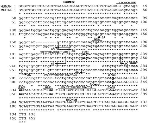
To characterize and compare the human and murine STAT3 genes, we cloned each from genomic libraries by using cDNA probes. Two human clones and one murine clone were isolated during genomic screening. Southern blotting, partial sequencing of inserts (90-120 kilobases) and comparison of the sequence with that published for the murine STAT3 gene12 confirmed their identities and delineated their exon-intron boundaries (Figure 1 and data not shown; GenBank accession numbers for the murine and human sequences, AF332507 and AF332508, respectively). Analysis of the sequence of each gene from exons 22 through 24 identified highly conserved 5′ donor sites, branch-point sequences (BPSs), polypyrimidine tracts (PPTs), and 3′ ASs in intron 22 for generation of STAT3α mRNA. Each sequence also contained in exon 23 a conserved alternative 3′ AS 50 nucleotides downstream of the α 3′ AS, as well as an alternative BPS and weaker PPT for generation of STAT3β mRNA. Two additional possible 3′ ASs (δ and ε) were also identified. The δ site was found to be located 12 nucleotides downstream of the α 3′ AS and the ε site was observed 144 nucleotides upstream of the α 3′ AS.
Sequence alignment and analysis of the human and murine STAT3 genes.
Exon 22 to exon 23 in the human and murine STAT3 genes was aligned. Sequence analysis identified the possible 5′ splice donor site and possible adenosine BPSs, PPTs, and 3′ ASs for generating STAT3α and STAT3β mRNA. The critical nucleotides in each element are in boldface type. In addition, cryptic δ and ε 3′ splice ASs were detected. The δ 3′ AS presumably uses the same BPS and PPT as α. For ε, the possible BPS and PPT are indicated. An asterisk indicates nucleotide homology. The regions of intron 22 deleted in the ΔA, ΔB, ΔC, and ΔD constructs are indicated. The GenBank accession numbers for the murine and human sequences are AF332507 and AF332508, respectively.
Only one STAT3 gene was identified in mice; it was mapped by 2 groups to chromosome 11.12,13 There is one report of mapping human STAT3 to chromosome 17q21 by using microclones from a 17q21 band–specific microdissection library and fluorescent in situ hybridization (FISH).14 We used the human STAT3 gene and phytohemagglutinin-stimulated lymphocytes in FISH experiments to establish definitively that both human STAT3 isoforms are derived from a single gene. We also more precisely mapped human STAT3 to 17q21.1-q21.2 (data not shown), a site of frequent translocations and unbalanced chromosomal abnormalities in acute lymphoblastic leukemia, non-Hodgkin lymphoma, and acute myeloid leukemia (not involving the retinoic acid receptor-α locus).15 The contribution of STAT3 to these malignant diseases remains to be investigated.
Targeting of the STAT3β 3′ splice AS eliminated STAT3β splicing
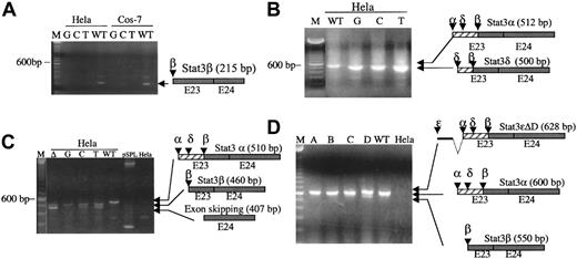
To determine whether the α and β cis regulatory sequences identified were functional, we generated a minigene construct composed of exon 22 (43 nucleotides), intron 22 (280 nucleotides), exon 23 (113 nucleotides), intron 23 (713 nucleotides), and exon 24 (137 nucleotides). The minigene construct was subcloned into the exon-trap vector pSPL3 and the eukaryotic expression vector pSG5. Transfection of the 2 minigene-containing vectors into human HeLa and monkey Cos-7 cell lines generated RNA transcripts corresponding to that predicted for STAT3α (Figure 2B-D, wild-type [WT] lanes; and data not shown) and STAT3β (Figure 2A, WT lanes), indicating that the minigene construct included the essential sequence information required for correct splicing.
Effect of mutations of
cis regulatory elements in the murine STAT3 gene on in vivo splicing of minigene constructs. (A) RT-PCR analysis using primers specific for the β splice product (pspl-b-F1 and pspl-E24-R) of RNA isolated from the indicated cells transfected with WT construct or construct containing the indicated point mutation (A to G, C, or T) in the β 3′ AS. (B) RT-PCR analysis using vector-derived primers duSD2 and duSA4 of RNA isolated from HeLa cells transfected with WT construct or constructs containing the indicated point mutation (A to G, C, or T) in the α 3′ AS. (C) RT-PCR analysis using vector-derived primers duSD2 and duSA4 of RNA isolated from untransfected HeLa cells or HeLa cells transfected with WT construct or construct in which the δ 3′ AS AG was replaced by TC and the α AS CAG was either deleted (Δ) or the A was mutated to G, C, or T. (D) RT-PCR analysis using vector-derived primers SD6 and SA2 of RNA isolated from untransfected HeLa cells or HeLa cells transfected with WT construct or construct containing the ΔA, ΔB, ΔC, or ΔD deletions. The 100-bp DNA ladder is indicated by M; the position of the 600-bp band is shown. The sequence-confirmed products of the RT-PCR are shown schematically to the right of each gel.
Of note, the expression level of the STAT3β splice product was much lower than that of the STAT3α splice product. STAT3β splice product could not be detected with amplimers capable of detecting both products (Figure 2B-D); instead, its detection required a STAT3β-specific amplimer (Figure 2A). Our inability to detect β splice product without using isoform-specific amplimers was somewhat surprising, since this had not been the case for endogenous STAT3β mRNA in myeloid cells.5 This finding suggests that there may be myeloid cell–specific, trans-acting proteins that interact with the basal splicing machinery to promote increased β splicing or that β splicing is enhanced with splicing of the complete transcript. This latter effect would not be observed when using minigene constructs containing only 3 exons and 2 introns.
To assess the contribution of the β 3′ splice AS site to STAT3β splicing more rigorously, the A in the β 3′ AS was replaced by G, C, or T in pSPL3 and pSG5 and the mutated minigenes were transiently transfected into HeLa and Cos-7 cells (Figure 2A and data not shown). Each point mutation eliminated β splicing (Figure 2A) but had no effect on α splicing (data not shown). To confirm that point mutation of the β 3′ AS did not affect α splicing, we conducted in vitro transcription and splicing. Minigene constructs were generated in pGEM-11Zf (+) that contained exon 22, intron 22, and either WT exon 23 or exon 23 with a point mutation in the β 3′ splice AS. In vitro RNA transcription was done with SP6 RNA polymerase. After exposure to HeLa cell nuclear extract, STAT3α splice product was detected in equivalent amounts, relative to unspliced mRNA, in the WT and in each of the mutated transcripts (data not shown). These results confirmed that point mutation in the β 3′ AS does not affect α splicing.
Targeting of the α 3′ AS
Targeting of the α 3′ AS reduced α splice-product formation and activated a cryptic 3′ AS, δ. Targeting of both α and δ 3′ ASs was necessary to increase STAT3β splice-product formation. To examine the contribution of the α 3′ AS to splicing, the A in the α splice AS was replaced by G, C, or T in pSPL3 and pSG5 and the mutated minigenes were transiently transfected into HeLa and Cos-7 cells (Figure 2B and data not shown). Surprisingly, the mutations of the α splice AS activated a cryptic splice site, δ, located 12 nucleotides downstream of the α 3′ AS site in exon 23, and shifted the predominant splice product from α to δ. Moreover, no β splice product was detected. Mutation of both α and δ 3′ splice ASs yielded 2 products—the β splice product and a splice product that skipped exon 23 entirely and used the 3′ AS of exon 24 (Figure 2C). The choice of generating the β splice product or the exon 24 splice product was sensitive to the nature of the point mutation in the α 3′ AS. Mutation of A to G resulted in detection of only the β splice product, whereas mutation of A to T or C resulted in detection of both products. These findings indicate that minimal mutations in the murine STAT3 gene alone may dramatically alter the ratio of STAT3α to STAT3β mRNA in cells.
The δ 3′ AS does not appear to be used in splicing of the endogenous STAT3 mRNA, since an isoform missing the first 12 nucleotides in exon 23 was not detected in human HeLa cells or murine 32Dcl3 cells screened with nested RT-PCR using a primer specific for this splice product (data not shown).
Effects of deletion of the α/δ BPS and PPT
Deletion of the α/δ BPS and PPT resulted in loss of α splice product, activation of a cryptic 3′ AS, ε, and increased STAT3β splice-product formation. To investigate the role of the other putative α cis regulatory sequences (BPS and PPT) in α splicing, we generated 4 constructs in pSPL3 (ΔA, ΔB, ΔC, and ΔD; Figure1A) in which a portion of intron 22 (35 to 83 nucleotides in length) was deleted by site-directed mutagenesis. RT-PCR analysis of RNA from HeLa cells transfected with the ΔA, ΔB, and ΔC constructs resulted in detection of α splice product, similar to findings in cells transfected with the WT STAT3 minigene construct (Figure 2D). In contrast, analysis of RNA from cells transfected with the ΔD construct, which is missing the putative α BPS and PPT, found no detectable α splice product, an increase in β splice product, and appearance of a new splice product, εΔD. Therefore, ciselements such as α BPS and PPT, contained in the region removed in the ΔD construct, are essential for α splicing.
To date, all successful strategies for targeting STAT3 (gene targeting, dominant-negative STAT3 constructs, and antisense methods) targeted both the α and β isoforms and consequently did not allow assignment of the effects of targeting to the specific isoform. In this study, we established for the first time the molecular basis for generation of the STAT3α and STAT3β splice isoforms in humans and mice. The information gained from mutation analysis of the murine gene can be used to generate mice deficient in only one isoform. Examination of such mice will permit determination of the specific roles of each isoform in mice generally.
We thank Mr Kevin F. Dyer for the initial preparation of the cDNA probes used for genomic screening and the initial characterization and sequencing of the murine and human STAT3 genomic clones, Dr Michael Cascio for providing temporary laboratory space for Dr Shao before her move to Baylor College of Medicine, and Dr Susan Berget (Baylor College of Medicine) and Dr Christine Milcarek (University of Pittsburgh School of Medicine) for helpful discussions and suggestions.
Supported in part by National Institutes of Health R01 grants CA72261 and CA86430.
The publication costs of this article were defrayed in part by page charge payment. Therefore, and solely to indicate this fact, this article is hereby marked “advertisement” in accordance with 18 U.S.C. section 1734.
References
Author notes
David J. Tweardy, Section of Infectious Diseases, Baylor College of Medicine, One Baylor Plaza, BCM 286, Rm 1319, Houston, TX 77030; e-mail: dtweardy@bcm.tmc.edu.


This feature is available to Subscribers Only
Sign In or Create an Account Close Modal