Key Points
There is a high level of ROS in bone marrow CD34posCD38low cells of MDS and sAML.
There is a specific antioxidant molecular signature in BM cells of MDS and sAML.
Abstract
Myelodysplastic syndromes (MDS) are a heterogeneous group of clonal stem cell disorders with an inherent tendency for transformation in secondary acute myeloid leukemia. This study focused on the redox metabolism of bone marrow (BM) cells from 97 patients compared with 25 healthy controls. The level of reactive oxygen species (ROS) was quantified by flow cytometry in BM cell subsets as well as the expression level of 28 transcripts encoding for major enzymes involved in the antioxidant cellular response. Our results highlight increased ROS levels in BM nonlymphoid cells and especially in primitive CD34posCD38low progenitor cells. Moreover, we identified a specific antioxidant signature, dubbed “antioxidogram,” for the different MDS subgroups or secondary acute myeloblastic leukemia (sAML). Our results suggest that progression from MDS toward sAML could be characterized by 3 successive molecular steps: (1) overexpression of enzymes reducing proteic disulfide bonds (MDS with <5% BM blasts [GLRX family]); (2) increased expression of enzymes detoxifying H2O2 (MDS with 5% to 19% BM blasts [PRDX and GPX families]); and finally (3) decreased expression of these enzymes in sAML. The antioxidant score (AO-Score) defined by logistic regression from the expression levels of transcripts made it possible to stage disease progression and, interestingly, this AO-Score was independent of the revised International Scoring System. Altogether, this study demonstrates that MDS and sAML present an important disturbance of redox metabolism, especially in BM stem and progenitor cells and that the specific molecular antioxidant response parameters (antioxidogram, AO-Score) could be considered as useful biomarkers for disease diagnosis and follow-up.
Introduction
Myelodysplastic syndromes (MDS) are a heterogeneous group of blood diseases with varying degrees of severity, which can be classified into several subgroups based on features of the abnormal cells. Progression of MDS to secondary acute myeloblastic leukemia (sAML) is a good example of the multistep theory of leukemogenesis in which a series of mutations occur in an initially normal cell and transform it into a cancer cell. The pathophysiology of MDS and its progression to sAML involve cytogenetic, genetic, and epigenetic aberrations of hematopoietic cells,1 as well as alterations of the microenvironment.2
There is growing awareness that redox metabolism plays a role in the successive stages of hematopoiesis, with a low amount of reactive oxygen species (ROS) in primitive progenitors, followed by an increasing concentration allowing cell differentiation.3 It is now well-established that a low ROS level is associated with quiescence and self-renewal, whereas an intermediate level is required for ROS signaling pathways and, finally, a higher level represents an oxidative stress and induces nonspecific damage and DNA modifications.3 The level of intracellular ROS is finely tuned by the expression of ROS-related enzymes suggested to be major players in the pathophysiology of human MDS/AML.4-6
The aim of this study was to determine the level of intracellular ROS in bone marrow (BM) cells from MDS and sAML patients and to identify an antioxidant signature of clinical interest in the various categories of the disease according to World Health Organization criteria,7 possibly reflecting a redox status associated to progression. By comparing these samples to BM from healthy controls, we observed an increase in intracellular ROS levels in CD34posCD38low cells and in the erythroblastic subpopulation of MDS with ring sideroblasts (MDS-RS), and determined specific antioxidant signatures of clinical interest related to MDS with <5% BM blasts, MDS with 5% to 19% BM blasts, and sAML.
Methods
Patients
A cohort of 97 patients (supplemental Table 1) classified according to World Health Organization criteria7 was analyzed: MDS with single-cell dysplasia (MDS-SLD), MDS with multilineage dysplasia (MDS-MLD), MDS-RS, and single lineage dysplasia, MDS-RS with multilineage dysplasia, and MDS with excess blasts (MDS-EB-1 and MDS-EB-2). All subjects provided informed consent according to a procedure approved by local ethical committees (CPP Tours and AFSSAPS identifier ID-RCB: 2011-A00262-39; CPP Ile-de-France III: 2753). sAML patients were characterized at diagnosis by the presence of blasts (≥20%) and morphologic dysplastic-related changes in BM samples. Gold standard sternal BM aspirates were obtained at the time of diagnosis, before treatment initiation (Tours University Hospital and Cochin University Hospital in Paris, France). They were compared with BM from 25 aged-matched healthy donors (HEALTHOX protocol, CPP Tours and AFSSAPS identifier ID-RCB: CPP Tours and AFSSAPS identifier ID-RCB: 2016-A00571-50, and ClinicalTrials.gov Identifier: NCT02789839). The sternal BM aspiration procedure was the same for all patients and healthy volunteers. The revised International Scoring System (IPSS-R) was calculated for each patient (supplemental Table 2).8 Samples were processed within 1 hour of BM aspiration for flow cytometry analyses. The antioxidant signature was specifically determined for the different subtypes of MDS and sAML. Because ring sideroblasts interfere with redox metabolism,9,10 MDS-RS were analyzed independently.
Flow cytometry quantification of ROS levels
Red blood cell lysis of BM samples was performed by addition of ammonium chloride (150 mM, 10-minute incubation at 37°C, and centrifugation at 250g for 5 minutes). White blood cells were washed with X-Vivo 10 (Lonza, Basel, Switzerland), incubated with 9.6 µM of the General Oxidative Stress Indicator CM-H2DCFDA (Molecular Probes, Waltham, MA), anti-CD45-APC-H7, anti-CD34-PE-Cy7, and anti-CD38-APC antibodies (BD Biosciences, Franklin Lakes, NJ). Cells were analyzed with a BD FACSCanto II (BD Biosciences) flow cytometer and FlowJo v7.6.2 software. Cell populations were gated using forward scatter, side scatter (SSC), and CD45 fluorescence.11 Briefly, CD45/SSC gating clearly separates mature and immature hematopoietic cell populations in the BM (supplemental Figure 1). Lymphocytic cells show the highest CD45 fluorescence intensity and the lowest SSC. Monocytic cells express slightly lower but still high amounts of CD45 and are distinguished from lymphocytes by their higher SSC. Granulocytic cells express low CD45 and broad SSC related to their granulations. Nucleated erythroid cells are characterized by reduced/absent CD45 expression and low SSC; this region can also contain mature (anucleate) red cells and cellular debris, which are eliminated by red blood cell lysis and forward scatter threshold. With this strategy, the earliest cells committed to each lineage occupy a position of low-medium SSC and CD45 dubbed the “bermudes” area of progenitors as per Arnoulet et al.12
Quantitative polymerase chain reaction assay for antioxidant signature
RNA extraction was performed after red blood cell lysis using a Maxwell 16 System RNA Purification kit (Promega, Fitchburg, WI) according to the manufacturer’s recommendations. RNA quality was checked using a 2100 Bioanalyzer (Agilent, Santa Clara, CA). RNA was reverse-transcribed using a SuperScript VILO cDNA Synthesis kit (Life Technologies, Carlsbad, CA). Transcripts quantification (Ensembl nomenclature; supplemental Table 3) was achieved by reverse transcription-quantitative polymerase chain reaction using the Universal Probe Library technology (https://lifescience.roche.com) on a LightCycler 480 (Roche, Rotkreuz, Switzerland). Assays were designed to quantify either only 1 or several transcript variants concomitantly (x,y,z…), reported as GENE(x,y,z…). All targets were analyzed simultaneously in triplicate and average values were used to determine relative quantification (RQ) values by the 2-ΔΔCt method.13
Statistical analyses
All statistical analyses were conducted using R v3.2.2 software. Differences in ROS levels between pathologies were tested using a Kruskal-Wallis test followed by Dunn's post hoc test. Differences in expression between patients and controls were tested using a Mann-Whitney U test on ΔCt, RQ, or calculated antioxidant score (AO-Score) values. Principal component analyses (PCA) were performed using the FactoMineR package on ΔCt values.14 The optimal model was estimated using a stepwise function (MASS package). Multinomial logistic regressions were performed using the multinom() function from the nnet package. A split-sample approach was used to determine optimal weighted coefficients.15 Significant differences were identified as changes of at least twofold and P < .05.
Results
Abnormal ROS abundance in BM CD34posCD38low cells
An original flow cytometry strategy was developed to standardize data between individual patients. ROS levels in each subpopulation of fresh BM samples was normalized to the monocytic fraction, these cells having the highest ROS level in healthy BM (Figure 1A),16 without significant variation (P = .7752) when considering BM samples from MDS/sAML patients (Figure 1B). Analyses of healthy BM samples clearly evidenced a low level of ROS in nonmyeloid cells (ie, lymphocytic [P = .0398] and especially erythroblastic [P = .0170] subpopulations). Moreover, this level was intermediate in CD34pos progenitors in which the lowest level was found in the CD34posCD38low fraction (P = .0134), as expected.17,18
ROS levels in bone marrow cell subpopulations of MDS/sAML patients. (A) Flow cytometry quantification of ROS in BM cells (ie, monocytic, granulocytic, lymphocytic, erythroblastic subpopulations and CD34pos, CD34posCD38low, and CD34posCD38high progenitors) of healthy controls. ROS levels were evaluated by DCFDA mean fluorescence intensity (DCFDA-MFI) and results are expressed as % of DCFDA MFI of monocytes (± standard error of the mean [SEM]). The highest level of ROS was found in monocytic cells in all samples, and the lowest level in the erythroblastic subpopulation. *Significant difference with monocytic cells, P < .05; #significant difference, P < .05. (B) Absolute ROS levels (DCFDA-MFI) in the BM monocytic subpopulation from healthy controls, MDS subgroups, and sAML are not statistically different (P = .77, Kruskal-Wallis test). (C) ROS levels in the BM subpopulations of MDS/sAML patients compared with healthy controls, after normalization with the respective DCFDA-MFI value of monocytes in each sample. Increased ROS levels were observed exclusively in myeloid and especially progenitor subpopulations. The most important oxidative stress was found in the erythroblastic subpopulation in MDS-SLD-RS and MDS-MLD with ring sideroblasts, enriched in ring sideroblasts, and in the CD34posCD38low progenitors in all pathologies, especially in sAML. Data are expressed as mean ± SEM (n = 65). *Significant difference with the healthy controls, P < .05. #Significant difference with healthy controls and with MDS subgroups, P < .05. ns, nonsignificant difference with the healthy controls.
ROS levels in bone marrow cell subpopulations of MDS/sAML patients. (A) Flow cytometry quantification of ROS in BM cells (ie, monocytic, granulocytic, lymphocytic, erythroblastic subpopulations and CD34pos, CD34posCD38low, and CD34posCD38high progenitors) of healthy controls. ROS levels were evaluated by DCFDA mean fluorescence intensity (DCFDA-MFI) and results are expressed as % of DCFDA MFI of monocytes (± standard error of the mean [SEM]). The highest level of ROS was found in monocytic cells in all samples, and the lowest level in the erythroblastic subpopulation. *Significant difference with monocytic cells, P < .05; #significant difference, P < .05. (B) Absolute ROS levels (DCFDA-MFI) in the BM monocytic subpopulation from healthy controls, MDS subgroups, and sAML are not statistically different (P = .77, Kruskal-Wallis test). (C) ROS levels in the BM subpopulations of MDS/sAML patients compared with healthy controls, after normalization with the respective DCFDA-MFI value of monocytes in each sample. Increased ROS levels were observed exclusively in myeloid and especially progenitor subpopulations. The most important oxidative stress was found in the erythroblastic subpopulation in MDS-SLD-RS and MDS-MLD with ring sideroblasts, enriched in ring sideroblasts, and in the CD34posCD38low progenitors in all pathologies, especially in sAML. Data are expressed as mean ± SEM (n = 65). *Significant difference with the healthy controls, P < .05. #Significant difference with healthy controls and with MDS subgroups, P < .05. ns, nonsignificant difference with the healthy controls.
By comparing BM samples from patients and healthy volunteers, a major increase in ROS level was found in the erythroblast subpopulation in MDS-RS (P = .0294 for MDS-SLD with ring sideroblasts [MDS-SLD-RS) and P = .0184 for MDS-MLD with ring sideroblasts) in accordance with the mitochondrial iron accumulation in ring sideroblasts (Figure 1C).19 This result validates the analytical strategy used. ROS levels were indeed not statistically increased in lymphocytic nor granulocytic cells in MDS/sAML samples. Conversely, ROS levels were highly increased in CD34pos progenitors, mainly in the CD34posCD38low fraction; this increase was the most important in sAML (supplemental Table 4).
MDS subtype/sAML-related disturbance in antioxidant enzymes gene expression
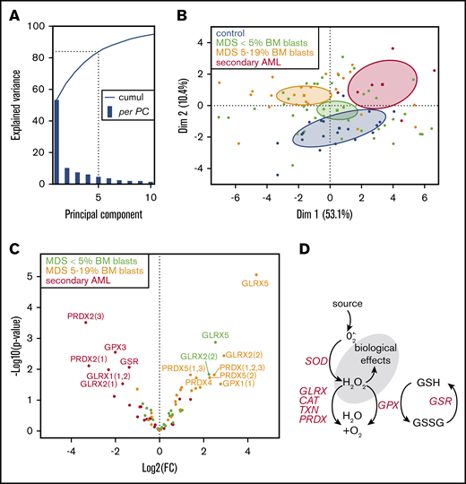
The antioxidant response in BM cells was studied at the molecular level by quantifying the expression of 28 transcripts encoding the most efficient antioxidant enzymes.20-22 Aiming to validate the pertinence of our approach and to obtain an overview of gene expression variations, PCA was computed in ΔCt values for MDS with <5% BM blasts, MDS with 5% to 19% BM blasts, sAML, and healthy controls. This highlighted that the 2 first principal components explained ∼63% of variance (Figure 2A). Genes related to H2O2 reduction such as the PRDX, GLRX, and TXN gene families dominated the expression changes associated with PC1, whereas PC2 was mainly correlated to GSR expression. This PCA analysis allowed the determination of almost nonredundant 95% confidence interval between controls, MDS with <5% BM blasts (including single lineage [MDS-SLD] and multiple lineage [MDS-MLD] dysplasia with or without RS), MDS with 5% to 19% BM blasts (MDS-EB-1 and −2) and sAML, validating the potential of discrimination between these 4 groups using this set of antioxidant genes (Figure 2B). Of note, even if these 95% confidence intervals were clearly characterized, the predictive potential of this descriptive approach is not sufficient. An overall analysis of statistical differences in antioxidant genes expression was thus performed, revealing a contrary antioxidant response in MDS and sAML, characterized respectively by exclusive over- and underexpression of antioxidant genes (Figure 2C).
Principal component analysis (PCA) of BM antioxidant transcripts of healthy controls and MDS/sAML patients. (A) Cumulative explained variance (blue line) and per PCA explained variance (blue bar) showing that PC1 explains 53.1% of the variance and that the 5 first PCs explain >80% of the total variance (dotted line). (B) PCA individual plot of patients: MDS with <5% BM blasts, MDS with 5% to 19% BM blasts, and sAML in green, orange, and red, respectively, and healthy controls (in black) highlighting the specific partition in a 2-dimensional PCA diagram (PC1, x-axis; PC2, y-axis). Barycenter and 95% confident ellipse of each group are represented and show that the expression profiles of antioxidant-related genes allow the discrimination between healthy controls, MDS with <5% BM blasts, MDS with 5% to 19% blasts, and sAML patients. (C) Volcano plot plotting fold change (FC) and P values between healthy controls and different MDS subgroups/sAML, highlighting an increased expression of different genes in MDS and a strongly decreased expression in sAML. (D) Schematic representation of antioxidant gene function.
Principal component analysis (PCA) of BM antioxidant transcripts of healthy controls and MDS/sAML patients. (A) Cumulative explained variance (blue line) and per PCA explained variance (blue bar) showing that PC1 explains 53.1% of the variance and that the 5 first PCs explain >80% of the total variance (dotted line). (B) PCA individual plot of patients: MDS with <5% BM blasts, MDS with 5% to 19% BM blasts, and sAML in green, orange, and red, respectively, and healthy controls (in black) highlighting the specific partition in a 2-dimensional PCA diagram (PC1, x-axis; PC2, y-axis). Barycenter and 95% confident ellipse of each group are represented and show that the expression profiles of antioxidant-related genes allow the discrimination between healthy controls, MDS with <5% BM blasts, MDS with 5% to 19% blasts, and sAML patients. (C) Volcano plot plotting fold change (FC) and P values between healthy controls and different MDS subgroups/sAML, highlighting an increased expression of different genes in MDS and a strongly decreased expression in sAML. (D) Schematic representation of antioxidant gene function.
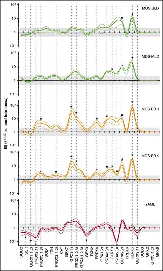
To precisely analyze the antioxidant response of all disease subtypes, relative gene expression levels of patients’ samples compared with healthy subjects (RQ) were plotted as antioxidant curves or antioxidograms (Figure 3), from the highest to the lowest levels in healthy subjects (supplemental Figure 2). Antioxidograms allowed for the identification of specific profiles depending on MDS subgroups or sAML. Antioxidant gene expression was not decreased in MDS. Conversely, genes encoding for glutaredoxins, enzymes reducing disulfide bonds in proteins, were overexpressed in all MDS subgroups. There was thus an increase of the GLRX5 (P = .0350) and GLRX2(2) transcripts (P = .0080), and a similar trend was found for GLRX3 (P = .0576). When considering MDS with <5% BM blasts, it is interesting to note that the overexpression was limited to antioxidant genes weakly expressed in BM from healthy controls. MDS with 5% to 19% BM blasts were characterized by the overexpression of additional genes encoding for peroxiredoxins and glutathione peroxidase 1, H2O2-reducing enzymes. Increases in PRDX(1,2,3) (P = .0015), PRDX2(3) (P = .0192), PRDX3(1,2) (P = .0284), PRDX4 (P = .0018), PRDX5(1,3) (P = .0016), PRDX5(2) (P = .0007), and GPX1 (P = .0001) transcripts were observed. Moreover, MDS-EB-2 presented a higher number of overexpressed genes compared with MDS-EB-1 (respectively, 10 and 6 genes). More generally, MDS progression appeared to be associated with overexpression of genes already strongly expressed in healthy BM. Contrarily to MDS, sAML were characterized by a collapse of the antioxidant response. This is illustrated by significant decreases of several transcripts: GLRX1(1-2) (P = .0291), GLRX2(1) (P = .0349), GSR (P = .0380), GPX3 (P = .0115), PRDX2(1) (P = .0014), and PRDX2(3) (P = .0007). This profile is in accordance with the higher level of ROS in sAML compared with MDS BM samples.
Antioxidant profiles of MDS/sAML patients. Expression levels of 28 transcripts of key antioxidant genes in the BM of patients MDS/sAML. Transcripts are ranked in decreasing order of expression in normal BM. Results are expressed as relative quantification (RQ) compared with normal BM (mean ± SEM). The horizontal black line (y = 1) represents the healthy control profile (n = 16), and green, yellow, and red lines represent patients’ data (thick and thin lines representing mean and SEM, respectively). The gray area corresponds to variations less than twice that of controls. Dashed vertical lines indicate variants with at least 1 significant difference between patients and healthy controls. MDS with <5% blasts (MDS-SLD and MDS-MLD) display an overexpression of only a few low-expressed transcripts (GLRX family), conversely to MDS with 5% to 19% blasts (MDS-EB-1 and MDS-EB-2), showing several overexpressions (PRDX and GPX families). The profile of sAML was different, with decreased expression of 6 transcripts, especially PRDX2 or GPX3. Significant difference with the healthy controls (ΔCt values), *P < .05; #P < .01.
Antioxidant profiles of MDS/sAML patients. Expression levels of 28 transcripts of key antioxidant genes in the BM of patients MDS/sAML. Transcripts are ranked in decreasing order of expression in normal BM. Results are expressed as relative quantification (RQ) compared with normal BM (mean ± SEM). The horizontal black line (y = 1) represents the healthy control profile (n = 16), and green, yellow, and red lines represent patients’ data (thick and thin lines representing mean and SEM, respectively). The gray area corresponds to variations less than twice that of controls. Dashed vertical lines indicate variants with at least 1 significant difference between patients and healthy controls. MDS with <5% blasts (MDS-SLD and MDS-MLD) display an overexpression of only a few low-expressed transcripts (GLRX family), conversely to MDS with 5% to 19% blasts (MDS-EB-1 and MDS-EB-2), showing several overexpressions (PRDX and GPX families). The profile of sAML was different, with decreased expression of 6 transcripts, especially PRDX2 or GPX3. Significant difference with the healthy controls (ΔCt values), *P < .05; #P < .01.
Because ring sideroblasts interfere with redox metabolism,9,10 MDS-SLD-RS were compared with MDS-SLD, all having a defective erythropoiesis (without isolated thrombocytopenia or neutropenia), to determine the specific signature resulting from RS. Indeed, the presence of RS was associated with an increased expression of 13 antioxidant enzyme genes (Figure 4) that detoxify H2O2 and reduce disulfide bounds, including the expected overexpression of GLRX5, because mutations of the SF3B1 gene have been identified in a high proportion of patients with RARS.23,24
Ringed sideroblast antioxidant signature. Expression levels of 28 transcripts of key antioxidant genes in the BM of MDS-SLD-RS patients, compared with MDS-SLD patients. Transcripts are ranked in decreasing order of expression in normal BM. Results are expressed as RQ compared with MDS-SLD (mean ± SEM). The horizontal black line (y = 1) represents the MDS-SLD profile (n = 10); blue lines represent the data of MDS-SLD-RS patients (thick and thin lines representing mean and SEM, respectively, n = 13). The gray area corresponds to variations less than twice the normal expression. Dashed vertical lines indicate variants with significant difference. The presence of RS was associated with overexpression of 13 transcripts, including GLRX5. Significant difference with the MDS-SLD (RQ values), *P < .05.
Ringed sideroblast antioxidant signature. Expression levels of 28 transcripts of key antioxidant genes in the BM of MDS-SLD-RS patients, compared with MDS-SLD patients. Transcripts are ranked in decreasing order of expression in normal BM. Results are expressed as RQ compared with MDS-SLD (mean ± SEM). The horizontal black line (y = 1) represents the MDS-SLD profile (n = 10); blue lines represent the data of MDS-SLD-RS patients (thick and thin lines representing mean and SEM, respectively, n = 13). The gray area corresponds to variations less than twice the normal expression. Dashed vertical lines indicate variants with significant difference. The presence of RS was associated with overexpression of 13 transcripts, including GLRX5. Significant difference with the MDS-SLD (RQ values), *P < .05.
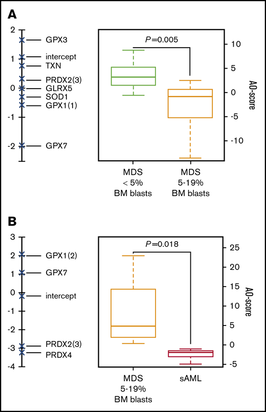
Antioxidogram genes do not all have the same importance in the observed variations between MDS with <5% BM blasts, MDS with 5% to 19% BM blasts, and sAML. To enhance the clinical potential of antioxidograms, their expression pattern was transformed into the AO-Score by logistic regression from the RQ value: AO-Score = , C(i) and RQ(i) being weighted coefficients and relative quantification of gene i, respectively, and n the number of genes in the model.15 The AO-score was computed using a split-sampling strategy (1000 iterations) allowing for internal cross-validation. This algorithmic approach increases the robustness of the score. It identified a set of 7 transcripts (GLRX5, GPX1(1), GPX3, GPX7, PRDX2(3), SOD1, and TXN) and weighted coefficients sufficient to statistically discriminate between MDS with <5% BM blasts and MDS with 5% to 19% BM blasts (Figure 5A) by using the AO-Score. A similar strategy identified 4 transcripts (GPX1(2), GPX7, PRDX2(3), and PRDX4) and weighted coefficients sufficient to statistically discriminate MDS with 5% to 19% BM blasts and sAML (Figure 5B). Based on oligogenic data, the AO-Score avoids potential bias in strategies based on monogenic predictions and allows for the discrimination between MDS grades and sAML.
Logistic regression of antioxidogram RQ values to define the AO-Score. AO-Score was calculated as follows: AO-Score = , C(i) and RQ(i) being weighted coefficients and relative quantification of gene i, respectively, and n the number of genes in the model. (A) Weighted coefficients of 7 transcripts allowed the discrimination between patients with MDS with <5% BM blasts or MDS with 5% to 19% BM blasts plotted in a vertical axis. (Right) Calculated weighted values (AO-Score) plot. (B) Weighted coefficients of 4 transcripts allowed the discrimination between MDS with 5% to 19% BM blasts and sAML patients. Significant difference (AO-Score values), P < .05.
Logistic regression of antioxidogram RQ values to define the AO-Score. AO-Score was calculated as follows: AO-Score = , C(i) and RQ(i) being weighted coefficients and relative quantification of gene i, respectively, and n the number of genes in the model. (A) Weighted coefficients of 7 transcripts allowed the discrimination between patients with MDS with <5% BM blasts or MDS with 5% to 19% BM blasts plotted in a vertical axis. (Right) Calculated weighted values (AO-Score) plot. (B) Weighted coefficients of 4 transcripts allowed the discrimination between MDS with 5% to 19% BM blasts and sAML patients. Significant difference (AO-Score values), P < .05.
Patients were risk-stratified according to the IPSS-R,8 which defines risk groups among patients with MDS by using refined cytogenetic risk assessment and a sophisticated categorization of BM blast percentages and cytopenias. Interestingly, the AO-Score was not correlated to IPSS-R (supplemental Figure 3), suggesting that this molecular signature could provide an additional prognostic value to IPSS-R.
Discussion
It is well established that leukemic stem cells have a low level of ROS,25 and we have reported the importance of the GPX3 antioxidant gene in this mechanism.4 The GPX3 gene is located at 5q33, on the long arm of chromosome 5, which frequently presents chromosomal abnormalities in MDS.26 On this basis, we postulated that the oxidative metabolism of preleukemic states could be disrupted. We quantified the level of ROS in BM subpopulations of MDS patients and concomitantly carried out a global approach to quantify the major antioxidant genes involved in the regulation of ROS levels.
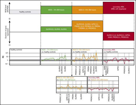
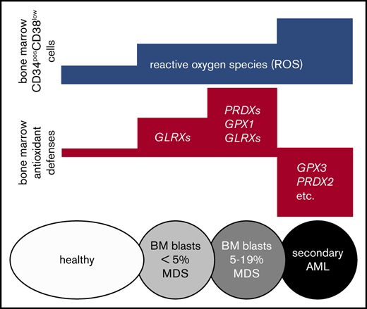
The original results presented here demonstrate major perturbations in redox homeostasis of BM CD34posCD38low cells in MDS and sAML when compared with age-matched healthy controls. It has been reported that ROS contribute to a dysfunction of BM hematopoietic stem cells in aged mice,27 but this study extended the concept to diseased human BM. A progressive increase in ROS levels was observed in CD34posCD38low progenitor cells from MDS patients compared with healthy controls, and higher levels in sAML compared with MDS patients (Figure 6). These data evidence a role for the oxidative stress in MDS pathogenesis, which has been suggested by studies reporting markers of oxidative stress in MDS patients. These include an increased plasma concentration of malondialdehyde,28 the presence of oxidized DNA bases in CD34pos BM cells29 and peripheral leukocytes,30 elevated ROS levels in red blood cells and platelets,31 and an increase in peroxide levels in the BM cells of MDS patients.32 In light of these findings, antioxidant treatments should be explored in MDS patients with low blast counts to avoid the risk of promoting transformation because a low ROS level has been described in leukemic stem cells.4,25
Evolutive profile of ROS levels in BM primitive progenitors and BM antioxidogram in MDS/sAML patients. (Top) Increasing ROS levels in CD34posCD38low progenitors during MDS evolution, from healthy controls (low ROS level) to MDS patients (intermediate ROS level), then sAML transformation (high ROS level), according to Figure 1. According to Figure 2, the middle panel shows the evolution of the antioxidogram from healthy controls to MDS with <5% BM blasts, MDS with 5% to 19% BM blasts, and sAML patients, successively. (Bottom) Variations of the antioxidant profile during transitions from MDS with <5% BM blasts to MDS with 5% to 19% BM blasts and sAML. Significant difference (ΔCt values), *P < .05.
Evolutive profile of ROS levels in BM primitive progenitors and BM antioxidogram in MDS/sAML patients. (Top) Increasing ROS levels in CD34posCD38low progenitors during MDS evolution, from healthy controls (low ROS level) to MDS patients (intermediate ROS level), then sAML transformation (high ROS level), according to Figure 1. According to Figure 2, the middle panel shows the evolution of the antioxidogram from healthy controls to MDS with <5% BM blasts, MDS with 5% to 19% BM blasts, and sAML patients, successively. (Bottom) Variations of the antioxidant profile during transitions from MDS with <5% BM blasts to MDS with 5% to 19% BM blasts and sAML. Significant difference (ΔCt values), *P < .05.
The abnormalities of oxidative metabolism observed in the different groups tested here suggest their evolution together with progression of the disease. In MDS with < 5% blasts, the oxidative stress is associated with a limited antioxidant response (GLRX), adapted but insufficient to limit the excess of ROS. When the blast count increases to 5% to 19%, there is an increased antioxidant response (GLRX, PRDX) which is still insufficient. Finally, at the time of acutisation in AML, a significant increase in ROS is observed, likely from a collapse of the antioxidant response (GLRX, GPX, PRDX, GSR).
Regarding MDS-SLD-RS, the specific BM antioxidant signature resulting from the presence of RS was characterized by the overexpression of numerous antioxidant genes, including the expected overexpression of GLRX5,23,24 in suitability with the very high level of ROS in these patients’ erythroblasts (Figure 1). These results are in accordance with the major disturbance in redox metabolism previously reported,10 with the antioxidant response observed in BM cells being adapted but insufficiently efficient.
The oxidative metabolism abnormalities described here may at least partially explain the benefits of recent therapeutic strategies for MDS/sAML, such as hypomethylating agents (HMAs), Bcl2 inhibitors (BT-199), and Smoothened (Smo) inhibitors. It is interesting to note that the expression of many antioxidant genes of the antioxidogram can be regulated via CpG islands, such as GLRX2,33 PRDX234 GPX1,35 and GPX3.36 Moreover, BCL2 is highly expressed in ROS-low AML leukemia stem cells resistant to chemotherapy.25 Of note, a combined treatment of BT-199 and HMA increases the induction of ROS compared with HMA alone.37 An antioxidant effect has been described through activation of the SHH pathway by overexpression of SOD and CAT.38 Finally, Smo inhibition induces oxidative stress in AML, notably via a secondary inhibition of the phosphatidylinositol 3-kinase and Akt pathways,39 involved in the redox metabolism of leukemic cells.40 Future studies are needed to precisely clarify the biological impact of these therapies in the redox metabolism of MDS/sAML.
In conclusion, this study establishes that MDS progenitors are characterized by oxidative stress and that antioxidant enzyme genes are overexpressed as an appropriate but ineffective response to normalize ROS levels. Our results suggest that progression from MDS toward sAML could be characterized by 3 successive molecular steps: (1) overexpression of disulfide-reducing enzymes (GLRX family), then (2) overexpression of those that reduce H2O2 (PRDX and GPX families), and finally (3) global enzymatic collapse during acutisation. Moreover, the antioxidograms and AO-Score could be interesting molecular biomarkers of clinical interest for diagnosis (ie, to discriminate MDS from the recently described group of indolent hematopoietic disorders, respectively, idiopathic cytopenia of unknown significance, idiopathic dysplasia of unknown significance, clonal hematopoiesis of indeterminate potential, and clonal cytopenia of unknown significance). They could also prove helpful for the longitudinal follow-up of MDS patients as well as to develop treatment decision algorithms.
Acknowledgments
This work was partly supported by SATT Grand-Centre, Novartis, the Ligue Nationale Contre le Cancer, the Cancéropôle Grand Ouest, the International Rotary Club of Blois (district 1720), and the French associations CANCEN and Les Sapins de l’Espoir Contre le Cancer. F.P. was supported by the grant 0010-UFR-SMD from SATT Grand Centre. The antioxidogram is patented (publication number: WO2012085188 A1).
Authorship
Contribution: O.H. designed the research; F.P., C.V., C.D., and S.L. performed the research; F.P., C.V., C.D., S.L., and O.H. analyzed data; N.G., A.F., M.-H.E., N.R., and J.D. provided technical support; O.K., E.G., and M.F. contributed study material; M.C.B. edited the manuscript; and F.P., C.V., and O.H. wrote the paper.
Conflict-of interest-disclosure: The authors declare no competing financial interests.
Correspondence: Olivier Herault, Service d’Hématologie Biologique, CHRU de Tours, 2 Blvd Tonnellé, 37000 Tours, France; e-mail: olivier.herault@univ-tours.fr.
References
Author notes
F.P. and C.V. contributed equally to this study.
The full-text version of this article contains a data supplement.

![ROS levels in bone marrow cell subpopulations of MDS/sAML patients. (A) Flow cytometry quantification of ROS in BM cells (ie, monocytic, granulocytic, lymphocytic, erythroblastic subpopulations and CD34pos, CD34posCD38low, and CD34posCD38high progenitors) of healthy controls. ROS levels were evaluated by DCFDA mean fluorescence intensity (DCFDA-MFI) and results are expressed as % of DCFDA MFI of monocytes (± standard error of the mean [SEM]). The highest level of ROS was found in monocytic cells in all samples, and the lowest level in the erythroblastic subpopulation. *Significant difference with monocytic cells, P < .05; #significant difference, P < .05. (B) Absolute ROS levels (DCFDA-MFI) in the BM monocytic subpopulation from healthy controls, MDS subgroups, and sAML are not statistically different (P = .77, Kruskal-Wallis test). (C) ROS levels in the BM subpopulations of MDS/sAML patients compared with healthy controls, after normalization with the respective DCFDA-MFI value of monocytes in each sample. Increased ROS levels were observed exclusively in myeloid and especially progenitor subpopulations. The most important oxidative stress was found in the erythroblastic subpopulation in MDS-SLD-RS and MDS-MLD with ring sideroblasts, enriched in ring sideroblasts, and in the CD34posCD38low progenitors in all pathologies, especially in sAML. Data are expressed as mean ± SEM (n = 65). *Significant difference with the healthy controls, P < .05. #Significant difference with healthy controls and with MDS subgroups, P < .05. ns, nonsignificant difference with the healthy controls.](https://ash.silverchair-cdn.com/ash/content_public/journal/bloodadvances/3/24/10.1182_bloodadvances.2019000677/3/m_advancesadv2019000677f1.png?Expires=1766176214&Signature=wPDV2s4xCugOzNcY~9argC-AxzaIMU-t8Nlu~XmXarOEFG68eyiIuMpgWOSN~VhFCH2~xr-R67SOdpQ6Q9bredySwhRE7eVjN8FJy~khd~uFV5Le~XK4S0hlp7NRIz3uNH4ChLqb-SXjNVRDEEVlN8AloURsRYj2DIZAeQavl~M9ps1ImDI1tJlZ42xPZfl~-yUqCkeZjWUn6QdngTu4FFvB5J2NmWH11YqSMMuDAhNwSWU5HiZf~KzhLGU1J0eed598MXiPGW7JZGQMT4QLTWV~NmMWGRlYmYAd9RiQ-ZBleqhy8qtMcCsKjLc5rxtlC526TgtfnITbObn8eCcVIw__&Key-Pair-Id=APKAIE5G5CRDK6RD3PGA)
