The molecular basis for the interaction between a prototypic non–I-domain integrin, αIIbβ3, and its ligands remains to be determined. In this study, we have characterized a novel missense mutation (Tyr143His) in αIIb associated with a variant of Glanzmann thrombasthenia. Osaka-12 platelets expressed a substantial amount of αIIbβ3(36%-41% of control) but failed to bind soluble ligands, including a high-affinity αIIbβ3-specific peptidomimetic antagonist. Sequence analysis revealed that Osaka-12 is a compound heterozygote for a single 521T>C substitution leading to a Tyr143His substitution in αIIb and for the null expression of αIIb mRNA from the maternal allele. Given that Tyr143 is located in the W3 4-1 loop of the β-propeller domain of αIIb, we examined the effects of Tyr143His or Tyr143Ala substitution on the expression and function of αIIbβ3 and compared them with KO (Arg-Thr insertion between 160 and 161 residues of αIIb) and with the Asp163Ala mutation located in the same loop by using 293 cells. Each of them abolished the binding function of αIIbβ3 for soluble ligands without disturbing αIIbβ3 expression. Because immobilized fibrinogen and fibrin are higher affinity/avidity ligands for αIIbβ3, we performed cell adhesion and clot retraction assays. In sharp contrast to KO mutation and Asp163AlaαIIbβ3, Tyr143HisαIIbβ3-expressing cells still had some ability for cell adhesion and clot retraction. Thus, the functional defect induced by Tyr143HisαIIb is likely caused by its allosteric effect rather than by a defect in the ligand-binding site itself. These detailed structure–function analyses provide better understanding of the ligand-binding sites in integrins.
Introduction
Integrins are a family of noncovalently associated αβ heterodimeric adhesion receptors that mediate cellular attachment to the extracellular matrix and cell–cell cohesion.1,2 Integrins are involved in a variety of physiological processes, including development, immune response, wound healing, and hemostasis.1 They are also involved in pathologic processes, such as tumor metastasis, thrombosis, and athelosclerosis.1
Although its expression is restricted to the megakaryocyte/platelet lineage, αIIbβ3 (GPIIb-IIIa) is a prototypic integrin that functions as a physiologic receptor for fibrinogen and von Willebrand factor and that plays a crucial role in platelet aggregation, normal hemostasis, and pathologic thrombosis.3 Indeed, Glanzmann thrombasthenia (GT) is an autosomal recessive bleeding disorder caused by a defect in the expression or the function of integrin αIIbβ3.4 Recent clinical studies have shown the beneficial effects of αIIbβ3 antagonists in patients undergoing coronary angioplasty and in those with unstable angina.5,6Integrin α subunits are grouped into 2 classes based on the presence or absence of an inserted domain of approximately 200 amino acid residues (I- or A-domain); αIIb and αvsubunits do not have the I-domain.7-9 Recently, the crystal structures of the extracellular segment of the other β3 integrin, αvβ3—in the presence or absence of its small ligand—have been described.10,11 As predicted, the putative ligand-binding head is primarily formed by a 7-bladed β-propeller domain from αv and a β I-domain from β3.10-12 The β-propeller domain contains 7 4-stranded β-sheets (W1-W7) arranged in a torus around a pseudosymmetry axis. However, the ligand-binding sites in the β-propeller domain of the αIIb subunit remain to be determined.
The characterization of molecular defects in GT from dysfunctional αIIbβ3 (variant GT) has succeeded in pinpointing ligand-binding sites and functionally important domains.13-15 We have demonstrated that 2-amino acid insertion (Arg-Thr) between amino acid residues 160 and 161 in αIIb is responsible for the ligand-binding defect in a Japanese variant GT known as KO.16 The insertion is located within the small loop (Cys146-Cys167) between W2 and W3 (W3 4-1 loop) located on the upper face of the β-propeller. Alanine substitutions further indicate that Asp163 within the Cys146-Cys167 loop is one of the critical residues for ligand binding.16In this context, 2 other naturally occurring missense mutations, Pro145Ala and Leu183Pro, which impair αIIbβ3 expression and its ligand-binding function, have been identified in the W3 4-1 loop and the W3 2-3 loop of the β-propeller, respectively.17 18
In this study we demonstrate that a novel naturally occurring missense mutation (Tyr143His) within the W3 4-1 loop in αIIb is responsible for a binding defect in αIIbβ3for soluble ligands. Because Tyr143His, KO, and Asp163Ala mutations are located within the same loop in αIIb, we further compared functions of these mutant αIIbβ3 by using cell adhesion and clot retraction assays. Compared with the KO mutation and Asp163AlaαIIbβ3, Tyr143HisαIIbβ3-expressing cells still had some ability for cell adhesion and clot retraction. Our results indicate that the KO mutant and Asp163AlaαIIbβ3 impair ligand-binding function in αIIbβ3 more severely than Tyr143HisαIIbβ3.
Patients, materials, and methods
Patient history
Patient Osaka-12, a product of nonconsanguineous parents, was a 21-year-old Japanese woman who had a history of moderate mucocutaneous bleeding in her childhood. Hematologic examinations revealed a prolonged bleeding time (more than 15 minutes) and an absence of platelet aggregation in response to adenosine diphosphate (ADP), epinephrine, and collagen but a normal response to ristocetin. Clot retraction using the MacFarlane method was 46% (normal range, 40%-70%).19 Informed consent for analyzing their molecular genetic abnormalities was obtained from Osaka- 12 and her parents.
Antibodies and synthetic ligands
AP1 (GPIb-specific monoclonal antibody [mAb]), AP2 (αIIbβ3-specific mAb), and AP5 (β3-specific mAb) were generously provided by Dr Thomas J. Kunicki (Scripps Research Institute, La Jolla, CA).20,21 AP3 (β3-specific mAb) was a generous gift from Dr Peter J. Newman (Blood Center of Southeastern Wisconsin, Milwaukee).22 OP-G2 is a ligand-mimetic αIIbβ3-specific mAb that binds to nonactivated and activated αIIbβ3.23 PAC-1, a ligand-mimetic αIIbβ3-specific mAb that binds specifically to activated αIIbβ3, was kindly provided by Dr Sanford J. Shattil (Scripps Research Institute).24 PT25-2 (αIIbβ3-specific mAb) activates αIIbβ3 and was a kind gift from Drs Makoto Handa and Yasuo Ikeda (Keio University, Tokyo, Japan).25PMI-1 (αIIb-specific mAb), anti-LIBS1 (β3-specific mAb), and anti-LIBS6 (β3-specific mAb) were generously provided by Dr Mark H. Ginsberg (Scripps Research Institute).26,27 PMI-1, AP5, anti-LIBS1, and anti-LIBS6 recognize ligand-induced conformational changes on αIIbβ3, termed ligand-induced binding sites (LIBS).21,26,27 TP80 (αIIb-specific mAb) and MOPC21 (mouse myeloma immunoglobulin G1 [IgG1]) were purchased from Nichirei (Tokyo, Japan) and Sigma Chemical (St Louis, MO), respectively. FK633 (αIIbβ3-specific peptidomimetic antagonist) was generously provided by Dr Jiro Seki (Fujisawa Pharmaceutical, Osaka Japan),28 and cyclo(RGDfV) [cyclo(-Arg-D-Gly D-Asp-D-Phe-L-Val-D-)] peptide (αvβ3-specific antagonist) was a generous gift from Merck KGaA (Darmstadt, Germany).29 Fibrinogen was purchased from Calbiochem-Novabiochem (San Diego, CA). MOPC21, AP1, AP2, TP80, AP3, PAC-1, and fibrinogen were labeled with fluorescein isothiocyanate (FITC), as previously described.28
Flow cytometry
Flow cytometric analysis using various mAbs was performed as previously described.30 FITC-labeled mAbs were used to quantify the expression levels of αIIbβ3 on 293 cells and on platelets.
Analysis of platelet messenger RNA
Total cellular RNA of platelets was isolated from 30 mL whole blood, and αIIb or β3 messenger RNA (mRNA) was specifically amplified by reverse transcription–polymerase chain reaction (RT-PCR), as previously described.30 Primers for the amplification of αIIb or β3 mRNA and conditions for RT-PCR were described elsewhere.30 31Nucleotide sequences of PCR products were determined by using Taq DyeDeoxy Terminator Cycle Sequencing kit and an ABI373A DNA sequencer (Applied Biosystems, Foster City, CA).
Allele-specific restriction enzyme analysis
Amplification of the region around exon 4 of the αIIb gene was performed by using primers IIbE3, 5′-GTCGGTCGTCAGCTGGAGC-3′ (sense, nucleotide [nt] 3940-3958 in the αIIb gene), and IIbE4, 5′-CAGGTCGTAGCTGGCGCTTAC-3′ (antisense, nt 4192-4172) and by using 250 ng DNA as a template. First-round PCR products were reamplified using primers IIbg3947S, 5′-GTCAGCTGGAGCGACGTCATTGTG-3′ (sense, nt 3947-3970) and IIbE4 and then were digested with restriction enzyme ScaI. Resultant fragments were electrophoresed in a 1.5% agarose gel.
Construction of αIIb expression vectors and cell transfection
Wild-type αIIb and β3 complementary DNAs (cDNAs) cloned into a mammalian expression vector pcDNA3 (Invitrogen, San Diego, CA) were generously provided by Dr Peter J. Newman. To construct the expression vector containing the521C (His143) form of αIIb, PCR-based cartridge mutagenesis was performed as previously described.16 Platelet αIIb cDNA from Osaka-12 was amplified by RT-PCR using primers IIb1 and IIb3αAS, 5′-CCCACGATCAGGTCTGGGTATCCG-3′ (antisense, nt 1407-1384). Second-round amplification was then performed using 1 μL first-round PCR products as a template with nested primers IIb187S, 5′-GAGAGTGGCCATCGTGGTGG-3′ (sense, nt 187-206), and IIb3αAS2C, 5′-CCGTTGTCATCGATGTCTACGGC-3′ (antisense, nt 1364-1386). To construct the expression vector for Tyr143Ala αIIb substitution, we carried out the overlapping extension PCR as previously described.16Mismatched sense primer IIb143Ala-S, 5′-GAGCGGCCGCCGCGCCGAGGCCTCCCCCTG-3′ (sense, nt 502-531, 2 bp mismatched), and antisense primer IIb143Ala-AS, 5′-CAGGGGGAGGCCTCGGCGCGGCGGCCGCTC-3′ (antisense, nt 531-502, 2 bp mismatched) were synthesized. PCR was performed by using αIIb cDNA as a template and primers IIb1 and IIb143Ala-AS, or primers IIb143Ala-S and IIb3αAS. The 2 individually amplified PCR products were mixed and used as a template of PCR using primers IIb1 and IIb3αAS. Amplified fragments were digested with restriction enzyme SacII and ClaI, and the resultant fragments were extracted using GeneClean II kit (Bio 101, La Jolla, CA). Fragments were introduced into the pcDNA3 that had been digested with SacII and ClaI. Inserted fragments were characterized by sequence analysis to verify the absence of any other substitutions and the proper insertion of the PCR cartridge into the vector. Expression vectors containing the KO mutant (2-amino acid [Arg-Thr] insertion between residues 160 and 161) αIIbcDNA and Asp163AlaαIIb cDNA were constructed as previously described.16
The wild-type or mutant αIIb construct was cotransfected into 293 cells with the wild-type β3 construct by the calcium phosphate method, as previously described.16 The 293 cells transiently expressing mutant αIIbβ3 were obtained and analyzed 2 days after transfection. In selected experiments, CD36 expression vector was cotransfected with αIIb and β3 constructs to monitor transfection efficiency.32 In addition, stable transfectants expressing wild-type, Tyr143HisαIIbβ3, or KO mutant αIIbβ3 were selected for G418 resistance (Gibco BRL, Grand Island, NY) and were cultured in Dulbecco modified Eagle medium (DMEM) with 10% heat-inactivated fetal calf serum (Life Technologies, Gaithersburg, MD).
Adhesion assays
Adhesion assays were performed as described by Faull et al.33 Wells from 96-well microtiter plates were coated with up to 0.5 μg fibrinogen per well in 100 μL phosphate-buffered saline (PBS) and were incubated at 4°C overnight. After washing with PBS, wells were blocked with PBS containing 1% bovine serum albumin (BSA) (Sigma) for 90 minutes at 22°C. To determine background adhesion, control wells were coated with 1% BSA. Then 293 cells transiently expressing wild-type or mutant αIIbβ3 were washed twice with PBS and resuspended in DMEM containing 0.1% BSA at a concentration of 1 × 10⁶ cells/mL, and 100 μL aliquots cell suspension were added to wells in triplicate. The plate was incubated in a humidified 37°C incubator for 60 minutes. After washing with PBS 3 times, the adherent cells were quantified by measuring endogenous cellular acid phosphate activity in an enzyme-linked immunosorbent assay.34
For morphologic analysis, 293 cells stably expressing wild-type or mutant αIIbβ3 were added on fibrinogen-coated glass coverslips for 60 minutes at 37°C. After they were washed with PBS, adherent cells were fixed in 3.7% formaldehyde for 10 minutes, permeabilized in 0.5% Triton X-100 in PBS for 5 minutes at room temperature, and washed twice with PBS. Cells on coverslips were stained with rhodamine-phalloidin (Sigma) and were analyzed under a fluorescence microscope (Olympus, Tokyo, Japan).35
Tyrosine phosphorylation of pp125FAK
Adherent cells on fibrinogen were lysed in Triton X-100 buffer (1% Triton X-100, 25 mM Tris-HCl, 100 mM NaCl, pH 7.4, 0.1 mg/mL leupeptin, 4 μg/mL pepstatin A, 1 mM phenylmethylsulfonyl fluoride, and 10 mM benzamide) containing sodium vanadate and were scraped into microcentrifuge tubes. Lysates were incubated on ice for 30 minutes, and clarified supernatants were processed for immunoprecipitation. Focal adhesion kinase (pp125FAK) was immunoprecipitated with 1 μg rabbit polyclonal antibody specific for FAK (Santa Cruz Biotechnology, CA), and protein-G Sepharose (Pharmacia, Uppsala, Sweden). Precipitates were separated on 7.5% sodium dodecyl sulfate–polyacrylamide gel electrophoresis (SDS-PAGE) and were transferred to a polyvinylidene difluoride (PVDF) membrane. Phosphotyrosine was detected with mAb 4G10. To monitor the loading of gel lanes, the blots were stripped (2% SDS, 62.5 mM Tris, pH 6.7, 100 mM 2-mercaptoethanol for 30 minutes at 70°C) and reprobed with anti-FAK.36
Clot retraction
Clot retraction of stable cell lines was performed based on the method described by Katagiri et al.37 In brief, 293 cells stably expressing αIIbβ3 in Tyrode/HEPES (N-2-hydroxyethylpiperazine-N′-2-ethanesulfonic acid) buffer (2 × 10⁶ cells/mL) were incubated with 10 mM tranexamic acid, 250 μg/mL fibrinogen, and 2 mM CaCl2 at 37°C in a siliconized glass tube. To block the effect of αvβ3 composed of endogenous αv and transfected β3, these stable transfectants were preincubated with 50 μM cyclo(RGDfV) (αvβ3-specific antagonist) at room temperature for 30 minutes. Then 1 U thrombin was added to 1 mL cell suspension. Fibrin gels began to form immediately after the addition of thrombin, and the tubes were kept at 37°C. Clot retraction was monitored by taking photographs every 30 minutes using a digital camera. Quantification of retraction was performed by an assessment of the clot area using the NIH Image 1.67e software (Bethesda, MD). Data were expressed as follows: % clot retraction = [(areat0 − area t)/areat0] × 100.
Results
Platelets from thrombasthenic patient Osaka-12 show impaired ligand-binding function
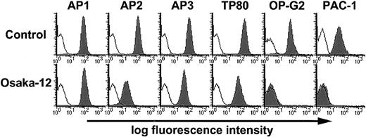
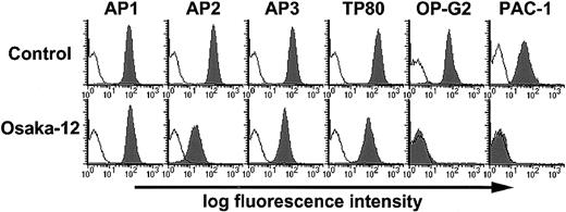
We first examined the surface expression of αIIbβ3 on platelets from patient Osaka-12 and 3 control subjects using flow cytometry. Although FITC-AP1 (GPIb-specific mAb) bound to Osaka-12 platelets slightly higher than to control platelets (113% of control value [mean value obtained from 3 control subjects]), FITC-AP3 (β3-specific mAb) and FITC-TP80 (αIIb-specific mAb) revealed a significant reduction in the expression of αIIbβ3 on Osaka-12 platelets (AP3 binding, 41% of control value; TP80 binding, 36% of control value; n = 2) (Figure1). Compared with AP3 and TP80 binding to Osaka-12 αIIbβ3, AP2 binding was markedly impaired (13% of control value; n = 2) probably because of a disturbance of AP2 epitope formation. Because Osaka-12 platelets expressed 36% to 41% of normal amounts of αIIbβ3 on their surfaces, we examined their ligand-binding function. OP-G2 and PAC-1 mAbs are activation-independent and activation-dependent, ligand-mimetic mAbs specific for αIIbβ3, respectively. Neither OP-G2 nor PAC-1 in the presence of an activating PT25-2 mAb bound to Osaka-12 platelets (Figure 1). Thus, Osaka-12 αIIbβ3 seems to have a ligand-binding defect and a quantitative defect.
Flow cytometric analysis of the surface expression and ligand-binding function of αIIbβ3 Osaka-12 platelets.
Washed platelets obtained from Osaka-12 or 3 control subjects were incubated with FITC-AP1 (GPIb-specific mAb), FITC-AP2 (αIIbβ3-specific mAb), FITC-AP3 (β3-specific mAb), and FITC-TP80 (αIIb-specific mAb) at a concentration of 10 μg/mL for 30 minutes at room temperature. FITC-MOPC21 (mouse myeloma IgG1) was used as a negative control. For OP-G2 binding (activation-independent ligand-mimetic αIIbβ3-specific mAb), bound antibodies were detected by FITC-conjugated goat F(ab′)2 antimouse IgG. Filled and open histograms represent the binding of specific and control antibodies, respectively. For PAC-1 binding, washed platelets were preincubated with 10 μg/mL PT25-2 (activating αIIbβ3-specific mAb) for 30 minutes, and then 10 μg/mL FITC-labeled PAC-1 was added. Closed and open histograms represent the PAC-1 binding in the absence and presence of 10 μM FK633 (αIIbβ3-specific antagonist), respectively. Results are representative of 2 separate experiments.
αIIbβ3 LIBS expression on Osaka-12 platelets by αIIbβ3-specific peptidomimetic antagonist FK633
PMI-1, AP5, anti-LIBS1, and anti-LIBS6 mAbs preferentially recognize LIBS on αIIbβ3, which are exposed following occupancy of the receptor by ligands. To further investigate the ligand-binding function of Osaka-12 αIIbβ3, we examined the effect of FK633, αIIbβ3-specific peptidomimetic antagonist on the expression of these LIBS. Compared with Arg-Gly-Asp-Trp peptide, FK633 has approximately 100-fold higher potency for LIBS induction.28 Although we could not simply compare the binding of mAbs recognizing LIBS between Osaka-12 and control platelets because of the different expression levels of αIIbβ3, even 10 μM FK633 showed a negligible effect on the expression of LIBS1 of Osaka-12 αIIbβ3 (Table1). On the other hand, Table 1 shows that Osaka-12 αIIbβ3 aberrantly expressed LIBS recognized by PMI-1, AP5, and anti-LIBS6. These data further suggested that the ligand-binding sites of Osaka-12 αIIbβ3 were markedly impaired.
Nucleotide sequence and allele-specific restriction enzyme analyses for Osaka-12 αIIbβ3
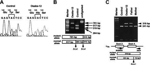
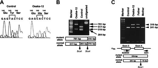
To identify the molecular genetic defect responsible for Osaka-12, all coding regions of αIIb and β3 cDNA amplified by RT-PCR from Osaka-12 and control platelet mRNA were analyzed. Direct sequence of the PCR fragments showed a single T>C substitution at nt 521 in αIIb cDNA that would lead to a Tyr143His substitution in exon 4 of αIIb (Figure2A). No other nucleotide substitutions were detected in the coding regions of either αIIb or β3 cDNAs. The T>C substitution abolished a restriction site for ScaI, and allele-specific restriction enzyme analysis for the amplified αIIb cDNA around exon 4 suggested that Osaka-12 might be homozygous for the substitution. However, allele-specific restriction enzyme analysis for the PCR fragments from genomic DNA revealed that Osaka-12 was heterozygous for the T>C substitution. The substitution was inherited from her father. The molecular genetic defect inherited from her mother remains determined. However, only the allele having 521C from her father was amplified by RT-PCR from Osaka-12 αIIb mRNA, suggesting that the expression levels of maternal αIIbmRNA seem to be extremely low in Osaka-12 platelets (Figure 2B-C).
Identification of the genetic defect responsible for Osaka-12.
(A) Nucleotide sequence analysis of αIIb cDNA from Osaka-12. Platelet αIIb mRNA was amplified by RT-PCR. Nucleotide sequence of the amplified fragments was determined by using Taq DyeDeoxy Terminator Cycle Sequencing kit and an ABI 373A DNA sequencer. (B) Allele-specific restriction enzyme analysis of αIIb cDNA. The region around exon 4 of the αIIb mRNA was amplified by PCR, followed by digestion with ScaI. ScaI digestion of the PCR products yielded 517-bp, 264-bp, and 316-bp fragments in the healthy allele. The T>C substitution abolished one of the restriction sites forScaI. Resultant fragments were electrophoresed in a 1.5% agarose gel. Marker indicates 100-bp DNA ladder. (C) Allele-specific restriction enzyme analysis of αIIb genomic DNA. The region around exon 4 of the αIIb gene was amplified by PCR using primers IIb3945S and IIbE4, followed by digestion withScaI. ScaI digestion of the PCR products yielded 241-bp and 75-bp fragments in the healthy allele. The T>C substitution abolished the restriction site for ScaI. Resultant fragments were electrophoresed in a 6% polyacrylamide gel. Marker indicates φX174 digested with HaeIII.
Effect of Tyr143His substitution in αIIb on αIIbβ3 expression and function
To confirm that the 521T>C substitution leading to Tyr143His substitution in αIIb is responsible for the functional defect, we constructed an expression vector that contained the wild-type or mutant His143 form of αIIb and cotransfected it with wild-type β3 vector into 293 cells. Effects of a Tyr143Ala substitution in αIIb on αIIbβ3 expression and function were also examined. Because Tyr143 is located within the W3 4-1 loop of the β-propeller domain, close to the KO mutation (the Arg-Thr insertion between 160 and 161 residues of αIIb) and the Asp163Ala αIIb mutation, we compared the expression and function of these mutant αIIbβ3 in parallel.
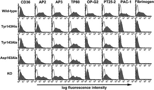
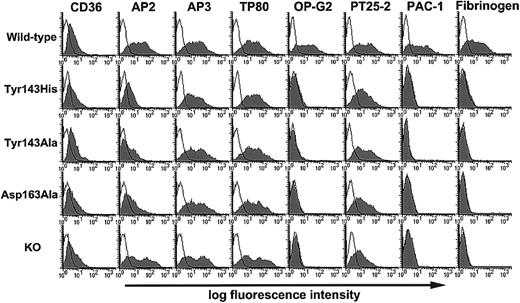
Transfection efficiency monitored by the cotransfected CD36 expression vector was essentially the same between wild-type and mutant αIIbβ3 (Figure3). Flow cytometric analysis using FITC-AP3 or FITC-TP80 mAb showed that the expression levels of Tyr143HisαIIbβ3 were essentially the same as wild-type αIIbβ3 (AP3 binding, 91% ± 15% of wild-type; TP80 binding, 102% ± 2% of wild-type; mean ± SD; n = 3). In sharp contrast, Tyr143HisαIIbβ3 failed to bind OP-G2 mAb, FITC-PAC-1 mAb, or FITC-fibrinogen in the presence of PT25-2 mAb (Figure 3). We also confirmed that the binding failure of these activation-dependent ligands was not caused by the binding failure of the activating mAb, PT25-2. In addition, FITC-AP2 binding was significantly impaired compared with TP80 binding (AP2 binding; 15% ± 3% of wild-type; mean ± SD; n = 3), which is consistent with the data obtained from Osaka-12 platelets (Figures 1and 3). Given that the expression levels of abnormal αIIbmRNA derived from her mother were so low that they were not detected in Osaka-12 platelets by RT-PCR, this maternal abnormality would reduce the expression of αIIbβ3 by 50%. As expected, the expression level of Tyr143HisαIIbβ3 on 293 cells was roughly twice as much as that on Osaka-12 platelets. These data indicate that the Tyr143His mutation is responsible for the variant GT phenotype in Osaka-12. The Tyr143Ala substitution induced essentially the same effects on αIIbβ3 as the Tyr143His mutation (AP3 binding, 117% ± 14% of wild-type; TP80 binding, 113% ± 8% of wild-type; AP2 binding, 14% ± 1% of wild-type; mean ± SD; n = 3). Because Tyr143Ala abolished the ligand binding, it is likely that the presence of Tyr143 is critical for αIIbβ3 function. Interestingly, the phenotype of Asp163AlaαIIbβ3 appears the same as that of Tyr143HisαIIbβ3. The phenotype of KO αIIbβ3 was essentially the same as wild-type αIIbβ3 except for the failure in OP-G2 and PAC-1 binding, as previously described.15 Thus, none of these mutant αIIbβ3 showed a binding ability for soluble ligands (Figure 3).
Effects of Tyr143HisαIIb, Tyr143AlaαIIb, Asp163AlaαIIb, or KO mutant on the expression and ligand-binding function of αIIbβ3 on 293 cells.
Wild-type αIIb or each mutant αIIb cDNA was transiently cotransfected with wild-type β3 cDNA into 293 cells. CD36 expression vector was cotransfected with αIIband β3 constructs, and CD36 expression was measured by FITC–anti-CD36 mAb to monitor transfection efficiency. The binding of FITC-AP2, FITC-AP3, FITC-TP80, OP-G2, and PT25-2 was analyzed by flow cytometry 2 days after transfection (filled histograms). FITC-MOPC21 was used as a negative control (open histograms). For OP-G2 or PT25-2 binding, bound antibodies were detected by FITC-conjugated goat F(ab′)2 antimouse IgG. For PAC-1 or fibrinogen binding, washed platelets were preincubated with 10 μg/mL PT25-2 (activating αIIbβ3-specific mAb) for 30 minutes, and then 10 μg/mL FITC-labeled PAC-1 or 150 μg/mL FITC-labeled fibrinogen was added. Closed and open histograms represent PAC-1 or fibrinogen binding in the absence and presence of 10 μM FK633 (αIIbβ3-specific antagonist), respectively. Results are representative of 3 separate experiments.
Adhesion of the cells to immobilized fibrinogen
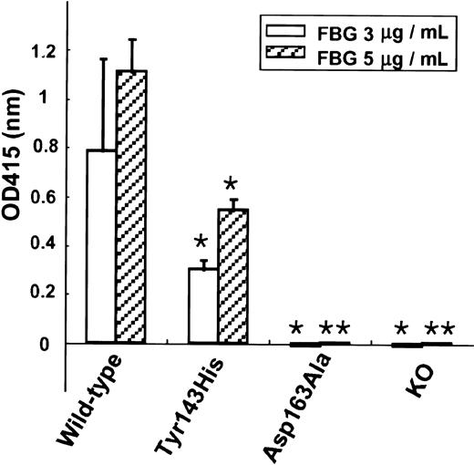
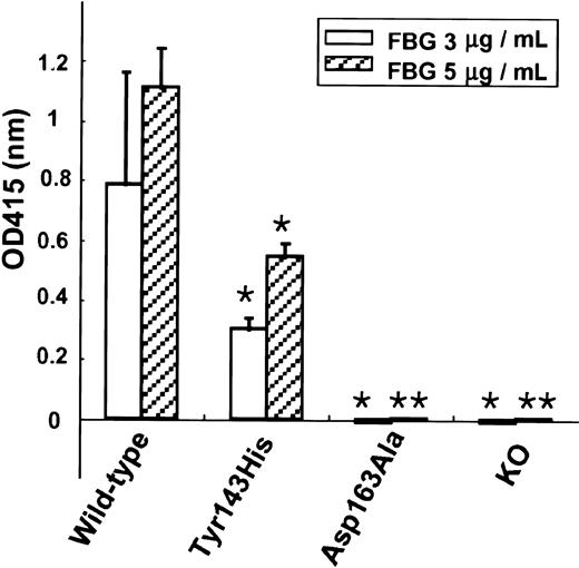
To further examine the functions of mutant αIIbβ3, we performed cell adhesion assay to immobilized fibrinogen. In contrast to parent cells, 293 cells transiently transfected with wild-type αIIbβ3 or mutant αIIbβ3 became adhesive to the immobilized fibrinogen. Given that endogenous αv in 293 cells can form αvβ3 with the exogenous β3 and may contribute to the adhesion, we preincubated the transfected cells with 50 μM cyclo(RGDfV) to block the αvβ3 function. Under these conditions, 293 cells transfected with wild-type β3 alone (data not shown), KO variant αIIbβ3, or Asp163AlaαIIbβ3 failed to adhere to immobilized fibrinogen (Figure 4). However, 293 cells expressing Tyr143HisαIIbβ3 showed a significant adhesion to immobilized fibrinogen, and the amounts of adherent cells were approximately 50% compared with 293 cells expressing wild-type αIIbβ3. The adhesion of 293 cells expressing Tyr143HisαIIbβ3 in the presence of 50 μM cyclo(RGDfV) was mediated solely by αIIbβ3 because 10 μM FK633 completely blocked the cell adhesion (data not shown).
Adhesion of αIIbβ3 mutants to immobilized fibrinogen.
Wild-type αIIb or each mutant αIIb cDNA was transiently cotransfected with wild-type β3 cDNA into 293 cells. Two days after transfection, wild-type or mutant αIIbβ3-transfected cells (1 × 10⁶ cells/well) were incubated with immobilized fibrinogen (FBG) in the presence of 50 μM cyclo(RGDfV) (αvβ3-specific antagonist) at 37°C. After washing with PBS, adherent cells were quantified with a colorimetric reaction using endogenous cellular acid phosphatase activity. Data represent the mean ± SD of triplicate measures of optical density at 415 nm. Statistical analysis (2-tailed P values for paired sample) was performed between 293 cells expressing wild-type αIIbβ3 and those expressing each mutant αIIbβ3 (**P < .01; *P < .05).
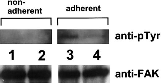
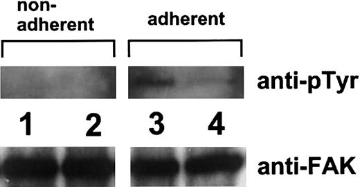
We then obtained the cells stably expressing wild-type, Tyr143His, and KO mutant αIIbβ3 with G418 selection for morphologic analysis of the adherent cells. Adhesive functions of 293 cells stably expressing Tyr143His αIIbβ3 or KO αIIbβ3 were the same as those transiently expressing these mutant αIIbβ3(data not shown). Although 293 cells stably expressing wild-type αIIbβ3 adhered firmly and showed complete spreading, 293 cells stably expressing KO mutant αIIbβ3 failed to adhere. In contrast to KO αIIbβ3, 293 cells stably expressing Tyr143HisαIIbβ3 moderately impaired cell spreading and cell adhesion (Figure 5). Because FAK, a 125-kDa cytoplasmic tyrosine kinase, is a component of focal adhesions and is a well-established component of integrin signaling pathways,38 the tyrosine phosphorylation of pp125FAK in these adherent 293 cells stably expressing wild-type αIIbβ3 or Tyr143HisαIIbβ3 was examined. As shown in Figure 6, pp125FAKphosphorylation was observed but impaired in cells expressing Tyr143HisαIIbβ3 compared with wild-type αIIbβ3 (Figure 6).
Morphologic analysis of the adherent cells stably expressing mutant αIIbβ3 to immobilized fibrinogen.
293 cells stably expressing (A) wild-type αIIbβ3, (B) Tyr143HisαIIbβ3, or (C) KO mutant αIIbβ3 were incubated with immobilized fibrinogen (5 μg/mL) at 37°C for 60 minutes in the presence of 50 μM cyclo(RGDfV) (αvβ3-specific antagonist). After gentle washing with PBS, adherent cells were fixed in 3.7% formaldehyde and permeabilized in 0.5% Triton X-100. Cells were stained with rhodamine–phalloidin and analyzed under a fluorescence microscope. Original magnification, × 100.
Tyrosine phosphorylation of pp125FAK.
Nonadherent or adherent 293 cells stably expressing wild-type αIIbβ3 (lanes 1 and 3) or Tyr143HisαIIbβ3 (lanes 2 and 4) on fibrinogen were lysed in Triton X-100 buffer containing sodium vanadate. pp125FAK was immunoprecipitated with 1 μg rabbit polyclonal antibody specific for FAK. Precipitates were separated on 7.5% SDS-PAGE and transferred to a PVDF membrane. Phosphotyrosine was detected with monoclonal antibody 4G10. To monitor the loading of gel lanes, the blots were reprobed with anti-FAK.
Clot retraction
We examined the capacity of Tyr143HisαIIbβ3 and KO mutant αIIbβ3 to mediate fibrin clot retraction by using stable transfectants. This process reflects the capacity of αIIbβ3 to transduce mechanical force between the intracellular cytoskeleton and the extracellular fibrin strands. Parent 293 cells failed to retract fibrin clots. Given that αvβ3 is involved in mediating fibrin clot retraction,37 we preincubated all stable transfectants with 50 μM cyclo(RGDfV) to block the αvβ3function. Under these conditions, 293 cells stably transfected with wild-type β3 alone failed to mediate fibrin clot retraction, whereas those with wild-type αIIb and β3 showed a marked retraction. Although 293 cells stably expressing Tyr143HisαIIbβ3 showed some ability for fibrin clot retraction (Figure7), 293 cells stably expressing KO mutant αIIbβ3 failed to mediate fibrin clot retraction.
Fibrin clot retraction mediated by the cells stably expressing mutant αIIbβ3.
293 cells (control), 293 cells stably expressing wild-type β3 alone, wild-type αIIbβ3, Tyr143HisαIIbβ3, or KO mutant αIIbβ3 suspended in Tyrode/HEPES buffer (2 × 10⁶ cells/mL) were incubated with 10 mM tranexamic acid, 250 μg/mL fibrinogen, and 2 mM CaCl2 at 37°C in a siliconized glass tube in the presence of 50 μM cyclo(RGDfV) (αvβ3-specific antagonist). Then 1 U thrombin was added to 1 mL cell suspension. Clot retraction was monitored by digital photography every 30 minutes. Data were expressed as % clot retraction = [(area t0 − areat)/area t0] × 100. Results are representative of 3 separate experiments.
Discussion
The molecular basis for the interaction between αIIbβ3 and its ligands remains to be determined. In this study, we have described a novel missense mutation (Tyr143His) in αIIb associated with a ligand-binding defect in this receptor. Because the Tyr143His mutation is located in the W3 4-1 loop of the β-propeller domain of αIIb, we compared the effects of Tyr143HisαIIb on the expression and function of αIIbβ3 with those of previously described KO and Asp163Ala mutations located in the same loop.16 Although GT patient analysis demonstrated that the surface expression level of Tyr143HisαIIbβ3on platelets from heterozygous Osaka-12 was 36% to 41% of control, this reduction in the expression resulted mainly from the null expression of αIIb mRNA from the maternal allele. Indeed, Tyr143HisαIIb mutation resulted in almost normal αIIbβ3 expression on 293 cells (91%-102% of wild-type αIIbβ3). In sharp contrast, this mutation abolished the interaction between αIIbβ3 and its macromolecular ligands OP-G2 mAb, PAC-1 mAb, and fibrinogen. The failure of LIBS expression by FK633 further suggested that Tyr143HisαIIbβ3 has a binding defect for high-affinity αIIbβ3-specific small ligand. The effects of Tyr143AlaαIIb on the expression and function of αIIbβ3 were essentially the same as Tyr143HisαIIb, suggesting that the presence of Tyr143, rather than the presence of a mutated residue such as His143, is critical for αIIbβ3 function. Interestingly, the defects for soluble ligands are essentially the same as those induced by KO or Asp163Ala mutation. However, cell adhesion and fibrin clot retraction assays revealed distinctive features among Tyr143HisαIIbβ3, KO αIIbβ3, and Asp163AlaαIIbβ3. Thus, KO αIIbβ3 and Asp163AlaαIIbβ3 show a more profound binding defect for immobilized fibrinogen and fibrin strands than Tyr143HisαIIbβ3, suggesting that Asp163 is located at or close to the ligand-binding sites in the β-propeller domain of αIIb.
Recently described crystal structure of the extracellular segment of αvβ3 shows that the ligand-binding head is formed by a 7-bladed β-propeller domain from αv and a β I-domain from β3.10 Moreover, there is mounting evidence that the W3 4-1 loop, W3 2-3 loop, and W4 4-1 loop of the β-propeller domain are important for ligand binding in non–I-domain α subunits.39-41 In terms of the W3 4-1 loop of αIIb, several residues critical for ligand binding have been suggested by alanine-scanning mutagenesis.16,41 In addition, KO, Pro145Ala, and Tyr143His (this study) are naturally occurring mutations responsible for the ligand-binding defect in patients with variant GT. Compared with soluble ligands, immobilized fibrinogen and fibrin are activation-independent ligands for αIIbβ3. Immobilized fibrinogen binding to αIIbβ3 initiates outside-in signaling that leads to cellular responses such as cell spreading. In addition to outside-in signaling, fibrin clot retraction requires inside-out transmission of the contractile forces from intracellular cytoskeleton to fibrin strand. Much higher concentrations of αIIbβ3-specific antagonists are needed to block cell adhesion to fibrinogen and fibrin clot retraction.42-44 Thus, these ligands show higher affinity/avidity for αIIbβ3 than soluble fibrinogen. Because some mutations in the W3 4-1 loop, such as Pro145Ala, impaired αIIbβ3 biosynthesis and its function,17 such mutations are likely to affect the conformation of this receptor, including the W3 4-1 loop. Therefore, we used these higher affinity/avidity ligands to further examine the residual function of αIIbβ3 mutants. Neither the KO mutant nor Asp163AlaαIIbβ3-expressing 293 cells adhered to immobilized fibrinogen or showed fibrin clot retraction. In contrast, Tyr143HisαIIbβ3-expressing 293 cells showed some ability for fibrin clot retraction and for cell adhesion to fibrinogen with a reduction in spreading. Consistent with these findings, the impaired tyrosine phosphorylation of pp125FAK mediated through αIIbβ3 was observed in Tyr143HisαIIbβ3-expressing 293 cells compared with wild-type αIIbβ3. These findings indicate that Tyr143HisαIIbβ3still has some ligand-binding function. Although a differential engagement of αIIbβ3 in fibrin clot retraction versus aggregation has been suggested,45 46 KO mutant and Asp163AlaαIIb abolished the interaction with soluble fibrinogen and immobilized fibrin(ogen), as shown in this study. These findings suggest that the integrin residues involved in these phenomena, at least in part, overlap. The close location of Tyr143 with Asp163 suggests that the defect in soluble ligand binding induced by the Tyr143HisαIIb mutation is likely the result of its allosteric effect rather than of a defect in the ligand-binding site itself.
More recently, the crystal structure of αvβ3 with cyclo(RGDfV) has been described.11 The Arg and Asp side chains exclusively contact the propeller and β I-domain, respectively, confirming the critical roles of these domains in ligand binding. The Arg side chain inserts into a narrow groove at the top of the propeller domain, formed primarily by the W3 4-1 loop and the W4 4-1 loop, and the Arg guanidinium group is held in place by a salt bridge to Asp218 in the W4 4-1 loop and to Asp150 in the W3 4-1 loop.11 However, our previous data showed that alanine-scanning mutagenesis in the W3 4-1 loop, including Asp150Ala, did not inhibit the ligand-binding function of αvβ3; this is not consistent with the crystal structure data.34 The role of the W3 4-1 loop appears to be different between non–I-domain α subunits. The W3 4-1 loops in αIIb and in α3 (Thr162 and Gly163) appear critical for ligand binding, whereas those in α4and in αv do not.16,34,39,40 Although alanine-scanning mutagenesis suggested several residues within the W3 4-1 and W3 2-3 loops of αIIb may be critical for ligand binding, the same procedure showed only Tyr178 in the W3 2-3 loop of αv as a critical residue.34,41 In addition, our recent study suggests that the expression and function of αIIbβ3 are more strictly regulated than αvβ3.47 Among non–I-domain α subunits, αIIb has the longest W3 4-1 loop, and it is possible that differences in the structure between αIIband αv may account for the distinctive role of the W3 4-1 loop in ligand binding between these β3 integrins. Unique features of the W3 4-1 loop of αIIbβ3 are also likely to account for the discrepancy between the ability for soluble ligand binding and cell adhesion. Detailed structure–function analyses of β3 integrins and of their crystal structure would provide a better understanding of integrin function and a new antagonist design for these integrins to prevent thrombosis.
We thank Dr Thomas Kunicki for mAbs AP1, AP2, and AP5; Dr Peter Newman for mAb AP3 and the αIIb and β3 cDNA cloned into a mammalian expression vector pcDNA3; Dr Sanford Shattil for mAb PAC-1; Drs. Makoto Handa and Yasuo Ikeda for mAb PT25-2; Dr Mark Ginsberg for mAbs PMI-1, anti-LIBS1, and anti-LIBS 6; Dr Jiro Seki for FK633; and Dr P. Raddatz for cyclo(RGDfV).
Prepublished online as Blood First Edition Paper, December 27, 2002; DOI 10.1182/blood-2002-07-2144.
Supported in part by a Grant-in Aid for Scientific Research from the Ministry of Education, Science and Culture in Japan, the Yamanouchi Foundation for Research on Metabolic Disorders, Tukuba, Japan, and Mitsubishi Pharma Research Foundation, Osaka, Japan.
The publication costs of this article were defrayed in part by page charge payment. Therefore, and solely to indicate this fact, this article is hereby marked “advertisement” in accordance with 18 U.S.C. section 1734.
References
Author notes
Yoshiaki Tomiyama, Department of Internal Medicine and Molecular Science, Graduate School of Medicine B5, Osaka University, 2-2 Yamadaoka, Suita Osaka 565-0871, Japan; e-mail:yoshi@hp-blood.med.osaka-u.ac.jp.


![Fig. 7. Fibrin clot retraction mediated by the cells stably expressing mutant αIIbβ3. / 293 cells (control), 293 cells stably expressing wild-type β3 alone, wild-type αIIbβ3, Tyr143HisαIIbβ3, or KO mutant αIIbβ3 suspended in Tyrode/HEPES buffer (2 × 10⁶ cells/mL) were incubated with 10 mM tranexamic acid, 250 μg/mL fibrinogen, and 2 mM CaCl2 at 37°C in a siliconized glass tube in the presence of 50 μM cyclo(RGDfV) (αvβ3-specific antagonist). Then 1 U thrombin was added to 1 mL cell suspension. Clot retraction was monitored by digital photography every 30 minutes. Data were expressed as % clot retraction = [(area t0 − areat)/area t0] × 100. Results are representative of 3 separate experiments.](https://ash.silverchair-cdn.com/ash/content_public/journal/blood/101/9/10.1182_blood-2002-07-2144/4/m_h80934230007.jpeg?Expires=1769817479&Signature=CUpkw9TFVNwlDmI6ZKJMYH~8S2zOjjK~DPfutzUTP4LLdQ9LQ2piGoNOmNfTz~vcvSEYDlM0yEYJLPcVz-bd-cn5FnCZOveT0vmpzABVJOZC2yLQQtkL9a2AR33KP46eA9W15yFrxrx~BFD~hS7GDafMprMKBQyvhHdhA8UNADJ-rfzJugyqLOSOkl5f1Pvt2dau~UydadmNHB9iLex0cKguVQYbwTo-LIzfgrtQmXQhnQgTa-v1tTj3LV2tgFHp18StztuHFnT7itgE5U0JIh2Y05mpj1rpsVX8lgWdtHenb07rfsnEKqTrxTMbvmKgWScHtLwwx7rCtFhAJCOWQA__&Key-Pair-Id=APKAIE5G5CRDK6RD3PGA)


![Fig. 7. Fibrin clot retraction mediated by the cells stably expressing mutant αIIbβ3. / 293 cells (control), 293 cells stably expressing wild-type β3 alone, wild-type αIIbβ3, Tyr143HisαIIbβ3, or KO mutant αIIbβ3 suspended in Tyrode/HEPES buffer (2 × 10⁶ cells/mL) were incubated with 10 mM tranexamic acid, 250 μg/mL fibrinogen, and 2 mM CaCl2 at 37°C in a siliconized glass tube in the presence of 50 μM cyclo(RGDfV) (αvβ3-specific antagonist). Then 1 U thrombin was added to 1 mL cell suspension. Clot retraction was monitored by digital photography every 30 minutes. Data were expressed as % clot retraction = [(area t0 − areat)/area t0] × 100. Results are representative of 3 separate experiments.](https://ash.silverchair-cdn.com/ash/content_public/journal/blood/101/9/10.1182_blood-2002-07-2144/4/m_h80934230007.jpeg?Expires=1770878277&Signature=nk66VtFmPL9wv2dudcjL29Md6lBbF6Z-r2kvDBh~VVIs11w5D~xJQlZQMC7DTIa7JzLm~mQXT7ZnGOQBWvmgcbRmeFeQrMZJDA7DcGqA~hgVMoIm1K1ZWkBOq5vxEmRR2f4TjV2aE-IyxCwGfyBnsLndnlHfO4BzjIfVU5fbCxztKITMde7a2VoHSQAUPvDxazAFI52MCINCNil~9oqZPqlywH1ya2rAWa~o3Bx3R1Gg2x5wACxKUvERMCJ5Q9ovMU87sUvLRqJd51xm-3dKL1O7ctCPtGj~4kzpGy6b7VXRB8H6zDbaPEf93UbhY778B0xejWoDr3~syDN~0sAoVA__&Key-Pair-Id=APKAIE5G5CRDK6RD3PGA)