Abstract
In B cells, expression of CIITA and resulting major histocompatibility complex II (MHCII) is mediated exclusively by promoter III (CIITA-PIII) activation. Recent studies have established that CIITA-PIII also participates in the expression of CIITA in activated human T cells, dendritic cells, and monocytes. In this study we characterized the various regulatory elements and interacting factors of CIITA-PIII that account for specific activation in B lymphocytes. We identified 2 E-box motifs and an Ets/ISRE-consensus element (EICE) in CIITA-PIII as playing a crucial role in the B-cell-specific transcriptional regulation of CIITA. Abolishment of factor binding to these elements resulted in a strong reduction of CIITA-PIII activation in B cells only, whereas it did scarcely affect or not affect the activity of CIITA-PIII in activated T cells and monocytes. We show that in B cells, E47 and PU.1/IRF-4 interact with the E-box motifs and the EICE, respectively, and act synergistically in the activation of CIITA-PIII. Moreover, functional inhibition of either E47 or IRF-4 resulted in strong reduction of CIITA-PIII activity in B lymphocytes only. The finding that PU.1, IRF-4, and E47 play an important role in the B-cell-mediated activation of CIITA-PIII provides a link between antigen presentation functions and activation and differentiation events in B lymphocytes.
Introduction
Major histocompatibility complex class II (MHCII) molecules play a central role in the presentation of antigens to CD4+ T lymphocytes during the initiation of the immune response.1,2 Constitutive expression of MHCII molecules is restricted to professional antigen presenting cells (APCs), like dendritic cells, macrophages, and B lymphocytes. All other cell types normally lack expression of MHCII molecules, but expression can be induced upon activation or in an environment rich in inflammatory cytokines.3-6 It is well established that the class II transactivator (CIITA) is the master regulator of MHCII and accessory gene expression (HLA-DM, invariant chain [Ii] and DO).7-10 The expression of MHCII molecules therefore is congruent with the expression of CIITA.
The transcriptional regulation of CIITA is controlled by a multipromoter region that harbors 4 independent promoter units each transcribing a unique first exon and which are located within a region upstream of the CIITA gene of approximately 14 kilobase (kb).11 Promoters PI and PIII are used for the constitutive expression in dendritic cells and B lymphocytes, respectively.11-14 Promoter PIV has been shown to be the promoter predominantly involved in interferon-γ (IFNγ)-inducible CIITA expression.11,15-17 The function of promoter PII is still poorly understood. Functional analysis of CIITA-PIII showed that a region ranging from position -322 to +124 base pairs (bp) is sufficient to confer B-lymphocyte-specific expression of CIITA.18 Although CIITA-PIII was first designated to be the promoter that controls CIITA expression in B lymphocytes, today several studies have shown that it also is used in dendritic cells, monocytes, and activated T cells,5,6,11,12,14 revealing hematopoietic lineage expression specificity. These observations suggest that CIITA-PIII can be activated by diverse cell type-specific compilations of transcription factors. However, the specific regulatory elements and interacting factors involved in cell type-specific transactivation of CIITA-PIII are not fully understood. In B cells, in vivo genomic footprint (IVGF) analysis of the CIITA-PIII core region revealed protein occupation at several DNA segments, for instance, activation response element (ARE) 1 and 2, Site-A, -B, and -C.5,18 Comparing the IVGF analyses of activated T and B cells exposed several differences in footprint patterns at the CIITA-PIII core promoter region, suggesting distinct elements and factors to be involved in its transcriptional regulation.5,19 Site-directed mutational studies indicated that ARE-1 and ARE-2 are essential regulatory elements in B- and T-cell lines.5,6,18,19 However, the 5′-UTR of CIITA-PIII appeared important for transcriptional activation of CIITA-PIII in B cells compared to T cells.19
Previously, we showed that CREB-1 and ATF-1 play an important role in the activation of CIITA-PIII in B cells. However, both factors are ubiquitously expressed, which makes it highly unlikely that they confer B-cell-specific activation of CIITA-PIII by themselves. Close examination of CIITA-PIII DNA sequence revealed a composite Ets/ISRE-consensus element (EICE) in Site-C and 2 potential E2A-binding sites upstream of Site-C. Both types of elements have been shown to play important roles in the transcriptional regulation of genes exclusively expressed in B cells, for instance, the immunoglobulins (Ig) and CD20.20-26 EICEs are coordinately regulated by the transcription factors PU.1 and IRF-4 or IRF-8, which form a ternary complex and synergistically activate transcription. PU.1 is a hematopoietic-specific transcription factor that belongs to the Ets family of DNA binding proteins and has been shown to be involved in the activation of genes essential for B-cell development.27,28 PU.1 can bind to a core Ets-motif by itself but can also interact with IRF-4/8 to regulate gene expression through an EICE.24 IRF-4 and IRF-8 activity require their physical interaction with PU.1 in order to bind the EICE.20,21,26 IRF-4 is specifically expressed in lymphoid and myeloid cells and was characterized to function as both a transcriptional repressor of interferon-induced genes and an activator of many B-cell-specific genes.21-23,25,29-31 The ability of IRF-4 to serve as both a transcriptional repressor and activator is mostly determined by the DNA context of its specific binding motif and the interactions with distinct transcription factors. IRF-8 can function similarly to IRF-4 as both a repressor of interferon-induced transcription or as an activator of transcription but can also interact with other interferon regulatory facts (IRFs).24,32-34 IRF-8 is also constitutively expressed in lymphoid and myeloid cells but can also be induced by IFNγ.32-35
Similarly to PU.1 and IRF-4, the transcription factors encoded by the E2A gene are known to be essential for B-lymphocyte development.36 The ubiquitously expressed E2A gene encodes 2 major proteins, E12 and E47, which are members of the basic helix-loop-helix (bHLH) family of transcription factors. Solely B cells can express E47 homodimer complexes, which is due to the absence of other bHLH factors that can interact with E47.36 B-cell-specific activation is mediated through the high-affinity interaction of E47-homodimers with E2A-specific E-box motifs.36,37 The absence of E47-homodimers in T cells and monocytes makes this factor a likely candidate in B-cell-specific activation of CIITA-PIII.36 In addition, many studies have revealed strong transcriptional synergy of E47-homodimers through interaction with PU.1 and/or IRF-4 in B cells.38-40 Notably, the transcriptional activation of genes mediated by bHLH proteins is inhibited by Id factors through formation of inactive Id/bHLH heterodimers.36,41 In particular, Id1 has been shown to selectively bind to and inhibit the function of one set of bHLH proteins, typified by E12, E47, E2-2, and HEB, which results in down-regulation of gene transcription controlled by these regulatory proteins.41 Given the observation of both an EICE and 2 potential E47 binding sites in CIITA-PIII, we have examined the role of these elements and their interacting factors in the activation of this promoter in B lymphocytes.
In this article we demonstrate that the EICE and the E-box motifs appear essential for the activation of CIITA-PIII in B cells. Moreover PU.1, IRF-4, IRF-8, and E47 interact with these elements in vivo, respectively, and most of all functional inhibition of either IRF-4 or E47 results in strong reduction to complete loss of activity of CIITA-PIII in B cells. Finally, we show that transfection of these factors together into HeLa cells results in synergistic activation of CIITA-PIII, which is ablated after site-directed mutation of either the EICE or the E47 binding sites.
Materials and methods
Cell culture and flow cytometric analysis
The following human cell lines (American Type Culture Collection, Manassas, VA) were used in this study: Raji, a human Epstein-Barr virus (EBV)-positive Burkitt lymphoma; Ramos, an EBV-negative Burkitt lymphoma; Jurkat clone E6-1, an acute T-cell leukemia cell line; U937 and THP-1 2 monocytic cell lines; U251, a glioma cell line; and HeLa, a cervical carcinoma. All cell lines were cultured in Iscove modified Dulbecco medium (IMDM) supplemented with 10% heat-inactivated fetal bovine serum (Greiner, Alphen a/d Rijn, the Netherlands) and 100 IU/mL streptomycin and penicillin. Primary B cells were purified from peripheral blood lymphocytes (PBLs) using a Ficoll gradient purification and CD19-specific Dynal beads (Dynal Biotech, Oslo, Norway). Alternatively, CD19-expressing B cells were enriched from PBL through fluorescence-activated cell sorting (FACS) using a CD19-PE-conjugated (345 777) antibody (Becton Dickinson, Mountain View, CA). MHCII expression was monitored with HLA-DR biotin or fluorescein isothiocyanate (FITC)-conjugated (347 361 and 347 363) and an FITC-conjugated isotype control (349 051).42 Streptavidin (SA)-PerCP conjugated (340 169) was used to visualize HLA-DR expression. All antibodies were from Becton Dickinson.
Reporter and expression plasmids
The pGL3-CIITA-PIII reporter plasmid (PIIIfl) used in this study encompasses the CIITA-PIII DNA region -545 to +123 (Figure 1A).17,19 Mutations in the Ets-box were introduced into PIIIfl by site-directed mutagenesis using the oligonucleotides CIITA-mut-Ets sense 5′-CAGTCCACAGTATCCTAGTGAAATTA-3′ and CIITA-mut-Ets antisense 5′-TAATTTCACTAGGATACTGTGGACTG-3′ in an overlapping extension polymerase chain reaction (PCR).43 The mutated CIITA-PIII fragment was cloned into pGL3 basic (Promega, Madison, WI) generating PIII-mut-Ets (Figure 1A). PIIIs and derived mutants that lack the most proximal E-box and in addition harbor an Ets box mutation were generated as follows: first, the PstI-HindIII PIII fragment of PIIIfl was cloned into pGL3-basic to generate PIIIs. Subsequently, we deleted 21 bp including the downstream E-box by digestion with the restriction enzymes KpnI and PvuII to generate PIIIs-DelE. Similar digestions and subcloning experiments were performed with PIII-mut-Ets generating, PIIIs-mut-Ets, and PIIIs-mut-Ets-DelE. All plasmids are depicted in Figure 1A and 6A. Id1 coding sequence was amplified from cDNA obtained from U251 cells with the following primers; Id1 sense 5′-AAGCTTCGCCAAGAATCATGAAAGTCGC-3′ and Id1 antisense 5′-TCTAGACTCTCCTCGCCAGTGCCTC3′. Subsequently, the PCR fragment was cloned into pRC/RSV, and its identity was confirmed by sequence analysis.
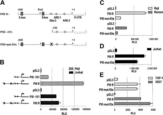
Identification of regulatory regions that are involved in B cell-specific activation of CIITA-PIII. (A) Schematic representation of the CIITA-PIII-derived reporter plasmids PIIIfl, PIII-151, and PIII-mut-Ets. Indicated are the 2 E-boxes, Site-C comprising the EICE, ARE-1, ARE-2, and the 5′-UTR. (B) Transient transfections of reporter plasmids pGL3 basic, PIIIfl, and PIII-151, which harbor a deletion as depicted in panel A, were performed in Raji B cells and Jurkat T cells. Transient transfection of reporter plasmids pGL3 basic, PIIIfl, and PIII-mut-Ets were performed in Raji and Ramos B cells (C), Jurkat T cells (D), and THP-1 and U937 monocytic cell lines (E). B and T cells were harvested after 48 hours and monocytic cells after 24 hours and analyzed for luciferase activity. Luciferase activity values were normalized with Renilla luciferase activity values and represent the means of 3 experiments. Error bars indicate SEM.
Identification of regulatory regions that are involved in B cell-specific activation of CIITA-PIII. (A) Schematic representation of the CIITA-PIII-derived reporter plasmids PIIIfl, PIII-151, and PIII-mut-Ets. Indicated are the 2 E-boxes, Site-C comprising the EICE, ARE-1, ARE-2, and the 5′-UTR. (B) Transient transfections of reporter plasmids pGL3 basic, PIIIfl, and PIII-151, which harbor a deletion as depicted in panel A, were performed in Raji B cells and Jurkat T cells. Transient transfection of reporter plasmids pGL3 basic, PIIIfl, and PIII-mut-Ets were performed in Raji and Ramos B cells (C), Jurkat T cells (D), and THP-1 and U937 monocytic cell lines (E). B and T cells were harvested after 48 hours and monocytic cells after 24 hours and analyzed for luciferase activity. Luciferase activity values were normalized with Renilla luciferase activity values and represent the means of 3 experiments. Error bars indicate SEM.
E47, PU.1 and IRF-4 synergistically activate CIITA-PIII in HeLa cells in a site-dependent manner. (A) Schematic representation of all CIITA-PIII reporter constructs tested. E-boxes and Site-C are indicated, and the mutated Ets-box of Site-C is depicted by an X. (B) The different mutant CIITA-PIII reporter constructs depicted panel A were cotransfected with expression plasmids coding for E47fd, PU.1, and IRF-4 or IRF-8 into HeLa cells as indicated. Cotransfections of PIIIs with expression plasmids coding for either E47fd, PU.1, IRF-4, or IRF-8 are indicated in lanes 15 to 18. Fold induction is indicated as “fold ind.” on top of panel B. HeLa cells were harvested after 48 and analyzed for luciferase activity. Luciferase activity values were normalized with Renilla luciferase activity values, and relative luciferase activities are indicated as mean SD of n = 4.
E47, PU.1 and IRF-4 synergistically activate CIITA-PIII in HeLa cells in a site-dependent manner. (A) Schematic representation of all CIITA-PIII reporter constructs tested. E-boxes and Site-C are indicated, and the mutated Ets-box of Site-C is depicted by an X. (B) The different mutant CIITA-PIII reporter constructs depicted panel A were cotransfected with expression plasmids coding for E47fd, PU.1, and IRF-4 or IRF-8 into HeLa cells as indicated. Cotransfections of PIIIs with expression plasmids coding for either E47fd, PU.1, IRF-4, or IRF-8 are indicated in lanes 15 to 18. Fold induction is indicated as “fold ind.” on top of panel B. HeLa cells were harvested after 48 and analyzed for luciferase activity. Luciferase activity values were normalized with Renilla luciferase activity values, and relative luciferase activities are indicated as mean SD of n = 4.
Transient transfections
All T- and B-cell lines were transfected by electroporation (Genepulser; Bio-Rad Laboratories, Hercules, CA) as described previously and harvested after 48 hours.19 The monocytic cell lines THP-1 and U937 were transfected at 210 V and 960 μF in medium without fetal calf serum (FCS) at 4°C. The following expression plasmids were used: pRc/RSV-IRF-4, pRc/RSV-IRF-8, pcDNA3.1-PU.1, pcDNA3-E47 forced dimer (E47fd), pcDNA3-FKBP52, and enhanced green fluorescence protein (EGFP) (Clontech, Leusden, Belgium) cloned into pcDNA3.1 and pRc/RSV-Id1.37,44,45 Expression plasmids were added as indicated. Transfections were performed in triplicate, and luciferase and Renilla luciferase activity was measured according to the manufacturer's instructions and normalized for transfection efficiency with the Renilla luciferase assay (Promega, Madison, WI). Primary B cells were transfected using the human B-cell Nucleofector Kit (Amaxa Biosystems, Cologne, Germany) according to manufacturer's instructions.
EMSA and probes
Nuclear extract (NE) preparations and electrophoretic mobility shift assays (EMSAs) were performed as described previously.19 NE (2 μL) was incubated with 2 ng [32P]-labeled dsDNA probe for 20 minutes at 4°C. The following oligonucleotides were used as a probe: PIII-EICE sense 5′-CAGTCCACAGTAAGGAAGTGAAATTA-3′and PIII-EICE antisense 5′-TAATTTCACTTCCTTACTGTGGACTG-3′; PIII-mut-EICE sense 5′-GTCCACAGTATCCTAGTGAATCAAATTTCA-3′ and PIII-mut-EICE antisense 5′-TGAAATTTGATTCACTAGGATACTGTGGAC-3′; 3′Eλ-EICE sense 5′-AAATAAAAGGAAGTGAAACCAAG-3′and 3′Eλ-EICE antisense 5′-CTTGGTTTCACTTCCTTTTATTT-3′; cons-E-box sense 5′-CAAACACCACCTGGGTAATC-3′and cons-E-box antisense 5′-GATTACCCAGGTGGTGTTTG -3′; 473-E-box sense 5′-GAAAATGACAGGTGGGCCACTTAT-3′ and 473-E-box antisense 5′-ATAAGTGGCCCACCTGTCATTTTC-3′; 298-E-box sense 5′-GATATTGGCAGCTGGCACCAGTGC-3′ and 298-E-box antisense 5′-GCACTGGTGCCAGCTGCCAATATC-3′. Supershift assays were performed by adding 1 μg of antibody to 10 μL of the NEs and probe mixture and incubating for 60 minutes at 4°C. The following antibodies were used for supershift assays: rabbit-anti-PU.1 (sc-352X), goat-anti-IRF-4 (sc-6059X), goat-anti-IRF-8 (sc-6058X), and rabbit-anti-E47 (sc-763X; Sanver Tech, Heer-hugowaard, the Netherlands).
Chromatin immunoprecipitation (ChIP) assay
ChIP assays were performed as described previously.19,46 Genomic DNA derived from formaldehyde-treated Raji cells was sonicated using a microtip until the average DNA fragments were approximately 600 bp. Immunoprecipitations were performed at 4°C overnight with 5 μg of primary antibody, and immune complexes were harvested with secondary sheep anti-rabbit antibodies linked to M-280 Dynalbeads (Dynal Biotech) or with protein-A-Agarose beads (Upstate Biotechnology, Campro Scientific, Veenendaal, the Netherlands). The following primary antibodies were used: anti-E47 (sc-763X), anti-PU.1 (sc-352), anti-STAT-1α (sc-345X) (Sanver Tech), goat-anti-IRF-4 (sc-6059X), goat-anti-IRF-8 (sc-6058X), and anti-acetylated H3 (ac-H3) (Upstate Biotechnology). Isolated immune complexes were elaborately washed and subsequently disrupted. DNA samples were precipitated and purified by proteinase K digestion, phenol/chloroform extraction, and ethanol precipitation. All samples were dissolved in 40 μLof H2O, and 5 μL was used as a template for PCR reactions using primers specific for the CIITA-PIII- and CIITA-PIV-core region as described previously.19
Results
Identification of potential B-cell-specific regulatory elements in CIITA-PIII
In order to identify cis-acting elements that contribute to B-cell-specific expression of CIITA, we initially inspected the DNA sequence of the CIITA-PIII region -545 to +124 for the presence of potential regulatory elements, which could confer B-cell-specific activation. Scrutiny of the DNA sequence revealed the presence of an EICE in Site-C and 2 E-box motifs that showed strong homology with E47 binding sites. Both motifs have been shown to play an important role in the activation of B-cell-specific genes.20-26 DNA sequence comparison of the EICE and E-box motifs of CIITA-PIII with those found in enhancers and promoters of various B-cell-specific genes revealed 100% core sequence homologies (Table 1).
To evaluate the role for these regulatory motifs in the B-cell-specific activation of CIITA-PIII, we compared the activity of various CIITA-PIII promoter reporters in B-, T-, and monocytic cell lines. Deletion of a large region including both putative E-box motifs and the EICE from PIIIfl (PIII-151, Figure 1A) resulted in a strong reduction of PIIIfl activity in Raji B cells, whereas in Jurkat T cells only a modest reduction in activity was observed (Figure 1B). When we evaluated PIIIfl and PIII-151 activity in the monocytic cell lines THP-1 and U937, again we observed that deletion of both putative E-boxes and the EICE did not affect PIII-151 activity when compared with PIIIfl (data not shown).
To specifically address the contribution of the EICE in CIITA-PIII in transcriptional activation, we mutated the core Ets-box in a similar fashion as previously explored for the EICE of the Eλ2-4 (AGGA→TCCT), which resulted in strong down-regulation of activity of the Eλ2-4 enhancer in mouse B cells.21 Raji and Ramos B cells, Jurkat T cells, and THP-1 and U937 monocytic cells were transiently transfected with the PIIIfl luciferase reporter plasmid that harbors the Ets-box mutation in Site-C (PIII-mut-Ets), and the activity of this mutated promoter-reporter was compared to the activity of PIIIfl. As shown in Figure 1C, mutation of the Ets-box of Site-C resulted in a severe reduction of reporter activity in both Raji and Ramos B cells. In contrast, in Jurkat T cells, or in THP-1 and U937 monocytic cells, this mutation yielded respectively very moderate or no reduction at all in PIIIfl activity (Figure 1D-E).
Several CIITA-PIII-reporter plasmids as depicted in Figure 2A were constructed to investigate the contribution of both E-box motifs and possible cooperation with the EICE of Site-C in CIITA-PIII activation. PIIIs was generated from PIIIfl by removal of a large 224-bp upstream region upstream of the E-box at position -298, which includes the E-box at position -473 of CIITA-PIII. Deletion of this upstream region did not reduce promoter activity in either Ramos B cells or U937 monocytes or Jurkat T cells (Figure 2B-D). This observation is in line with previous studies showing that PIIIs (-322 to +124) represents the core promoter of CIITA-PIII, which confers full activation in B cells.11 However, subsequent removal of a 26-bp fragment that harbors the second E-box at position -298 of PIIIs (PIIIs-DelE) strongly reduced PIIIs activity in Ramos and Raji B cells (not shown) but had no such effect in U937 monocytic or Jurkat T cells (Figure 2B-D). Noteworthy, a full-length CIITA-PIII reporter that harbored a specific mutation solely in the E-box at position -298, which abolished binding of E47, was equally active as PIIIfl in B cells, indicating that the upstream E-box (at position -473) can replace the function of the E-box at position -298 (data not shown). Similar to previous results with the PIIIfl reporter, mutation of the Ets-box in site-C of PIIIs yielded again strongly reduced reporter activity in Ramos B cells and had no such effect in U937 monocytic cells and Jurkat T cells (Figure 2B-D). Together, these findings demonstrate an important role for both the putative E-box motif at -298 and the EICE in transcriptional activation of CIITA-PIII in B cells.
The E-box motifs of CIITA-PIII confer high promoter activity in B cells. (A) Schematic illustration of CIITA-PIII reporter plasmids used in transient transfection assays with Ramos B cells (B), U937 monocytic cells (C), and Jurkat cells (D). Ramos and Jurkat cells were harvested after 48 hours, monocytic cells were harvested after 24 hours, and the cells were analyzed for luciferase activity. Luciferase activity values were normalized with Renilla luciferase activity values, represent the means of 3 experiments, and were related to the activity of PIIIfl. Error bars indicate SEM.
The E-box motifs of CIITA-PIII confer high promoter activity in B cells. (A) Schematic illustration of CIITA-PIII reporter plasmids used in transient transfection assays with Ramos B cells (B), U937 monocytic cells (C), and Jurkat cells (D). Ramos and Jurkat cells were harvested after 48 hours, monocytic cells were harvested after 24 hours, and the cells were analyzed for luciferase activity. Luciferase activity values were normalized with Renilla luciferase activity values, represent the means of 3 experiments, and were related to the activity of PIIIfl. Error bars indicate SEM.
PU.1, IRF-4/IRF-8, and E47 interact with CIITA-PIII in vivo and bind in vitro to their cognate recognition sequences
PU.1, IRF-4, IRF-8, and E47 are factors known to interact with EICEs and E-box motifs, respectively. To investigate the interaction of these factors with the EICE and E-boxes of CIITA-PIII in vivo, we performed ChIP analyses with chromatin from Raji B cells using specific antibodies as described in “Materials and methods.” As negative and positive controls, we used antibodies specific for STAT-1 and ac-H3, respectively.
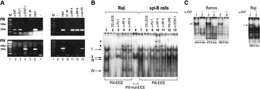
As shown in Figure 3A, CIITA-PIII-specific products were clearly detected with the PU.1, IRF-4, IRF-8, E47, and ac-H3-purified chromatin. No CIITA-PIII-specific products were detected with STAT-1 or no-antibody purified chromatin. CIITA-PIV-specific products could only be detected with STAT-1-purified chromatin and not with the other chromatin purifications. These observations reveal the in vivo interaction of PU.1, IRF-4, IRF-8, and E47 with CIITA-PIII in B cells.
In vivo and vitro interactions of the EICE and E-boxes of CIITA-PIII in B cells. (A) ChIP analysis of CIITA-PIII and CIITA-PIV in Raji B cells with antisera specific for E47 (lane 2), PU.1 (lane 3), STAT-1 (lane 4), IRF-4 (lane 11), IRF-8 (lane 12), and ac-H3 (lane 13). PCR products specific for both CIITA-PIII (top panels) or CIITA-PIV (bottom panels) are indicated. PCR products obtained with input DNA are shown in lanes 6 and 9, and PCR products obtained without the addition of a primary antibody are shown in lanes 5 and 10. The 1-kb marker and the negative water control are indicated in lanes 1, 7, and 8, respectively. (B) EMSA with NE derived from Raji B cells (lanes 1-6) and splenic B cells (lanes 7-12). EICE-interacting factors were visualized using the CIITA-derived PIII-EICE as a probe and compared to the pattern obtained with a PIII-mut-EICE encoding probe. Supershift antibodies and competition probe are indicated on the top of each plot, and supershifts were performed with antibodies specific for PU.1 (lanes 3 and 9), IRF-4 (lanes 4 and 10), IRF-8 (lanes 5 and 11), and STAT-1 (lane 13). Competition was performed using a 3′Eλ-EICE probe (lanes 2 and 8) that encodes the consensus EICE sequence of the 3′Eλ2-4 and the PIII-CRE (ARE-2 motif) probe (lane 12), as described previously.19,21 Supershifted complexes are indicated with an asterisk, and complexes reduced or eliminated after incubation with the PU.1 are indicated by arrows I-IV, of which complexes II and III are also reduced after incubation with IRF-4/8 specific antibodies. (C) EMSA analysis for E-box-interacting factors were performed using a consensus E-box (cons-E-box) and both E-box motifs of CIITA-PIII (473-E-box and 298-E-box) as probes with NE extracts derived from Ramos B cells and Raji B cells. Supershift analysis with E47-specific antibody is indicated on the top of the plots, and E47 complex is indicated with an arrow. Probes are indicated below all EMSA plots.
In vivo and vitro interactions of the EICE and E-boxes of CIITA-PIII in B cells. (A) ChIP analysis of CIITA-PIII and CIITA-PIV in Raji B cells with antisera specific for E47 (lane 2), PU.1 (lane 3), STAT-1 (lane 4), IRF-4 (lane 11), IRF-8 (lane 12), and ac-H3 (lane 13). PCR products specific for both CIITA-PIII (top panels) or CIITA-PIV (bottom panels) are indicated. PCR products obtained with input DNA are shown in lanes 6 and 9, and PCR products obtained without the addition of a primary antibody are shown in lanes 5 and 10. The 1-kb marker and the negative water control are indicated in lanes 1, 7, and 8, respectively. (B) EMSA with NE derived from Raji B cells (lanes 1-6) and splenic B cells (lanes 7-12). EICE-interacting factors were visualized using the CIITA-derived PIII-EICE as a probe and compared to the pattern obtained with a PIII-mut-EICE encoding probe. Supershift antibodies and competition probe are indicated on the top of each plot, and supershifts were performed with antibodies specific for PU.1 (lanes 3 and 9), IRF-4 (lanes 4 and 10), IRF-8 (lanes 5 and 11), and STAT-1 (lane 13). Competition was performed using a 3′Eλ-EICE probe (lanes 2 and 8) that encodes the consensus EICE sequence of the 3′Eλ2-4 and the PIII-CRE (ARE-2 motif) probe (lane 12), as described previously.19,21 Supershifted complexes are indicated with an asterisk, and complexes reduced or eliminated after incubation with the PU.1 are indicated by arrows I-IV, of which complexes II and III are also reduced after incubation with IRF-4/8 specific antibodies. (C) EMSA analysis for E-box-interacting factors were performed using a consensus E-box (cons-E-box) and both E-box motifs of CIITA-PIII (473-E-box and 298-E-box) as probes with NE extracts derived from Ramos B cells and Raji B cells. Supershift analysis with E47-specific antibody is indicated on the top of the plots, and E47 complex is indicated with an arrow. Probes are indicated below all EMSA plots.
Because the ChIP analyses indicated that PU.1, IRF-4, IRF-8, and E47 are associated with CIITA-PIII chromatin, we examined the in vitro interaction of PU.1, IRF-4, IRF-8, and E47 with their specific binding motif in CIITA-PIII by EMSA. Using the PIII-EICE probe and NE from the B-cell line Raji and CD40-activated primary splenic B lymphocytes, the supershift analyses confirmed the in vitro interaction of PU.1, IRF-4, and IRF-8 in both Raji B cells and activated splenic B lymphocytes (Figure 3B, lanes 3-5, 9-11). No supershifts were visible using an unrelated STAT-1-specific antibody. Mutation of both the Ets-box and the interferon-stimulated response element (ISRE) in the EICE resulted in loss of all PU.1-(I, II, III, IV) and IRF-specific complexes (II and III) as indicated by arrows and gave rise to a complex slightly larger than complex II, which could not be supershifted with any of the mentioned antibodies (Figure 3B; data not shown). Likewise, competition with the consensus EICE probe coding for the 3′Eλ-EICE resulted in reduction or loss of all specific complexes, whereas a nonrelated probe PIII-CRE had no effect at all (Figure 3B). Interaction of PU.1, IRF-4, and IRF-8 with PIII-EICE also was observed using NE derived from THP-1 and U937 (data not shown). As previously explored, such complexes cannot be detected in Jurkat T cells or activated T cells (HLA-DR+) established from peripheral blood mononuclear cells (PBMCs).5
Using a consensus E47-binding site (cons-E-box) and the -473-E-box and -298-E-box of CIITA-PIII as probes, the EMSA confirmed the binding of E47 to both E-box motifs using NEs of both Raji and Ramos B cells. A single protein-DNA complex was detected that reacted with an E47-specific antibody as indicated by the arrow (Figure 3C). No such complexes were observed in Jurkat T cells, THP-1, and U937 monocytic cells or HeLa cells (data not shown).
Taken together, these data show that in B cells, PU.1, IRF-4, IRF-8, and E47 interact with CIITA-PIII in vivo, and this interaction is conferred through binding of PU.1 with either IRF-4 or IRF-8 to the EICE, and of E47 to the E box-motifs of CIITA-PIII.
IRF-4 is involved in the activation of CIITA-PIII in B cells in vivo
Next we evaluated the in vivo role of IRF-4 in the regulation of MHCII expression in primary B lymphocytes. Since RNAi constructs that reduce IRF-4 transcription resulted in poor down-modulation of IRF-4 protein expression within 24 hours (not shown), we directly blocked IRF-4 DNA binding and protein function by coexpression of FK506-binding protein 52 (FKBP52), an inhibitor of IRF-4 activity as reported by several studies.44,47,48 For this purpose we examined CD19-expressing B lymphocytes from peripheral blood by performing transfections with EGFP with and without the addition of FKBP52. Subsequently, we monitored cell surface HLA-DR expression in CD19+ B lymphocytes to evaluate the impact of FKBP52 on CIITA expression. Using this approach, about 4% of the CD19+ B lymphocytes became transfected with EGFP (Figure 4Ai-ii), and coexpression of FKBP52 abolished HLA-DR expression in about 30% of the transfected, EGFP-positive B lymphocytes (Figure 4Aiv). No such population was observed in B cells transfected with EGFP alone (Figure 4Aiii). In order to exclude any effect of EGFP on HLA-DR expression, we performed similar transfections of CD19-enriched peripheral blood B cells using only FKBP52 or the empty expression vector. Although we cannot distinguish the transfected B-cell population from the untransfected cells, again, the addition of FKBP52 resulted in a clear population of HLA-DR-negative cells within the pool of CD19-positive B lymphocytes, which was not observed with the empty vector (Figure 4A, compare v and vi). Because FKBP52 targets IRF-4 function, these results indicate that IRF-4 is involved in the in vivo activation of HLA-DR expression in B cells, which could be mediated through the activation of CIITA transcription.44,47,48
Inhibition of IRF-4 by FKBP52 specifically reduces CIITA-PIII activity in B cells and abolishes HLA-DR expression in primary B lymphocytes. (A) CD19+-enriched B cells were transfected with EGFP with either addition of empty expression vector (i,iii) or addition of FKBP52 (ii,iv), respectively. (v-vi) Transfections without EGFP but solely with empty expression vector (v) or FKBP52 (vi). FACS analysis was performed after 18 hours of transfection, and in all plots cells are gated for CD19 expression and lymphocyte-specific cell scatter pattern. Cells were stained as described in “Materials and methods” and expression of CD19, EGFP, and HLA-DR is indicated on the axes. (B-C) Transient transfection of CIITA-PIII reporter plasmids pGL3 basic, PIIIfl, and PIII mut-Ets were performed in (B) Raji and Ramos B cells, and (C) Jurkat T cells, THP-1, and U937 monocytic cell lines. Five and 10 μg FKBP52 expression plasmid was added to the B cells (PIIIfl+), panel B, lanes 3 and 4, respectively, and 10 μg FKBP52 expression plasmid was added to the Jurkat T cells (PIIIfl+), THP-1, and U937 monocytic cell lines (PIIIfl+) panel C, lane 3. B and T cells were harvested after 48 hours and monocytic cells after 24 hours and analyzed for luciferase activity. Luciferase activity values were normalized withRenilla luciferase activity values and represent the means of 3 experiments.
Inhibition of IRF-4 by FKBP52 specifically reduces CIITA-PIII activity in B cells and abolishes HLA-DR expression in primary B lymphocytes. (A) CD19+-enriched B cells were transfected with EGFP with either addition of empty expression vector (i,iii) or addition of FKBP52 (ii,iv), respectively. (v-vi) Transfections without EGFP but solely with empty expression vector (v) or FKBP52 (vi). FACS analysis was performed after 18 hours of transfection, and in all plots cells are gated for CD19 expression and lymphocyte-specific cell scatter pattern. Cells were stained as described in “Materials and methods” and expression of CD19, EGFP, and HLA-DR is indicated on the axes. (B-C) Transient transfection of CIITA-PIII reporter plasmids pGL3 basic, PIIIfl, and PIII mut-Ets were performed in (B) Raji and Ramos B cells, and (C) Jurkat T cells, THP-1, and U937 monocytic cell lines. Five and 10 μg FKBP52 expression plasmid was added to the B cells (PIIIfl+), panel B, lanes 3 and 4, respectively, and 10 μg FKBP52 expression plasmid was added to the Jurkat T cells (PIIIfl+), THP-1, and U937 monocytic cell lines (PIIIfl+) panel C, lane 3. B and T cells were harvested after 48 hours and monocytic cells after 24 hours and analyzed for luciferase activity. Luciferase activity values were normalized withRenilla luciferase activity values and represent the means of 3 experiments.
To examine whether IRF-4 plays a role in activation of CIITA-PIII in B cells, we performed transient coexpression assays with PIIIfl and FKBP52. Inclusion of increasing amounts of FKBP52 together with PIIIfl in Raji and Ramos B cells resulted in strong down-regulation of PIIIfl activity (Figure 4B). The impact of FKBP52 on PIIIfl also was evaluated in Jurkat T cells, which do not express IRF-4 (Figure 4C) but display high CIITA-PIII activity. No reduction in PIIIfl activity was observed in Jurkat T cells, indicating specificity for IRF-4 of FKBP52 (Figure 4C). Next we investigated whether IRF-4 was important for the activation of CIITA-PIII in the monocytic cell lines THP-1 and U937. As shown in Figure 4C, cotransfection of PIIIfl and FKBP52 did not result in a reduction in PIIIfl activity in both monocytic cell lines, which is similar to the lack of reduction observed with the Ets-box mutant reporter (PIII-mut-Ets) in these cells. Together, these results indicate that solely in B cells IRF-4 participates in the activation of CIITA-PIII. Moreover, these observations suggest that mainly IRF-4, and not IRF-8, which harbors no obvious homology with the IRF-4-FKBP52 interaction domain, mediates the activation of CIITA-PIII in Raji and Ramos B cells.44
Id1 reduces activation of CIITA-PIII by endogenous E47 in B-cell lines
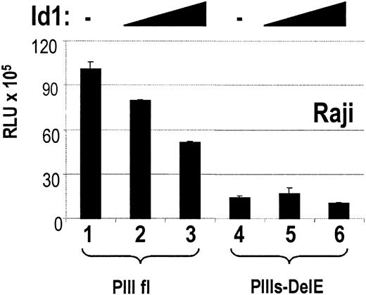
To investigate the in vivo role of E47 homodimers in the transcriptional activation of CIITA-PIII, we cotransfected different amounts of Id1 expression plasmids with CIITA reporters PIIIfl and PIIIs-DelE. Transient cotransfections using increasing amounts of Id1 expression plasmids resulted in a dose-dependent repression of PIIIfl activity (Figure 5), whereas deletion of the E-box in PIIIs-DelE abrogated further repression of promoter activity. Together with the previous observations, this again indicates that E47 is involved in the activation of CIITA-PIII in B cells.
Cotransfection of Id1 reduces CIITA-PIII activity in Raji B cells. Transient cotransfections of CIITA reporter plasmids PIIIfl and PIIIs-DelE with either 4 μg empty expression plasmid pRC/RSV (lanes 1 and 4) or 2 μg (lanes 2 and 5) and 4 μg (lanes 3 and 6) of Id1 expression plasmid were performed with Raji B cells. B cells were harvested after 48 hours and analyzed for luciferase activity. Luciferase activity values were normalized with Renilla luciferase activity values and represent the means of 3 experiments.
Cotransfection of Id1 reduces CIITA-PIII activity in Raji B cells. Transient cotransfections of CIITA reporter plasmids PIIIfl and PIIIs-DelE with either 4 μg empty expression plasmid pRC/RSV (lanes 1 and 4) or 2 μg (lanes 2 and 5) and 4 μg (lanes 3 and 6) of Id1 expression plasmid were performed with Raji B cells. B cells were harvested after 48 hours and analyzed for luciferase activity. Luciferase activity values were normalized with Renilla luciferase activity values and represent the means of 3 experiments.
Coexpression of E47, PU.1, and IRF-4 but not IRF-8 synergistically induces high CIITA-PIII activity in HeLa cells
The previous studies indicate that the E47 binding sites and the EICE and their interacting factors are essential for CIITA-PIII activity in B cells. To study the cooperation between E47, PU.1, and IRF-4 and the possible role of IRF-8 in the activation of CIITA-PIII, transactivation experiments were performed in HeLa cells. HeLa cells, which do not to express CIITA, were transfected with different CIITA-PIII reporter plasmids as depicted in Figure 6A in combination with either E47, PU.1, and IRF-4 or E47, PU.1, and IRF-8 expression constructs. Given that HeLa cells can express other bHLH- and Id proteins, we used a homodimeric E47 fusion protein, called E47 forced-dimer (E47fd), as described previously to study the function of solely E47 homodimers and exclude effects of E47 heterodimers.37
Cotransfection of either E47fd, PU.1, IRF-4, or IRF-8 with PIIIs showed a modest increase in PIII activity by IRF-4 or PU.1 only and not by IRF-8 or E47 (Figure 6B, compare lane 3 with lanes 15 to 18). The IRF-4-mediated induction could not be enhanced by inclusion of PU.1 (results not shown). However, a strong increase in activation of PIIIs, up to 54-fold, was achieved when E47fd, PU.1, and IRF-4 were simultaneously included in the reporter assay (Figure 6B, compare lanes 3 and 7). Such a strong enhanced activation of PIIIs was not noted with IRF-8, together with PU.1 and E47fd (Figure 6B, compare lane 3 with 11). The specificity of this interaction and resulting activation of PIIIs were revealed from the mutation studies because mutation of either the E-box or the EICE severely reduced the synergistic activation of PIIIs by E47fd, PU.1, and IRF-4 (Figure 6B, lanes 8-10). Together, these observations indicate that E47, PU.1, and IRF-4 can collaborate in the activation of CIITA-PIII and mediate their activity through interaction with both the E-box motif and the EICE of CIITA-PIII. Furthermore, in B cells IRF-4, and not IRF-8, appears to be the most potent interferon regulatory fact (IRF) in the activation of CIITA-PIII. This is in line with previous observations concerning B-cell-expressed genes.20-26
Discussion
CIITA and resulting MHCII expression in several hematopoietic cell lineages is mediated by CIITA-PIII. Considering the overall strong diversity in transcription factor expression profiles within the different hematopoietic cell lineages, activation of CIITA-PIII is most likely due to cell type-specific promoter assembly of regulatory proteins.
In this study we have investigated the factors and elements that confer B-cell-specific activation of CIITA-PIII. Mutational analyses and transient cotransfection experiments revealed that the EICE present in Site-C and the E-box motifs (at -473 or -298) upstream of site-C cooperate to activate CIITA-PIII in B cells mainly through synergistic interaction with PU.1, IRF-4, and E47.
ChIP analysis confirmed the in vivo interaction of E47, PU.1, IRF-4, and IRF-8 with CIITA-PIII in B cells. Interestingly, specific inactivation of IRF-4 by FKBP52 resulted in down-regulation of CIITA-PIII activity in Raji and Ramos B cells, suggesting that mainly IRF-4 and not IRF-8 is involved in the B-cell activation of CIITA-PIII. Analogous cotransfection of these factors in HeLa cells revealed that IRF-4 induced a stronger activation of CIITA-PIII together with PU.1 and E47 than IRF-8. Moreover, inhibition of IRF-4 abrogated cell surface MHCII expression in about 30% of PBMC-derived B lymphocytes. Considering that IRF-4 does not interact with the MHCII promoter itself, these observations again suggest that mainly IRF-4 is involved in the activation of CIITA-PIII in B cells.
Similar to studies concerning the transcriptional regulation of Ig light chains through the EICE located in the 3′Eκ and the 3′Eλ, mutation of the Ets-box in the EICE of CIITA-PIII abolished the interaction of both PU.1 and IRF-4 and resulted in strong reduction in CIITA-PIII activity in both Raji and Ramos B-cell lines. No strong decrease in CIITA-PIII activity was observed in THP-1 and U937 monocytes and Jurkat T cells. This is in line with the observation that both in Jurkat and activated primary T cells, no PU.1 and IRF-4 containing complexes binding to a PIII-EICE probe are detected.5 It indicates that other factors are important for CIITA-PIII promoter assembly and resulting CIITA-PIII activation in activated T cells. In contrast, monocytic cells, including THP-1 and U937 cells, do express moderate levels of IRF-4 and high levels of PU.1 and IRF-8, and bandshift analyses revealed that both factors could interact in vitro with the EICE. However, the lack of inhibition of CIITA-PIII activity in monocytic cell lines by FKBP52 or after mutation of the Ets-box within the EICE clearly indicated that IRF-4 and PU.1 do not confer CIITA-PIII activation in monocytic cell lines in vivo. Moreover, we could not detect expression of the E47 homodimers, which were shown to interact and mediate the activation of CIITA-PIII through the E-box motifs in B cells. Together, it suggests that the activation of CIITA-PIII in monocytic cells is under the control of a different set of regulatory promoter elements and interacting factors. In addition, it emphasizes the cooperative role of E47 homodimers with the EICE interacting factors in the activation of CIITA-PIII in B cells.
IRF-4 expression is strictly regulated and is strongly increased after activation during B-cell development. Although plasma cells continue to express high levels of IRF-4, CIITA expression is completely abolished. Coherently, many other B-cell-specific genes also are lost upon plasma cell differentiation. Recent studies have demonstrated that this repression is mediated through positive regulatory domain I-binding factor 1 (PRDI-BF1), the human homolog of murine B-lymphocyte-induced maturation protein (BLIMP-1).49,50 Interestingly, PRDI-BF1 interacts with the same ISRE half of the EICE in CIITA-PIII as IRF-4 does. Moreover, BLIMP-1 has been shown to physically interact with the DNA binding domain of IRF-4.51 Although repression of cMyc by BLIMP-1/PRDI-BF1 is mediated though recruitment of Groucho and histone deacetylases, this was not the case for CIITA expression in plasma B cells.49,51 Therefore, it could be envisaged that repression of CIITA-PIII activity by BLIMP-1/PRDI-BF1 upon plasma cell differentiation is due to inhibition of IRF-4-mediated activation of this promoter.
In eukaryotes, cell type-specific expression is regulated by the assembly of distinct and cell type-specific combinations of transcription factors and the recruitment of chromatin remodeling factors at promoters and/or enhancers. The formation of such a cell type-specific enhanceosome enables activation of different regulatory units through a relatively small set of transcription factors. Importantly, accessibility of a gene through the action of chromatin remodeling factors is crucial for such activation. The exact kinetics in gene recruitment of the specific transcription factors and chromatin remodelers is currently still under debate; however, it is definite that both events are crucial for proper activation of a gene. In this study, we addressed the B-cell-specific enhanceosome involved in the activation of CIITA-PIII. Several previous studies have designated the combination of PU.1, IRF-4, and E47 to mediate synergistic activation of genes exclusively expressed in B cells.23,38,39,52 Although CIITA-PIII-mediated CIITA expression is not limited to B cells and occurs in various hematopoietic cells, this study clearly reveals differential and cell type-specific recruitment of specific transcription factors to activate this promoter. This divergence in CIITA-PIII activation implies that different sets of regulatory proteins can govern the activation of the same promoter in a cell type-specific fashion.
Apart from cell type-specific transcription factors, ubiquitously expressed transcription factors also are involved in transcriptional regulation. This is illustrated in the 3′Eκ, which harbors several cAMP-responsive elements (CREs) in addition to the EICE and E2A motifs.53 Previous studies have shown that the PU.1/IRF-4 complex bound to the 3′Eκ indeed physically interacts with the more generally expressed CRE binding factors ATF-1 and CREM.54 These interactions resulted in a synergistically increase in the level of gene transcription and, importantly, transcription could not be mediated after mutation of either element.39,54 Furthermore, E47 and PU.1 interact with the general coactivator CBP, revealing an indirect link with the basal transcription apparatus.55,56 In this respect, we have shown in a previous study that CIITA-PIII contains several CREs namely in the 5′UTR and in ARE-2 that interact with CREB-1 and ATF-1, which play an important role in the transcriptional activation of CIITA-PIII in B cells.5,19 These observations suggest that similar to the 3′Eκ, the PU.1, IRF-4, and E47 complexes could interact with promoter-bound CREB/ATF factors and/or with CBP and establish high levels of B-cell expression.
Together, our data reveal that in B cells CIITA-mediated expression of the MHCII antigen presentation pathway is in part under control of the same factors that govern B-cell differentiation and activation. Therefore, factors that can enhance B-cell antigen receptor (BCR) expression will coherently enhance CIITA-induced genes, which implies that an increase in antigen uptake can result in a strong increase in antigen presentation and subsequent B- and T-cell activation. In general, this observation provides a transcriptional link between antigen presentation functions, and B-cell activation events mediated through signal transduction processes of the BCR complex, CD40, and other B-cell-stimulatory molecules.
Prepublished online as Blood First Edition Paper, July 8, 2004; DOI 10.1182/blood-2004-03-0790.
Supported by a grant from the Dutch Cancer Society (RUL 98-1732) and by the Dr Gisela Thier Foundation. N.v.d.S. is a Research Fellow of the Royal Netherlands Academy of Arts and Sciences.
The publication costs of this article were defrayed in part by page charge payment. Therefore, and solely to indicate this fact, this article is hereby marked “advertisement” in accordance with 18 U.S.C. section 1734.
The authors would like to thank Dr J.-Y. Ting, Dr J. Hiscott, Dr M. Liang, Dr M. Fenton, and Dr M. Sigvardsson for providing the pGL2-CIITA-PIIIDEL1 reporter plasmid and the expression plasmids of FKBP52, PU.1, IRF-8, IRF-4, and E47fd, respectively. We thank Dr I. L. C. van Dinten and Dr F. Koning for critically reading the manuscript.


