Key Points
Reactivations of multisystem Langerhans cell histiocytosis (MS-LCH) are reduced by prolonging initial chemotherapy.
The previously high mortality of high-risk (risk-organ–positive) MS-LCH in children has been markedly reduced.
Abstract
Langerhans cell histiocytosis (LCH)-III tested risk-adjusted, intensified, longer treatment of multisystem LCH (MS-LCH), for which optimal therapy has been elusive. Stratified by risk organ involvement (high [RO+] or low [RO–] risk groups), >400 patients were randomized. RO+ patients received 1 to 2 six-week courses of vinblastine+prednisone (Arm A) or vinblastine+prednisone+methotrexate (Arm B). Response triggered milder continuation therapy with the same combinations, plus 6-mercaptopurine, for 12 months total treatment. 6/12-week response rates (mean, 71%) and 5-year survival (84%) and reactivation rates (27%) were similar in both arms. Notably, historical comparisons revealed survival superior to that of identically stratified RO+ patients treated for 6 months in predecessor trials LCH-I (62%) or LCH-II (69%, P < .001), and lower 5-year reactivation rates than in LCH-I (55%) or LCH-II (44%, P < .001). RO– patients received vinblastine+prednisone throughout. Response by 6 weeks triggered randomization to 6 or 12 months total treatment. Significantly lower 5-year reactivation rates characterized the 12-month Arm D (37%) compared with 6-month Arm C (54%, P = .03) or to 6-month schedules in LCH-I (52%) and LCH-II (48%, P < .001). Thus, prolonging treatment decreased RO– patient reactivations in LCH-III, and although methotrexate added no benefit, RO+ patient survival and reactivation rates have substantially improved in the 3 sequential trials. (Trial No. NCT00276757 www.ClinicalTrials.gov).
Introduction
Langerhans cell histiocytosis (LCH) is characterized by a reactive clonal proliferation and accumulation of dendritic cells,1,2 with a wide range of clinical presentations.3 Localized or “single-system” disease has an excellent survival rate.4 Multisystem disease (MS-LCH), ie, involving two or more organs or systems, however, has an unpredictable course, especially when there is involvement of risk organs, frequently associated with a poor prognosis.5-7 Because of this wide disease spectrum, many different treatment approaches, ranging from minimal conservative to intensive combination chemotherapy, have been used.8-13
To improve outcome, the Histiocyte Society embarked upon a series of randomized international clinical trials using standardized diagnostic criteria, uniform disease assessment,6,7,13-15 and a treatment backbone of vinblastine+steroid. LCH-I (1991-1995) compared 6-month therapy of vinblastine (VBL) or etoposide, together with an initial 3-day pulse of prednisone (PDN), in all patients with MS-LCH.6 The 2 treatments were equivalent with respect to response, survival, disease reactivation, permanent consequences and toxicity. However, early response and prevention of disease reactivation were inferior to results of the more aggressive (five drug combination) and longer (12 months) DAL-HX83/90 protocols,16,17 suggesting a need to intensify treatment.
In LCH-II (1996-2001), MS-LCH patients were stratified by risk. Treatment was intensified but not lengthened for high-risk patients (<2 years old and/or with risk organ involvement [RO+], ie, liver, spleen, hematopoietic system, and lung). They were randomized to either the standard combination of PDN and VBL alone or the same combination intensified by adding etoposide. 6-mercaptopurine (6-MP) was added to the continuation therapy of both arms, and total therapy duration was again 6 months.7 In both treatment arms, these RO+ patients showed faster disease resolution and a higher survival rate than those in LCH-I,6 suggesting that the somewhat intensified therapy in MS-RO+ patients reduced mortality. However, the 44% reactivation rate was still high. The fact that there was not a significant difference in outcome between the 2 arms suggested that the addition of etoposide in this trial did not improve outcome. Together with its reported leukemogenic potential,18 this caused us to discontinue study of this drug in MS-LCH in LCH-III and instead to consider methotrexate (MTX). The intensified use of prednisone and the addition of 6-MP, on the other hand, may have been beneficial, and these were retained in LCH-III.
In this successor study, LCH-III (2001-2008), we tested the efficacy of increasing intensity by adding methotrexate in RO+ patients, all of whom received increased total treatment duration (12 months) and prolonged initial intense therapy if only partially responding by 6 weeks. In RO– patients we tested the impact of 6 vs 12 months of therapy.
Methods
Study design and objectives
LCH-III (Trial No. NCT00276757), a prospective, open, multicenter, randomized clinical trial for MS-LCH, had 2 risk-stratified components. High risk was defined by involvement of one or more “risk organs” (RO+). Low risk patients were those without such involvement (RO–).5,7
Risk organs and their involvement were defined according to modified Lahey criteria5 as follows: hematopoietic system—anemia (hemoglobin <100 g/L, infants <90g/L) and/or leukopenia (white blood cell count <4.0 × 10⁹/L) and/or thrombocytopenia (platelets <100 × 109/L); liver—enlargement of more than 3 cm below the costal margin and/or dysfunction (hypoproteinemia, hypoalbuminemia, hyperbilirubinemia, and/or increased liver enzymes), spleen—enlargement to >2 cm below the costal margin); and lung—typical changes on high-resolution computed tomography and/or histopathological diagnosis. In RO– patients with multisystem disease, the systems involved can include skin, bone, lymph nodes, thymus, hypophysis, and/or mucous membranes (ie, oral, gastrointestinal, genital).
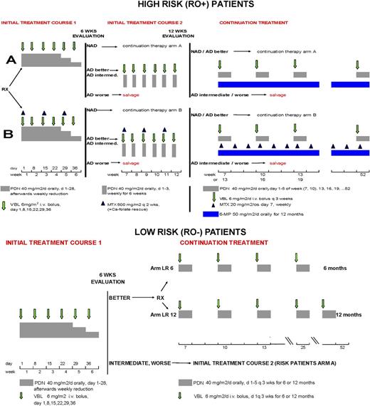
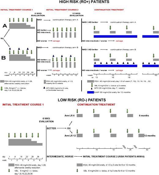
The objectives of the RO+ trial were (a) to improve initial response and survival by randomized addition of MTX to standard therapy (PDN+VBL) and by prolonging initial intensive therapy if the early response was inadequate (discussed later), and (b) to reduce morbidity and disease reactivations by prolonging therapy to a total of 12 months (Figure 1).
In RO– patients, the primary goal was to critically test whether prolonging continuation therapy in responders by 6 months (from a total of 6-12 months) reduces the reactivation rate (Figure 1).
Response to treatment was assessed by the International LCH Study Group criteria13 and, in RO+ patients, reflected response in risk organs. The categories were nonactive disease (NAD), defined as complete resolution, active disease (AD)/better (continuous regression of disease), AD/intermediate (unchanged disease or regression with some new involvement), and AD/worse (progression). Treatment decisions in RO+ patients were based on the response in risk organs and in RO– patients based on regression of lesions in the systems involved. The definition of response in RO+ patients is defined as being the response in risk organs (rather than “overall” response, which would include nonrisk organs) because it is known that the response in risk organs may be noted much earlier than the slower processes of resolution of skin disease or the healing of bone lesions. Particularly the latter may require many more weeks or even months after actual resolution of the histiocytic lesion itself and therefore much longer than required to observe hematopoietic system recovery, for example. For this reason we have used resolution of risk organ involvement as the criterion for response evaluation in RO+ patients.
Patient entry, randomization, and therapy
Eligibility criteria for both RO+ and RO– trials were age <18 years, definitive diagnosis of LCH,14 multisystem involvement, no prior systemic treatment of LCH, and informed consent obtained in accordance with the Declaration of Helsinki. Institutional review board approval was obtained at the individual institutions that enrolled patients in the trial. Six RO+ and 14 RO– patients lacked central review of the histopathology but were included in the analysis according to the intention-to-treat principle.19 Patients were enrolled through 9 study subcenters representing national/multinational study groups. The randomization in both RO+ and RO– trials was done in blocks stratified according to these subcenters. RO– randomizations were additionally stratified on age at diagnoses (≤2 years, >2 years). The randomization lists were provided by the study reference center in Vienna to each subcenter coordinator, who executed the randomization and informed the participating hospitals.
RO+ patients were randomized to standard Arm A or experimental Arm B, beginning with one or two 6-week courses of intensive initial therapy (according to response) followed by continuation therapy (Figure 1). Arm A initial treatment was VBL and continuous oral PDN. MTX was added in Arm B. Patients with NAD at week 6 proceeded to continuation of therapy. Patients with AD/better or AD/intermediate received a second course of intense initial therapy before receiving continuation therapy. Continuation therapy in Arm A consisted of daily 6-MP and VBL+PDN pulses every 3 weeks. In Arm B, weekly oral MTX was added. The overall duration was 12 months. Patients who were AD/worse after the first course (6 weeks) or AD/intermediate or AD/worse after the second course (12 weeks) were excluded from the study and shifted to salvage options.
RO– patients received the same initial 6-week treatment as those in Arm A of the RO+ group (Figure 1). Then they were evaluated. If they were NAD or AD/better, they were randomized to continuation therapy. This consisted of pulses of VBL and oral PDN, without 6-MP, for a total treatment of either 6 (Arm C) or 12 (Arm D) months. If patients were AD/intermediate or AD/worse at week 6, they were ineligible for randomization and were excluded from the study.
Statistical analysis
The primary end point for RO+ patients was a combined end point that includes early progression at week 6 and response at week 12, analyzed according to the intention-to-treat principle, ie, patients were analyzed in their allocated treatment group, even in case of noncompliance or protocol violations. This was done following the LCH guidelines19 because this is a conservative approach, providing the most reliable statistical significance. Patients who were not randomized were not included in the analyses. Nonresponse to initial therapy was defined as progression (AD/worse) in risk organs at week 6, or lack of response in risk organs (AD/intermediate or AD/worse) or death within 12 weeks of treatment initiation. Fisher’s exact test was used for the comparison.20 A power >80% required a sample size of 202 patients to show this 20% difference (2-sided α = 5%, NQuery 3.0, Fisher’s exact test). Statistical analyses of the secondary end points were exploratory. Five interim-analyses were planned and performed. Early stopping was implemented to either reject the null hypothesis of no difference between the 2 randomized arms or to retain the null hypothesis according to the design of O’Brien and Fleming.21
The primary end point in the RO– patients was the assessment of reactivations (progression in any organ or system). In the unexpected case of a death, this also would be considered as an event. Intervals to reactivation were calculated starting 6 months after initiation of therapy, when patients in Arm C stopped therapy and patients in Arm D received 6 additional months of the same therapy. Patients with reactivations within the first 6 months after randomization were excluded (prospectively planned departure from intention-to-treat analysis) because all patients received identical treatment during these 6 months, making any difference in reactivation rates during this time not attributable to the assigned treatments. The interval for censored patients was calculated to the date of the last response evaluation. Primary statistical evaluation was by the log rank test.20 Secondary exploratory analyses were performed to directly investigate long-term reactivation rates22 and cure-models23 ; 38 reactivations would be needed to show a difference in the 1-year reactivation rate of 35% vs 15% with a power of 80% and a 2-sided significance level of 5%.24 With an exponential distributed loss to follow-up rate (λ = 0.664) this event rate would be reached with a sample size of 148 patients.
Exploratory historical comparison
To assess progress over the course of multiple trials on MS-LCH conducted by the Histiocyte Society, we undertook a historical comparison. For the historical analysis of survival, only RO+ patients from LCH-III and identically matched patients from the predecessor studies LCH-I and LCH-II were considered. Thus, because in LCH-III age was excluded as a risk factor for death on the basis of previous findings,7 patients younger than 2 years old, but RO– (100% survival), were excluded from these historical comparisons. Other parameters changed between studies were that LCH I (1991-1995) involved centers from Austria, Germany, the UK, Italy, Scandinavia, and the US. LCH II (1996-2001) added centers from France and Argentina and LCH III (2001-2008) also involved Spain. Although VBL and PDN (given only as an initial 3-day pulse in LCH-I) comprised a common backbone of all 3 trials, only in LCH-III was the duration of continuation therapy increased, and a second initial therapy given when the early response was inadequate.
Results
Patient accrual and treatment
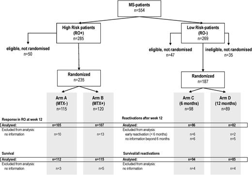
From April 2001 to February 2008, 285 RO+ and 269 RO– MS-LCH patients were enrolled (Figure 2). Seventy-five percent of RO+ and 47% of RO– patients were younger than 2 years old (Table 1). Demographic and clinical characteristics of both RO+ and RO– patients were equally distributed over their respective treatment arms, and involvement of the different risk organs among patients with RO+ MS-LCH was nearly equal in the 2 treatment arms (Table 1). Two-hundred thirty-five RO+ and 187 RO– patients were randomized. Six RO+ and 14 RO– patients lacked central review of the histopathology but were randomized and included in the analysis according to the intention-to-treat principle. The analysis cutoff date was December 31, 2010; median follow-up was 4.2 years.
RO+ trial
Response in risk organs at weeks 6 and 12
Response after 6 weeks of treatment was evaluable in 216 patients (Table 2); 41 patients achieved NAD in risk organs and were eligible directly for continuation treatment. Thirty patients were AD/worse or died and were excluded from the study. One-hundred forty-five patients (equally distributed between Arms A and B) were AD/better or AD/intermediate, and 112 (77%) received the prescribed second 6-week initial intense therapy. Their response rate (NAD or AD/better) at week 12 was 81%. Of the remaining 33 of 145 patients, by physician decision, 23 directly entered continuation treatment and 6 received other therapy, and 4 lacked adequate data.
Two-hundred twelve of 235 randomized patients (90%) with complete data at week 12 were eligible for analysis (105 in Arm A, 107 in Arm B; Table 2 and Figure 2). Response rates in risk organs at week 12, calculated according to the intention-to-treat principle, were 74/105 (70%) in Arm A and 77/107 (72%) in Arm B (P = .81). Thus, with 151 responding patients, both arms were similarly highly effective in inducing a rapid response in risk organs. Subsequently, 121 of the 151 responding patients (59 in Arm A, 62 in Arm B) received continuation therapy for a total treatment of 12 months (19 patients received other therapy, 3 stopped therapy, and 8 lacked information).
Survival
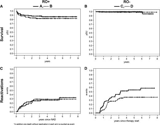
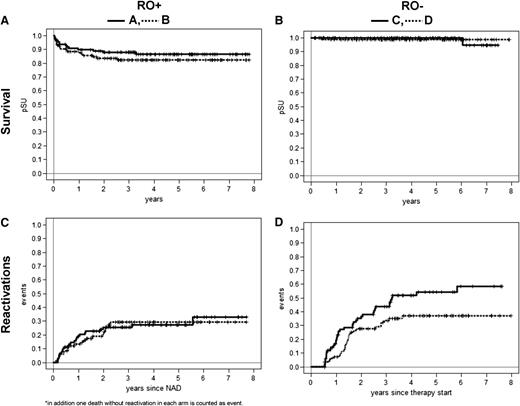
The overall 5-year survival probability of RO+ patients in LCH-III (84%) was very similar, and high in both arms (87% in Arm A, 82% in Arm B, P = .36; Figure 3A, Table 2). Several other important findings emerged in LCH-III: The degree of response at week 6 was predictive of survival: 95% when the response was NAD or AD/better, 83% when it was AD/intermediate, and only 57% when it was AD/worse (P < .001 for all pair-wise comparisons). Age at diagnosis was confirmed to not affect survival significantly; the 5-year probability of survival (pSU) was 84% and 91% in patients ≤2 and >2 years respectively (P = .16). In addition, of all deaths—14/112 (13%) in Arm A, 19/115 (17%) in Arm B (Table 2)—half occurred relatively early (12 deaths by week 6; 4 in Arm A, 8 in Arm B) and 2 additional occurred in each arm by week 12), emphasizing the critical importance of the initial period of treatment. Deaths were caused by disease progression with septic complications (12 in Arm A, 16 in Arm B) or organ failure (2 in Arm A, 3 in Arm B).
Survival and reactivations in LCH-III. (A) Survival in RO+ patients. (B) Survival in RO– patients. (C) Reactivations in RO+ patients. (D) C reactivations in RO– patients.
Survival and reactivations in LCH-III. (A) Survival in RO+ patients. (B) Survival in RO– patients. (C) Reactivations in RO+ patients. (D) C reactivations in RO– patients.
Disease resolution and reactivation
The median time to NAD in risk organs was similar (1 year) in both arms. The cumulative incidence of achieving NAD in risk organs was 19% in Arm A and 17% in Arm B at 6 weeks; 34% and 31%, respectively, at 12 weeks; and 57% and 63%, respectively, at 1 year (Table 2). Complete overall resolution of disease (ie, including complete response in nonrisk organs) was also evaluated (Table 2). At 5 years, the cumulative incidence of achieving overall NAD was 87% in Arm A and 85% in Arm B.
Forty-four of 173 patients who achieved NAD had disease reactivation, with a similar overall incidence in the 2 arms (Table 2, Figure 3C). Ten patients in Arm A and 5 patients in Arm B had reactions in one or more risk organs (7 lung, 6 spleen, 5 liver, 1 hematopoietic system). In nonrisk systems, there were 13 reactivations in Arm A and 16 in Arm B—predominantly bone lesions, skin lesions, and diabetes insipidus (DI). The 3- and 5-year reactivation rates with 12 months of treatment were 25% in Arm A and 29% in Arm B. Mortality after reactivation was very low, occurring in only 3 patients (1 in Arm A, 2 in Arm B). The 3-year cumulative incidence of DI in RO+ patients was 8% and 9% in Arms A and B (Table 2).
Toxicity
Toxicity at week 6 was generally transient. Overall, World Health Organization (WHO) grade 3/4 toxicity occurred in 30% of patients in Arm A and was 1.6 times higher (∼47%) in Arm B, which included MTX (P < .02); details by WHO grade are shown in Table 3. Prominent toxicities (WHO grade 3/4) were infections (48%), bone marrow dysfunction (28%), hepatotoxicity (25%), gastrointestinal symptoms (less often), and transient neurotoxicity (rarely). The specific toxicities that were significantly (P < .05) more frequent (always in arm B) were hepatotoxicity, thrombocytopenia, oral mucositis, and nausea and vomiting. It should be noted that distinguishing between therapy-related and disease-induced findings in RO+ MS-LCH is not always possible. For example, pancytopenia may be either primary disease involvement and/or an effect of therapy.
RO– trial
Patient flow and response at 6 weeks
Two-hundred sixty-nine RO– MS-LCH patients received the initial 6-week treatment (Figure 1). The 6-week response rate (NAD or AD/better) was 234 patients (86%), and these patients were the initial cohort, eligible for randomization in the trial. Achievement of NAD/AD better (Table 4) was thus similar to that in RO+ patients. One-hundred eighty-seven of the 234 patients were randomized. Thirty-five patients were ineligible for randomization because of inadequate response by week 6 (AD/intermediate or worse), whereas 47 patients were not randomized, 28 either by physicians’ choice or for lack of consent (Figure 2), and 19 because complete response data were not available. Further analyses excluded 8 randomized patients, according to the study design because of reactivation within 6 months of treatment, and 11 because of missing follow-up beyond 6 months. The final analyses included 168/187 patients (86 in Arm C, 82 in Arm D, Figure 2).
Survival
Disease reactivation and toxicity
The key issue was reduction of reactivations. When comparing the 5-year probability of reactivation between shorter treatment Arm C (41/86, 54%) and longer Arm D (28/82, 37%, P = .03), the effect of longer treatment is clear (Figure 3D, Table 4); the Kaplan-Meier curves for both arms show a plateau and these long-term reactivation rates differ between the 2 arms. Specifically, looking at long-term reactivation rates at 5 years, the 17% lower reactivation rate in the 12-month arm vs the 6-month arm reaches statistical significance (P = .04). Similar results are seen by an independent analysis (Cure-model).23 This analysis showed that the impact of treatment prolongation on time to reactivation was not significant (P = .775, HR = 0.9), whereas and importantly, there was a significant impact on long-term reactivation rates (P = .009, OR for “cure” = 0.44). Only 3 RO– patients had reactivation in a risk organ (liver, spleen, and hematopoietic system). The cumulative incidence of DI in RO– patients in each arm was 12% (Table 4). Toxicity in Arms C and D was similar (mostly mild hematopoietic) and less severe than in patients with RO+ involvement.
Discussion
Progress in treating LCH has benefitted from the adoption of standard diagnostic criteria, standard evaluation of disease extent, and stratified treatment. This has been particularly important for MS-LCH, with its varied manifestations and severity. Two major continuing problems in MS-LCH are (1) disease mortality and (2) disease reactivation, with its attendant risk of permanent consequences. The sequential Histiocyte Society clinical trials LCH-I, LCH-II, and LCH-III, address these issues.
Following the suggestion in LCH-I and LCH-II that survival was linked to speed of disease resolution in patients with RO+ MS-LCH, LCH-III tested a more intensive treatment of RO+ patients; these patients were randomized to receive an additional drug, MTX. In addition, patients with persistent active disease at week 6 received prolonged initial therapy (course 2) in a nonrandomized manner. The addition of MTX unfortunately neither accelerated disease resolution nor increased survival (Table 2), but it was associated with higher levels of toxicity (Table 3). Therefore, it will be excluded from the subsequent treatment protocol, LCH-IV.
Prolonged intense initial treatment, compared with the earlier trials, may have contributed to the increased survival in LCH-III that was associated with both the rapidity and degree of response. Deaths from RO+ MS-LCH occurred relatively early, half within 12 weeks, defining this time period as critical for patient outcome. What was heartening was that aggressive salvage therapy was quite effective in patients not responding to the protocol therapy, even though it was more toxic and therefore not suitable as a first-line treatment. Among the 37 patients receiving salvage therapy, the survival rate was 75%. There were 9 deaths (6/27 patients who received 2-CDA-based regimens and 3/10 who were transplanted). A comprehensive analysis of salvage therapy is in progress (Donadieu et al, in preparation), as is an analysis of possible differences in mortality among the national subcenters.
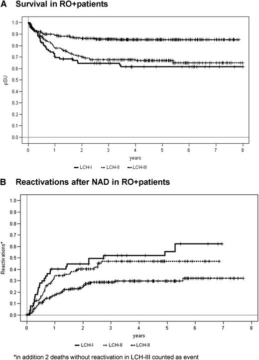
We conducted an exploratory historical comparison of the response and outcome of RO+ patients in the 3 LCH trials conducted by the Histiocyte Society. This exploratory analysis considered only exactly matched patients (with respect to diagnostic and stratification criteria). Likewise, the overall 5-year survival probability of RO+ patients in LCH-III, 84%, which was very similar in the two arms (87% in Arm A, 82% in Arm B), was substantially higher than in the corresponding (historical) RO+ patients in the predecessor trials LCH-I (62%) and LCH-II (69%),6,7 P < .001 (Figure 4A). Underscoring the importance of treatment duration on reactivation frequency, LCH-III RO+ patients (12-month treatment) had a 27% 5-year risk of reactivation, much lower than that of the comparable (RO+) historical controls treated in LCH-I (55%) and LCH-II (44%), who received only 6 months of therapy (Figure 4B).
Survival and reactivations in RO+ MS-LCH. RO+ patients in LCH-I, LCH-II, and LCH-III were analyzed, considering only the identically matched (ie, RO+) LCH-I and LCH-II patients. Both RO+ arms (A and B) of LCH-III are included in the curves shown for LCH-III. RO– patients younger than 2 years (age alone no longer a risk factor) were not included. (A) Survival: LCH-III vs LCH-II, LCH-III vs LCH-I, and LCH-III vs combined LCH-I and LCH-II; all are significant at P < .001. (B) Reactivations after NAD: LCH-III vs LCH-II, P < .004; LCH-III vs LCH-I, P < .005; and LCH-III vs combined LCH-I and LCH-II, P < .001.
Survival and reactivations in RO+ MS-LCH. RO+ patients in LCH-I, LCH-II, and LCH-III were analyzed, considering only the identically matched (ie, RO+) LCH-I and LCH-II patients. Both RO+ arms (A and B) of LCH-III are included in the curves shown for LCH-III. RO– patients younger than 2 years (age alone no longer a risk factor) were not included. (A) Survival: LCH-III vs LCH-II, LCH-III vs LCH-I, and LCH-III vs combined LCH-I and LCH-II; all are significant at P < .001. (B) Reactivations after NAD: LCH-III vs LCH-II, P < .004; LCH-III vs LCH-I, P < .005; and LCH-III vs combined LCH-I and LCH-II, P < .001.
We conclude that optimism regarding survival in RO+ MS-LCH is now clearly warranted, recognizing that the reduction in mortality of RO+ patients in our 3 sequential studies may be a result of multiple factors, including a second course of initial treatment, prolonged continuation therapy, effective salvage therapy in progressing patients, continuing improvement in supportive care, and possibly other unmeasured factors.
The strict test of the hypothesis regarding treatment duration and reactivation risk was in the RO– patients in LCH-III, randomized to 6 vs 12 months of therapy. The differences were striking: reactivation frequency was significantly reduced by the longer mild continuation therapy. Further reduction in reactivations is a goal for LCH-IV.
The LCH-I, II, and III trials underscore that clinical questions in rare diseases such as LCH can be adequately addressed only through rigorous controlled prospective trials in the setting of international cooperation. The upcoming LCH-IV study will need to address several remaining problems in MS-LCH management. The most urgent one is to eliminate the remaining mortality of about 15% among patients with risk organ involvement. Two-step stratification based on risk organ involvement at diagnosis and lack of response to standard initial treatment (eg, at week 6) will allow for early identification of patients with extremely poor prognosis. This particular “very-high-risk” group will be eligible for treatment with either a combination of cladribine and cytarabine27 or with hematopoietic stem cell transplantation after reduced-intensity conditioning.28
A reactivation rate of 30% to 40% is also still unacceptably high, because it results in both acute morbidity and permanent consequences.29 The LCH-III data and historical controls showed significant advantage of a 12-month over a 6-month treatment duration. A “2 × 2” factorial design of the LCH-IV Study will test the role of further prolongation of the therapy and the value on oral 6-mercaptopurine in the continuation treatment of MS-LCH.
A final consideration is that of the treatment of adults with LCH, an increasingly recognized diagnosis but for which the treatment has not yet been studied systematically. Because the majority of the patients treated in LCH-III (open to children and adolescents up to age 18) were young children, the results should not be extrapolated to adults.
The etiology and pathogenesis of MS-LCH remain enigmatic. Making sense of the many and sometimes quite disparate molecular and immunologic findings30 may ultimately identify either a neoplastic or an immune cause of LCH (2 prominent hypotheses). Clonality of the LCH cells1 (also seen in many non-neoplastic conditions), elevated (but not mutated) p53 levels,31 and a high incidence of BRAF V600E point mutations32 may support a neoplastic origin. Conversely, the inflammatory nature of LCH lesions, possible cytokine abnormalities,33,34 and a myeloid cell maturation defect in the LCH cell35 suggest dysfunctional immune responses. Overall, recent research now gives hope for targeted therapy and possibly a rational cure of LCH.32,35 Ultimately, better understanding could lead to a striking transformation of therapy. A caution is that whatever new drugs and regimens appear appropriate and justified for testing in LCH, future prospective trials must be carefully designed to reflect the clinical diversity and biological heterogeneity of MS-LCH to avoid being misled concerning therapeutic efficacy. In the interim, to continue to reduce mortality and decrease reactivation frequency warrants continued participation in clinical trials of MS-LCH.
There is an Inside Blood commentary on this article in this issue.
The publication costs of this article were defrayed in part by page charge payment. Therefore, and solely to indicate this fact, this article is hereby marked “advertisement” in accordance with 18 USC section 1734.
Acknowledgments
This work was supported by the Children’s Cancer Research Institute (Vienna, Austria) and Anthony House; the Histiocytosis Associations of America, Belgium, Italy, and Canada; Histiozytosehilfe (Germany); Programme Hospitalier de Recherche Clinique 2001 and CHU, Nantes (France); the Swedish Childhood Cancer Foundation and Swedish Research Council; and the Children’s Research Institute (Washington, DC).
Authorship
Contribution: H.G. and S.L. wrote the paper; U.P. performed the statistical analysis; M.M., N.G., and E.T. contributed to study coordination, data collection, and analysis; M.A., J.B., J.D., I.A., J.-I.H., G.J.-S., K.M., S.W., and K.W. made clinical contributions and provided critical manuscript review. All authors were involved in study design and final approval of the paper.
Conflict-of-interest disclosure: The authors declare no competing financial interests.
Correspondence: Stephan Ladisch, Center for Cancer and Immunology Research, Children’s Research Institute, Children’s National Medical Center, Washington, DC 20010; e-mail: sladisch@cnmc.org; Reprints: Helmut Gadner, St. Anna Children’s Cancer Research Institute, Zimmermann Platz 10, A-1090 Vienna, Austria; e-mail: helmut.gadner@ccri.at; or Stephan Ladisch, Children’s National Medical Center, 111 Michigan Ave. NW, Washington, DC 20010; e-mail: sladisch@cnmc.org.

