Key Points
Ebf1 regulates DNA repair in a dose-dependent manner.
Combined heterozygote loss of Ebf1 and Pax5 predisposes for leukemia development.
Abstract
Early B-cell factor 1 (Ebf1) is a transcription factor with documented dose-dependent functions in normal and malignant B-lymphocyte development. To understand more about the roles of Ebf1 in malignant transformation, we investigated the impact of reduced functional Ebf1 dosage on mouse B-cell progenitors. Gene expression analysis suggested that Ebf1 was involved in the regulation of genes important for DNA repair and cell survival. Investigation of the DNA damage in steady state, as well as after induction of DNA damage by UV light, confirmed that pro-B cells lacking 1 functional allele of Ebf1 display signs of increased DNA damage. This correlated to reduced expression of DNA repair genes including Rad51, and chromatin immunoprecipitation data suggested that Rad51 is a direct target for Ebf1. Although reduced dosage of Ebf1 did not significantly increase tumor formation in mice, a dramatic increase in the frequency of pro-B cell leukemia was observed in mice with combined heterozygous mutations in the Ebf1 and Pax5 genes, revealing a synergistic effect of combined dose reduction of these proteins. Our data suggest that Ebf1 controls DNA repair in a dose-dependent manner providing a possible explanation to the frequent involvement of EBF1 gene loss in human leukemia.
Introduction
One of the central proteins in B-lymphocyte development is the transcription factor early B-cell factor 1 (Ebf1).1 Ebf1 is critical for the activation of B-lineage restricted genes in the earliest B-lineage progenitors2,3 and for restriction of lineage fate options.4,5 The activity is highly dependent on functional Ebf1 dose because mice carrying a heterozygous deletion of the Ebf1 gene display reduced numbers of CD19+CD43– B-cell progenitors, whereas the CD19+CD43+ pro-B cell compartment remains intact.6-8 Ebf1 levels are also of relevance in leukemia because mutations resulting in reduced functional EBF1 dose9,10 and increased expression of post-transcriptional inhibitors of EBF1, ZNF521,11 or ZNF42312 are found in B-cell acute lymphoblastic leukemia (B-ALL).13,14 A direct role for Ebf1 dose in malignant transformation was supported by the findings that combined expression of constitutively active Stat5 (caStat5) and heterozygous loss of either Ebf1 or Pax5 results in B-cell leukemia in mice.15 Heterozygote deletion of PAX5 is a rather common genetic alteration in human B-ALL9,10 and a genetic polymorphism causing reduced functional PAX5 activity has been found in families with a high incidence of leukemia.16 The finding that the developmental block observed in Pax5-deficient leukemia cells can be reversed on restoration of Pax5 expression suggests that the reduction in Pax5 function results in a reversible disruption of differentiation.17 A similar mechanism of action has been proposed for Ebf1; however, reduced amounts of Ebf1 in normal cells appear to result in reduced proliferation and expansion of B-cell progenitors,4,6,18,19 indicating that the involvement of EBF1 in malignant transformation is more complex.
To increase our understanding of the functions of Ebf1 in malignant transformation, we identified dose-dependent processes regulated by Ebf1 in early B-cell development, revealing changes in DNA repair and cell survival. Because these data suggested that Ebf1 functions differently than what has been reported for Pax5 in the transformation process,16 we investigated the functional collaboration between Ebf1 and Pax5 in leukemogenesis, revealing a strong functional synergy on the development of leukemia. Together, our data suggest that reduced levels of Ebf1 may contribute to malignant transformation by a combination of impaired DNA repair and increased cell survival rather than simply by a differentiation block.
Experimental procedures
Animal models
Pax5+/−,20 Ebf1+/−,3 and vav-BCL221 were all on a C57BL/6 (CD45.2) background. Adoptive transfers were performed by tail vein injection of 2 million live lymph node (LN) cells into sublethally irradiated (4.5 Gy) or nonconditioned CD45.1 mice. Animal procedures were performed with consent from the local ethics committee at Linköping University (Linköping, Sweden).
Western blot
Western blots were made using extracts from cultured primary pro-B cells from bone marrow (BM) of wild-type (Wt) and Ebf1+/− mice. The membranes were incubated with the relevant primary and secondary antibodies (for details, please see supplemental Methods, available on the Blood Web site), and signals were measured with the Odyssey Infrared Imaging System (LI-COR, Lincoln, NE). The band intensities was quantified using ImageJ software.
Immunofluorescence analysis
Cultured Wt and Ebf1+/− pro-B cells from BM of mice or UV-induced pro-B cells22 were fixed and incubated with the relevant antibodies and mounted using VECTA SHEILD containing 4′,6 diamidino-2-phenylindole (DAPI; for details, see supplemental Methods). The fluorescence expression of stained proteins was viewed using an LM Zeiss upright confocal microscope (Zeiss). The integrated density of the protein expression was monitored using ImageJ software.
In vitro phospho flow
Cultured Wt and Ebf1+/− pro-B cells or ex vivo-sorted pro-B cells were fixed and stained with the relevant antibodies (for details, see supplemental Methods). The stained cells were analyzed using the BD fluorescence-activated cell sorter (FACS) Canto II analyzer (BD Biosciences). Gates were set based on the antibody isotype control. All gates were set according to fluorescence minus 1 control. All analysis was performed using FlowJo software.
Annexin V staining
Cultured Pro-B Wt and Ebf1+/− cells were washed and incubated with Annexin V-allophycocyanin (BD Pharmingen) along with 5 μL 7-aminoactinomycin D, and the cells were analyzed using BD FACS Canto II (BD Biosciences). All the gates were set according to the unstained control, Annexin V-allophycocyanin alone, and 7-aminoactinomycin D alone.
Cell cycle analysis
Cultured Pro-B Wt and Ebf1+/− cells were fixed and incubated with antibodies for Ki67 (BD Pharmingen), and DAPI after which G0 cells were scored as Ki67−DAPIlow (equal to that of a diploid cell), G1 as Ki67+DAPIlow, and SG2M as Ki67+DAPIhigh as in Åhsberg et al.6
Comet assay
The comet assay was performed using the reagents from the OxiSelect Comet assay kit (Cell Biolabs) according to the manufacturer’s instructions (see supplemental Methods for details). Comets were viewed using a Zeiss upright confocal microscope in the fluorescein isothiocyanate channel using a ×20 objective.
FACS staining and sorting of hematopoietic cells
Cell sorting of and analysis of primary cells was performed as in Åhsberg et al6 (for details, see supplemental Methods). The cellular composition of the primary tumors was analyzed using frozen single-cell suspensions from the LNs, BM, and spleen, whereas the analysis of the composition of the organs of transplanted mice was performed on freshly isolated tissues and blood.
Gene expression analysis with quantitative reverse transcription-polymerase chain reaction and Affymetrix
VDJ-recombination and exome sequencing analysis
Live cells were sorted, and DNA was extracted and subjected to PCR-based variable diversity joining (VDJ) analysis.24 For exome sequencing, the DNA was fragmented and enriched for coding regions, followed by sequencing to gain an average cover of ×100, and the obtained data were analyzed using Mutech software25 (see supplemental Methods for details).
Statistical analysis
The statistical analysis is described in the corresponding figure legends.
Results
Reduced amounts of Ebf1 increase DNA damage in pro-B cells
Ebf1 is a crucial regulator of cell differentiation; however, conditional deletion of the Ebf1 gene in B-cell progenitors reveals additional roles for this transcription factor in proliferation and cell survival.4,18,19 To understand more about how Ebf1 dose impacts cellular functions in B-cell development, we analyzed gene expression data from primary sorted CD19+ immunoglobulin (Ig)M– B-cell progenitors from Wt and Ebf1+/− mice.6 Gene expression data from CD43low/neg progenitors revealed reduced expression of Rad51, Rad51ap1, and Smc2, all coding for proteins with functions in DNA repair, whereas the mRNA levels for the antiapoptotic Bcl2 protein were increased.
To investigate whether Ebf1 directly regulates DNA repair genes in progenitor B lymphocytes, we transduced 230-238 pre-B cells with a dominant-negative Ebf1 protein. Here, the carboxy-terminal trans-activation domain of Ebf1 was replaced by the repressor domain of the Drosophila protein engrailed.26 Expression of Ebf1-engrailed resulted in downregulation of a large number of genes involved in DNA repair processes in 230-238 cells (supplemental Table 1).
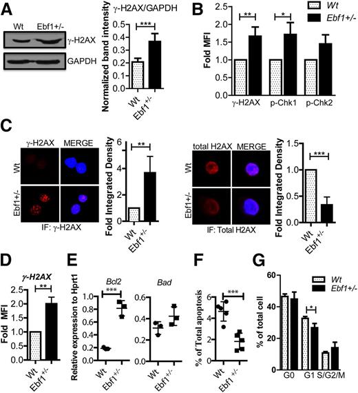
To investigate whether the reduction in DNA repair genes result in increased DNA damage in Ebf1+/− pro-B cells, we analyzed the levels of phosphorylated H2AX (γH2AX) in cultured Wt and Ebf1+/ cells. Western blot (Figure 1A), FACS (Figure 1B), and immunohistochemical (IHC) analysis (Figure 1C) determined that levels of γH2AX are higher in Ebf1+/− pro-B cells than in Wt cells, even though the overall levels H2AX protein were reduced (Figure 1C). Similar differences were detected on FACS analysis of γH2AX levels in B-cell progenitors ex vivo (Figure 1D). Median fluorescent intensities (MFIs) revealed significant differences between γH2AX in Wt and Ebf1+/−CD19+CD43+IgM− pro-B cells, supporting the conclusion that Ebf1 heterozygosity increases the level of DNA damage in early B-cell progenitors.
Heterozygous loss of Ebf1 results in increased levels of phosphorylated H2AX in B-cell progenitors. (A) Representative western blot out of 3 detecting γH2AX and glyceraldehyde-3-phosphate dehydrogenase from in vitro cultured Wt or Ebf1+/− pro-B cells. (Lower) Quantification of the signal intensity normalized to the Wt control and represents 3 experiments. The data shown in band intensity for mean ± standard deviation (SD) and statistical analysis was performed using an unpaired Student t test: ***P < .001 (B) Graphs over the relative MFI obtained by flow cytmetric analysis to detect γH2AX and indirect flow analysis of phosphorylated Chk1 (pChk1) and phosphorylated Chk2 (pCHK2) in in vitro-cultured Wt or Ebf1+/− pro-B cells. The data are normalized toward the Wt control and expressed in fold MFI that represents 3 independent experiments. The statistical analysis was done using an unpaired t test. Mean ± SD; **P < .01 and *P < .05 (C) Immunofluorescence analysis of phosphorylated and nonphosphorylated H2AX (total H2AX) in in vitro-cultured Wt or Ebf1+/− pro-B cells detected by using rabbit γH2AX monoclonal antibody or rabbit monoclonal total H2AX antibody followed by a specific anti-Alexa Fluor 594 secondary antibody. The nuclei were subsequently stained with DAPI, and the images were captured using an LM Zeiss upright confocal microscope. The quantification panel next to the image displaying the signal intensity was collected from 3 experiments. The statistical analysis was performed using an unpaired t test, and results are plotted as change in fold integrated density compared with Wt; **P < .01 and ***P < .001. (D) Diagram with relative MFI obtained by flow cytometric analysis to detect γH2AX, in ex vivo-isolated Wt or Ebf1+/− pro-B cells. The data are normalized toward the Wt control and represent 3 experiments, and an unpaired t test was performed. Mean ± SD; **P < .01 (E) Diagrams displaying quantitative RT-PCR data from in vitro-cultured Wt or Ebf1+/− pro-B cells. The data are normalized to the expression of HPRT in triplicate PCR reactions. An unpaired Student t test was performed for statistical analysis. ***P < .001. Data represent 3 independent experiments as mean ± SD. (F) Frequency of AnnexinV+ cells as estimated by flow cytometric analysis from in vitro-cultured Wt or Ebf1+/− pro-B cells. The cells were gated on a lymphoid gate for live cells. The data represent 5 experiments. The error bar in the panels indicate mean ± SD, and statistical analysis was performed using the Student t test. ***P < .001. (G) Cell cycle status of Wt or Ebf1+/− pro-B cells using Ki67 and DAPI staining. G0 cells were scored as Ki67−DAPIlow, G1 Ki67+ DAPIlow, and SG2M as Ki67+DAPIhigh. Mean ± SD and statistical analysis was performed using the Student t test. *P < .05.
Heterozygous loss of Ebf1 results in increased levels of phosphorylated H2AX in B-cell progenitors. (A) Representative western blot out of 3 detecting γH2AX and glyceraldehyde-3-phosphate dehydrogenase from in vitro cultured Wt or Ebf1+/− pro-B cells. (Lower) Quantification of the signal intensity normalized to the Wt control and represents 3 experiments. The data shown in band intensity for mean ± standard deviation (SD) and statistical analysis was performed using an unpaired Student t test: ***P < .001 (B) Graphs over the relative MFI obtained by flow cytmetric analysis to detect γH2AX and indirect flow analysis of phosphorylated Chk1 (pChk1) and phosphorylated Chk2 (pCHK2) in in vitro-cultured Wt or Ebf1+/− pro-B cells. The data are normalized toward the Wt control and expressed in fold MFI that represents 3 independent experiments. The statistical analysis was done using an unpaired t test. Mean ± SD; **P < .01 and *P < .05 (C) Immunofluorescence analysis of phosphorylated and nonphosphorylated H2AX (total H2AX) in in vitro-cultured Wt or Ebf1+/− pro-B cells detected by using rabbit γH2AX monoclonal antibody or rabbit monoclonal total H2AX antibody followed by a specific anti-Alexa Fluor 594 secondary antibody. The nuclei were subsequently stained with DAPI, and the images were captured using an LM Zeiss upright confocal microscope. The quantification panel next to the image displaying the signal intensity was collected from 3 experiments. The statistical analysis was performed using an unpaired t test, and results are plotted as change in fold integrated density compared with Wt; **P < .01 and ***P < .001. (D) Diagram with relative MFI obtained by flow cytometric analysis to detect γH2AX, in ex vivo-isolated Wt or Ebf1+/− pro-B cells. The data are normalized toward the Wt control and represent 3 experiments, and an unpaired t test was performed. Mean ± SD; **P < .01 (E) Diagrams displaying quantitative RT-PCR data from in vitro-cultured Wt or Ebf1+/− pro-B cells. The data are normalized to the expression of HPRT in triplicate PCR reactions. An unpaired Student t test was performed for statistical analysis. ***P < .001. Data represent 3 independent experiments as mean ± SD. (F) Frequency of AnnexinV+ cells as estimated by flow cytometric analysis from in vitro-cultured Wt or Ebf1+/− pro-B cells. The cells were gated on a lymphoid gate for live cells. The data represent 5 experiments. The error bar in the panels indicate mean ± SD, and statistical analysis was performed using the Student t test. ***P < .001. (G) Cell cycle status of Wt or Ebf1+/− pro-B cells using Ki67 and DAPI staining. G0 cells were scored as Ki67−DAPIlow, G1 Ki67+ DAPIlow, and SG2M as Ki67+DAPIhigh. Mean ± SD and statistical analysis was performed using the Student t test. *P < .05.
Increased DNA damage is often associated with increased apoptosis; however, analysis of mRNA levels in the pro-B cells by quantitative RT-PCR suggested that, although the levels of Bad mRNA were comparable between Ebf1 heterozygous and Wt cells, expression of Bcl2 message was increased (Figure 1E). In support of this, analysis of annexinV expression on the cultured cells suggested that frequencies of apoptotic cells were lower in the Ebf1+/− than in the Wt cells (Figure 1R). Analysis of the cell cycle status of the cultured pro-B cells suggested a slight reduction of cells in the G1 stage (Figure 1G). Hence, even though the Ebf1+/− cells showed more DNA damage as estimated by H2AX phosphorylation, this did not result in increased apoptosis.
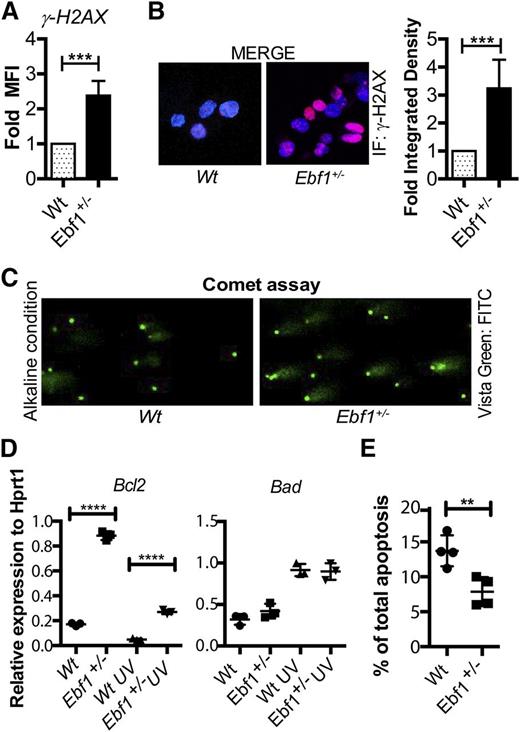
To investigate how Ebf1+/− cells respond to induced DNA damage, we exposed primary cultured Wt and Ebf1+/− pro-B cells to UV light and quantified the amounts of γH2AX 16 hours after UV exposure by FACS and IHC (Figure 2A-B). Both assays suggested that γH2AX was more abundant in Ebf1+/− cells than in Wt cells. Increased DNA damage in Ebf1+/− pro-B cells compared with Wt cells was also detected using comet assays (Figure 2C), supporting our hypothesis that reducing Ebf1 dose results in increased DNA damage. UV exposure resulted in increased Bad mRNA, as estimated by quantitative RT-PCR analysis in both Wt and Ebf1+/− cells (Figure 2D). Although expression of Bcl2 was downregulated in both Wt and Ebf1+/− cells after exposure, Bcl2 levels remained higher in the Ebf1+/− cells (Figure 2D). In line with this finding, FACS analysis estimating the fraction of apoptotic cells based on annexinV staining revealed significantly less apoptosis in Ebf1+/− pro-B cells relative to Wt cells (Figure 2E). These data suggest that Ebf1+/− cells possess an imbalance in DNA repair and cell survival that may drastically increase both the frequency of mutation and the continued survival of cells carrying damaged DNA.
Heterozygous loss of Ebf1 results in increased DNA damage after UV exposure of B-cell progenitors. (A) Relative MFI obtained by flow cytometric analysis to detect γH2AX in in vitro-cultivated primary Wt or Ebf1+/− pro-B cells 16 hours after UV exposure. The data are normalized toward the Wt control and represent 3 experiments, The statistics were performed using an unpaired t test; the error bar represents mean ± SD. ***P < .001. (B) Immunohistochemical staining of phosphorylated H2AX (γH2AX) in in vitro-cultured Wt or Ebf1+/− pro-B cells 16 hours after UV exposure. DAPI was used to stain the nucleus. (Lower) Quantification of the signal intensity collected from 3 experiments. The representation of fold-integrated density is based on an unpaired t test with error bars representing mean ± SD. ***P < .001. (C) Representative pictures of comet assays performed 16 hours after UV exposure of in in vitro-cultivated primary Wt or Ebf1+/− pro-B cells. (D) Quantitative RT-PCR data from in vitro-cultured Wt or Ebf1+/− pro-B cells before and 16 hours after UV exposure. The data are normalized to the expression of HPRT in triplicate PCR reactions and represent 3 independent experiments. The Student t test represents statistical analysis. ****P < .0001. (E) Frequency of annexinV+ cells as estimated by flow cytometric analysis from in vitro-cultured Wt or Ebf1+/− pro-B cells. The cells were gated on a lymphoid gate for live cells. The error bars in the diagrams indicate mean ± SD, and statistical analysis was performed using an unpaired Student t test. **P < .01.
Heterozygous loss of Ebf1 results in increased DNA damage after UV exposure of B-cell progenitors. (A) Relative MFI obtained by flow cytometric analysis to detect γH2AX in in vitro-cultivated primary Wt or Ebf1+/− pro-B cells 16 hours after UV exposure. The data are normalized toward the Wt control and represent 3 experiments, The statistics were performed using an unpaired t test; the error bar represents mean ± SD. ***P < .001. (B) Immunohistochemical staining of phosphorylated H2AX (γH2AX) in in vitro-cultured Wt or Ebf1+/− pro-B cells 16 hours after UV exposure. DAPI was used to stain the nucleus. (Lower) Quantification of the signal intensity collected from 3 experiments. The representation of fold-integrated density is based on an unpaired t test with error bars representing mean ± SD. ***P < .001. (C) Representative pictures of comet assays performed 16 hours after UV exposure of in in vitro-cultivated primary Wt or Ebf1+/− pro-B cells. (D) Quantitative RT-PCR data from in vitro-cultured Wt or Ebf1+/− pro-B cells before and 16 hours after UV exposure. The data are normalized to the expression of HPRT in triplicate PCR reactions and represent 3 independent experiments. The Student t test represents statistical analysis. ****P < .0001. (E) Frequency of annexinV+ cells as estimated by flow cytometric analysis from in vitro-cultured Wt or Ebf1+/− pro-B cells. The cells were gated on a lymphoid gate for live cells. The error bars in the diagrams indicate mean ± SD, and statistical analysis was performed using an unpaired Student t test. **P < .01.
Reduced Ebf1 dose causes impaired assembly of Rad51 complexes after induction of DNA damage
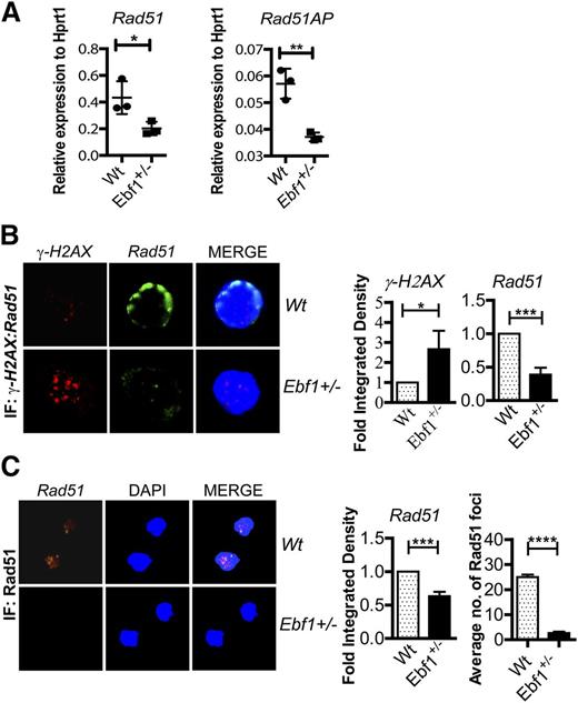
Although Ebf1 is involved in the regulation of expression of several genes in the DNA repair machinery (supplemental Table 1), a limited number of genes were significantly downregulated in freshly isolated Ebf1+/− progenitor B cells.6 These genes included Rad51 and Rad51ap, and quantitative PCR analysis of both primary cultured and ex vivo-isolated pro-B cells confirmed the lower expression levels in the Ebf1+/− cells (Figure 3A; supplemental Figure 1A). To investigate whether the reduced RNA levels translated to reduced steady-state expression of Rad51 protein, we stained cultured Wt and Ebf1+/− pro-B cells with antibodies for γH2AX and Rad51. Rad51 is a largely cytoplasmic protein, which on DNA damage, is recruited to the nucleus and the single-stranded ends at double-strand breaks.27 IHC staining of Wt cells resulted in what appeared to be a perinuclear staining likely as a result of a rather small cytoplasm of pro-B cells (Figure 3B-C). The Rad51 staining in Ebf1+/− cells was reduced, supporting the idea that the levels of Rad51 protein is reduced in Ebf1+/– cells relative to Wt cells, whereas the level of γH2AX displayed an opposite pattern. These results suggest that loss of 1 allele of Ebf1 proportionally decrease levels of Rad51 protein (Figure 3B).
Heterozygous loss of Ebf1 results in lower levels of Rad51 in B-cell progenitors. (A) Quantitative RT-PCR data from in vitro-cultured Wt or Ebf1+/− pro-B cells. The data are normalized to the expression of HPRT in triplicate PCR reactions and represent 3 independent experiments. The Student t test was performed to check the statistical significance. **P < .01 and *P < .05. (B) Immunofluorescence staining of γH2AX (rabbit) and mouse Rad51 in in vitro-cultured Wt or Ebf1+/− pro-B cells followed by the specific anti-Alexa Fluor 594 (γH2AX) and anti-Alex Fluor 488 secondary antibody (Rad51). DAPI was used to stain the nucleus. The panel adjacent to each IF image shows a quantification of the signal intensity collected from 3 experiments, and quantification is represented as fold integrated density with statistical significant value: *P < .05 for γH2AX and ***P < .001 for Rad51. (C) Immunohistochemical staining of Rad51 in in vitro-cultured Wt or Ebf1+/− pro-B cells 16 hours after UV exposure. DAPI was used to stain the nucleus. The data were collected from 3 experiments, and foci formation was counted from 3 cells from 3 different experiments. The error bars in the diagrams indicate mean ± SD, and statistical analysis was performed using the Student t test, with P < .001.
Heterozygous loss of Ebf1 results in lower levels of Rad51 in B-cell progenitors. (A) Quantitative RT-PCR data from in vitro-cultured Wt or Ebf1+/− pro-B cells. The data are normalized to the expression of HPRT in triplicate PCR reactions and represent 3 independent experiments. The Student t test was performed to check the statistical significance. **P < .01 and *P < .05. (B) Immunofluorescence staining of γH2AX (rabbit) and mouse Rad51 in in vitro-cultured Wt or Ebf1+/− pro-B cells followed by the specific anti-Alexa Fluor 594 (γH2AX) and anti-Alex Fluor 488 secondary antibody (Rad51). DAPI was used to stain the nucleus. The panel adjacent to each IF image shows a quantification of the signal intensity collected from 3 experiments, and quantification is represented as fold integrated density with statistical significant value: *P < .05 for γH2AX and ***P < .001 for Rad51. (C) Immunohistochemical staining of Rad51 in in vitro-cultured Wt or Ebf1+/− pro-B cells 16 hours after UV exposure. DAPI was used to stain the nucleus. The data were collected from 3 experiments, and foci formation was counted from 3 cells from 3 different experiments. The error bars in the diagrams indicate mean ± SD, and statistical analysis was performed using the Student t test, with P < .001.
To investigate whether the reduced levels of Rad51 impact the formation of Rad51 foci after DNA damage, we exposed Wt and Ebf1+/− cells to UV radiation prior to staining of Rad51. Although Rad51 foci were easily detected in Wt cells 16 hours after UV exposure, few were formed in Ebf1+/− cells as estimated by IHC (Figure 3C). Hence, reduced levels of Ebf1 impair the assembly of Rad51 foci, with potential consequences for the efficiency of homologous DNA repair in Ebf1+/− pro-B cells.
To investigate whether Rad51 is a direct target of Ebf1, we took advantage of published chromatin immunoprecipitation sequencing (ChIP-Seq)28 and chromosome capture data from pro-B cells29 to identify putative regulatory elements for the Rad51 gene interacting with Ebf1. This suggested that ≥1 of the Rad51 regulatory elements contains an Ebf1 binding site (supplemental Figure 1A). ChIP analysis suggested that the transcription factor Tcfe2a (E2A) binds in the proximity of several putative enhancer regions. This is of interest because the gene expression analysis of primary Ebf1+/− progenitors suggested that Tcfe2a transcription was downregulated (supplemental Figure 1A).6 Hence, reduced Ebf1 dose may impact Rad51 transcription both directly and indirectly by reduction of Tcfe2a levels that also may impact expression of Rad51ap1, because the Rad51ap1 promoter contains a binding site for Tcfe2a (supplemental Figure 2).
Combined heterozygous deletion of Pax5 and Ebf1 predisposes for development of pro-B cell leukemia
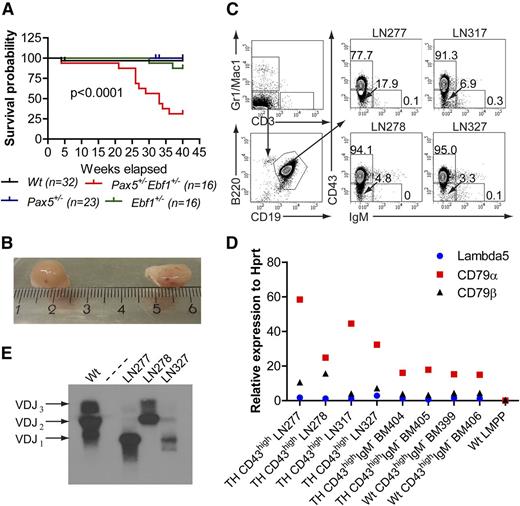
Our data suggest that normal Ebf1 dosage is important for the regulation of DNA repair. This would suggest that reduced amounts of Ebf1 contribute to the transformation process in a manner that differs from reductions of Pax5, which results in impaired differentiation of progenitor cells.17 Therefore, we hypothesized that combined reduction of Pax5 and Ebf1 dose synergistically could drive malignant transformation. To investigate this possibility, we generated mice carrying combined heterozygous deletion of Ebf1 and Pax5 genes. Although we did not detect malignant disease in Wt or Pax5+/− mice, 1 of 16 Ebf1+/− mice and 9 of 16 Pax5+/−Ebf1+/− mice presented with severely swollen LNs around week 30 (Figure 4A). Examination of lymphoid organs confirmed dramatically enlarged LNs reaching a diameter of several millimeters (Figure 4B). FACS analysis of LN cells from affected mice (Figure 4C; Table 1) detected a high frequency of B220+CD19+ cells (89% ± standard deviation [SD]), the majority of which were CD43high pro-B cells. Frequencies of CD43− pre-B cells varied from 3% to 36%, and the frequencies of IgM+ cells ranged from 0.04% to 15%. This indicates that LN enlargement is associated with a dramatic peripheral expansion of B-cell progenitors. We further analyzed the cellular composition of the BM and spleens of 4 sick Pax5+/−Ebf1+/− mice (supplemental Table 2), revealing that the LN enlargement was associated with a massive increase of B-cell progenitors in peripheral hematopoietic organs. Quantitative RT-PCR analysis using total live cells from 4 mice with enlarged LNs revealed expression of Cd79a, Cd79b, and Igll1 at levels comparable to, or higher than, sorted ex vivo-analyzed Wt CD19+CD43high pro-B cells (Figure 4D), verifying the identity of the cells and revealing that the transcriptional program of a pro-B cells is intact in the transformed cells.
Combined heterozygous loss of Ebf1 and Pax5 results in development of disease and increased mortality. (A) Kaplan-Meier curves describing the 40-week survival of Wt, Ebf1+/−, Pax5+/−, and Pax5+/−Ebf1+/− mice. A drop in the curve describes unknown cause of death or development of swollen lymph nodes and subsequent euthanasia due to animal protection regulations. Mice euthanized for known reasons other than swollen lymph nodes are censored in the curves. Overall log-rank (Mantel-Cox test) P value is displayed. (B) Two enlarged, one inguinal and one brachial, lymph nodes from a Pax5+/−Ebf1+/− mouse. (C) Representative flow cytometric analysis of the cellular content of a representative lymph node (left) and the fraction of CD43high or IgM+CD19+ cells in 4 analyzed nodes (right). (D) Quantitative RT-PCR data over gene expression analysis of sorted live cells in 4 analyzed lymph nodes. Sorted Pax5+/−Ebf1+/− BM pro-B cells and lymphoid primed multipotent progenitors were used as positive and negative controls. Expression levels are presented in relation to HPRT. Data represent 1 of 2 quantitative PCR experiments using triplicate PCR reactions for estimation of expression values. (E) Immunoglobulin heavy chain VDJ analysis from 3 of the analyzed live cell populations from nodes of mice displaying peripheral pro-B cell expansion. Cultured Wt pro-B cells were included to show the rearrangement signature of a polyclonal population.
Combined heterozygous loss of Ebf1 and Pax5 results in development of disease and increased mortality. (A) Kaplan-Meier curves describing the 40-week survival of Wt, Ebf1+/−, Pax5+/−, and Pax5+/−Ebf1+/− mice. A drop in the curve describes unknown cause of death or development of swollen lymph nodes and subsequent euthanasia due to animal protection regulations. Mice euthanized for known reasons other than swollen lymph nodes are censored in the curves. Overall log-rank (Mantel-Cox test) P value is displayed. (B) Two enlarged, one inguinal and one brachial, lymph nodes from a Pax5+/−Ebf1+/− mouse. (C) Representative flow cytometric analysis of the cellular content of a representative lymph node (left) and the fraction of CD43high or IgM+CD19+ cells in 4 analyzed nodes (right). (D) Quantitative RT-PCR data over gene expression analysis of sorted live cells in 4 analyzed lymph nodes. Sorted Pax5+/−Ebf1+/− BM pro-B cells and lymphoid primed multipotent progenitors were used as positive and negative controls. Expression levels are presented in relation to HPRT. Data represent 1 of 2 quantitative PCR experiments using triplicate PCR reactions for estimation of expression values. (E) Immunoglobulin heavy chain VDJ analysis from 3 of the analyzed live cell populations from nodes of mice displaying peripheral pro-B cell expansion. Cultured Wt pro-B cells were included to show the rearrangement signature of a polyclonal population.
To investigate whether the expansion of B-cell progenitors in Pax5+/−Ebf1+/− mice was due to a hyperproliferative syndrome with a polyclonal expansion or a result of malignant transformation of a few or single cells, we analyzed VDJ recombination products of immunoglobulin heavy chain gene (Igh) loci in LN cell preparations from mice with indications of disease. Although we were able to detect VDJ rearrangements to all the 3 examined J segments in Wt BM pro-B cells (Figure 4E), we detected rearrangements of 1 or 2 of the J-segments in cells derived from LN tumors obtained from Pax5+/−Ebf1+/− mice. This indicates that the accumulation of pro-B cells in the LNs was the result of an expansion of a single or a few B-cell progenitors.
To investigate the malignant potential of the accumulated pro-B cells in vivo, we transplanted 2 million CD19+CD43high cells from LNs of 3 different leukemic Pax5+/−Ebf1+/− mice into the tail veins of sublethally irradiated congenic (CD45.1) mice. Approximately 3 weeks after transplantation, the recipients injected with each of the cell preparations presented with swollen LNs. The median frequency of CD45.2+ cells varied from 10% to 42% of total live cells from LNs, with the absolute majority of the cells representing B-cell progenitors (supplemental Figure 3; Table 2). We also detected CD45.2+ pro-B cells in the BM (74-98% of the CD45.2+ population), peripheral blood (51-89%), and spleens (37-92%) (supplemental Figure 3; supplemental Table 3). Similar results were obtained 3 weeks after transplantation of LN cells to nonirradiated mice (Table 2; supplemental Table 3). These data support the idea that the pro-B cell expansion observed in several of the Pax5+/−Ebf1+/− mice is caused by malignant transformation resulting in progenitor B-cell leukemia.
Ectopic expression of Bcl2 in combination with heterozygous deletion of Pax5 results in pro-B cell expansion but does not cause leukemia
Knowing that combined reductions of Pax5 and Ebf1 result in malignant disease, we wanted to assess the potential contribution of increased Bcl2 expression per se in the transformation process. This could be of potential interest because it has been reported that homozygous deletion of Pax5 genes in combination with transgenic expression of Bcl2 results in B-cell leukemia.30 To this end, we crossed Pax5+/− mice to animals expressing human BCL2 under the regulatory elements of the vav-1 gene.21 Analysis of the B-cell compartments suggested a stage-specific increase in pro-B cell numbers in Pax5+/−BCL2Vav mice compared with wtBCL2Vav mice (supplemental Figure 4). However, even though we followed 16 of the Pax5+/−BCL2Vav mice for 40 weeks, we noted only a single case of leukemia. Therefore, although increased Bcl2 expression cooperates with reduction in Pax5 dose to expand the pro-B cell compartment, this is insufficient to cause malignant disease.
Tumors in Pax5+/−Ebf1+/− mice display a high molecular heterogeneity
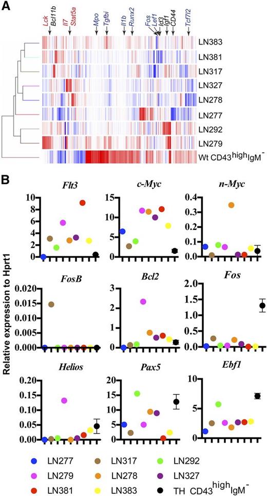
The expansion of a limited number of leukemic clones in each of the mice carrying tumors and late onset of disease are consistent with a molecular mechanism involving secondary genetic events, well in line with what could be anticipated to be a result of impaired DNA repair. Increased DNA damage could also be presumed to drive molecular heterogeneity, because damage-induced mutations would increase the number of possible pathways to a malignant stage. To investigate the molecular heterogeneity of the generated tumors, we performed gene expression microarray and quantitative RT-PCR analysis from sorted live LN cells from 8 primary Pax5+/−Ebf1+/− tumors, as well as sorted Lin−CD19+CD43highIgM− BM cells from healthy 10- to 12-week-old mice. All the tumors expressed the basic gene expression patterns of B-lineage progenitors including expression of Pax5, Ebf1, and their target genes. Even though gene expression analysis identified a set of downregulated genes, including the transforming growth factor β-responsive Tgfbi gene and genes encoding the transcription factors Runx2, Lef1, and Id3 (Figure 5A) in all of the tumors, the populations were heterogeneous with regard to changes in gene expression patterns. Quantitative RT-PCR analysis of genes indicated as differentially expressed from the microarray analysis verified a high degree of heterogeneity (Figure 5B). To investigate this further, we performed exome sequencing and used Mutech-based analysis25 with normal ex vivo-sorted Pax5+/−Ebf1+/− pro-B cells as a reference to identify unique mutations in the individually generated tumors. This indicated that most of the tumors contained in the range of 29 to 53 unique mutations, with an average allele depth of 0.16 to 0.29 (Table 3). Two of the tumors presented with a higher degree of unique changes and, although tumor LN327 carried a major part of the alterations on chromosome 11, the alterations in LN317 were more interspersed in the genome. In conclusion, the tumor populations arising after combined heterozygous loss of Ebf1 and Pax5 similarly represent early B-cell progenitors but display a substantial molecular heterogeneity consistent with the idea that tumor formation involves secondary events as could be anticipated by a defect in DNA repair.
Pax5+/−Ebf+/− tumor cells display molecular heterogeneity. (A) Heat map over gene expression patterns in primary sorted CD19+CD43highIgM− ex vivo-analyzed BM pro-B cells from 14-week-old mice and 8 pro-B cells tumors from Pax5+/−Ebf1+/− mice. The expression pattern is based on genes differentially expressed >10-fold in any of the tumor samples compared with normal pro-B cells. Red indicates high and blue indicates low expression of mRNA. The data are collected from 2 array experiments from each cell population. (B) Quantitative PCR data generated from primary sorted live lymph node cells from a set of independently generated tumors and primary sorted BM pro-B cells. The data were normalized to the expression of Hprt, and the diagrams present 1 of 2 independent quantitative RT-PCR experiments based on triplicate quantitative RT-PCR reactions.
Pax5+/−Ebf+/− tumor cells display molecular heterogeneity. (A) Heat map over gene expression patterns in primary sorted CD19+CD43highIgM− ex vivo-analyzed BM pro-B cells from 14-week-old mice and 8 pro-B cells tumors from Pax5+/−Ebf1+/− mice. The expression pattern is based on genes differentially expressed >10-fold in any of the tumor samples compared with normal pro-B cells. Red indicates high and blue indicates low expression of mRNA. The data are collected from 2 array experiments from each cell population. (B) Quantitative PCR data generated from primary sorted live lymph node cells from a set of independently generated tumors and primary sorted BM pro-B cells. The data were normalized to the expression of Hprt, and the diagrams present 1 of 2 independent quantitative RT-PCR experiments based on triplicate quantitative RT-PCR reactions.
Discussion
Several types of proto-oncogenes and oncogenic events have been defined as contributors to malignant transformation. In leukemogenesis, these events prominently include the loss or reduction of transcription factors that control differentiation.9 In this regard, heterozygous loss of transcription factors including Ebf1 and Pax5 has been suggested to cause a disruption in B-cell differentiation predisposing cells for transformation. Because developmental arrest is a hallmark of pro-B and pre-B ALL cells, this represents a logical explanation and a reasonable hypothesis; however, it is not obvious how a partial developmental arrest, as imposed by a reduction in transcription factor dosage, would directly contribute to malignant transformation. Developmental changes observed due to heterozygous loss of Pax5 in mouse models are modest6,20 and even though the loss of 1 allele of Ebf1 results in a more pronounced disturbance in the generation of B cells,6,8 this may be explained by a reduced expansion of the pre-B cell compartment due to the role of Ebf1 in cell growth and survival.6,8 Furthermore, a developmental block imposed by Rag deficiency did not result in the same degree of synergistic generation of leukemia when combined with ectopic expression of activated Stat5 as did either Pax5 or Ebf1 heterozygousity.15 Hence, it is reasonable to presume that the reduced transcription factor dose impacts the transformation process by multiple mechanisms.
Even though the most direct approach to identify molecular explanations to malignant transformation is a comparison of normal and leukemic cells, this provides limited information regarding the transformation process per se. Our analysis is based on how Ebf1 dose impacts gene expression in nontransformed cells, representing an early preleukemic stage, allowing us to investigate events upstream of the terminal transformation process. Our work reveals that one consequence of reduced Ebf1 levels in these nontransformed cells is impaired DNA repair. In contrast, gene expression analysis from leukemia cells generated from Ebf1+/− mice did not reveal a reduction in expression of DNA repair genes. This may reflect a post-transformation selection process because, although impaired DNA repair may be crucial for tumor formation, this could represent a growth disadvantage once the malignancy is established. Even though the reduced level of Ebf1 appears to modulate the expression of several genes involved in DNA repair (supplemental Table 1), the most obviously effected genes in normal Ebf1+/− cells ex vivo was Smc2, Rad51ap1, and Rad51. Rad51 expression is differentially regulated during the cell cycle, being expressed from late G1 to M phase,31 but even though we noted some changes in cell cycle dynamics, we could not detect any accumulation of cells in G0 that could not explain the observed changes in Rad51 expression. This in combination with the ChIP analysis suggests that the Rad51 gene is a direct target for Ebf1. Rad51 has crucial functions for homologous DNA repair and preservation of DNA integrity,27 and in myeloid leukemia, reduced Rad51 expression results in increased sensitivity to DNA-damaging drugs.32 Hence, even though we have not established any direct link between the reduction of Rad51 levels and an increase in γH2AX foci formation, we believe that the effect on expression of Rad51 and Rad51ap1 may contribute to the observed increase in DNA damage. Negative selection against a molecular mechanism active in a preleukemic state generates a challenge in establishing a direct link between the early event in the preleukemic cell and the observed characteristics of the fully developed leukemia cells. However, low clonal complexity and late onset of disease in Pax5+/−Ebf1+/− mice argues for a need of secondary genetic alterations to generate malignant cells, a process that should be facilitated by impaired DNA repair. Furthermore, molecular heterogeneity among the tumors reflected in both unique gene expression patterns and mutations is well in line with what could be expected from defects in DNA repair. Additionally, the mutation range of 30 to 50 unique alterations in the exomes (Table 3) is high compared with what has been reported from matched human B-ALL samples.33 Hence, there are several lines of support for the idea that reduced Ebf1 dose could impact malignant transformation via impairments in DNA repair. Although Ebf1 may contribute to malignant transformation by other mechanisms of action (ie, impaired differentiation and deregulated expression of Bcl2), increased Bcl2 levels were not sufficient for the development of leukemia when combined with a heterozygote mutation in Pax5 (supplemental Figure 4). However, this does not preclude that the high expression of Bcl2 is of importance in the transformation process, because Bcl2 may promote the survival of cells carrying DNA damage in a premalignant stage.
Our data demonstrate that combined heterozygous deletions of Ebf1 and Pax5 are preconditioned for the development of lymphoid leukemia with phenotypic features resembling B-ALL in humans. To investigate whether combined heterozgoous loss of PAX5 and EBF1 is a feature of human B-ALL, we analyzed a single nucleotide polymorphism data set collected from 242 human childhood B- and T-cell–ALLs.9 Deletion of PAX5 was detected in 57 of the 192 (30%) B-ALLs, and deletion of EBF1 was detected in 8 of these (4%) (supplemental Figure 5A-C). Of the 8 leukemias carrying EBF1 mutations, 3 were detected in combination with deletions of PAX5. Although the sample size is too small for reliable statistical analysis, these data reveal that combined heterozygote deletions of PAX5 and EBF1 are a feature of human B-ALL.
Taken together, our data suggest that Ebf1 contributes to malignant transformation via its crucial functions in the regulation of genes involved in DNA repair. A role for a lineage- and stage-specific transcription factor in such basic cellular functions appears counterintuitive. However, epigenetic dynamics in cell differentiation may result in that stage-specific transcription factors come to regulate genes involved in basic cellular functions and requirements for Ebf1 in VDJ recombination and other processes required for B-cell identity, and commitment may include the need for increased surveillance of genomic integrity.
The online version of this article contains a data supplement.
There is an Inside Blood Commentary on this article in this issue.
The publication costs of this article were defrayed in part by page charge payment. Therefore, and solely to indicate this fact, this article is hereby marked “advertisement” in accordance with 18 USC section 1734.
Acknowledgments
The authors thank our colleagues for help with transgenic mice and cell lines and Liselotte Lenner, Linda Bergström, and Anne Halden Waldermarsson for advice and assistance.
This work was supported by grants from the Swedish Cancer Society, the Swedish Research Council, including a Center grant to Hematolinne, Barncancerfonden, and by Linköping University. J.H. is supported generously by National Institutes of Health, National Institute of Allergy and Infectious Diseases grant AI081878.
Authorship
Contribution: J.Å., T.S., J.U., R.S., M.A.J.P., and M.S. conducted, designed, and interpreted the analysis of the mouse model; T.F. and H.L. analyzed the human SNP data; R.M. and A.D.P. analyzed High-C data; J.H. contributed essential reagents and experimental design; and all authors participated in the writing of the manuscript.
Conflict-of-interest disclosure: The authors declare no competing financial interests.
Correspondence: Mikael Sigvardsson, Faculty of Health Sciences, Linköping University Lab1, Level 13, SE-581 85 Linköping, Sweden; e-mail: mikael.sigvardsson@liu.se.
References
Author notes
M.A.J.P., J.U., and J.Å. contributed equally to this work.

