Abstract

Introduction:
Hepatitis E Virus (HEV) is one of the leading causes of acute infectious hepatitis worldwide; while usually a self-limiting, sub-clinical illness, genotype 3 HEV infection may become chronic in immunosuppressed patients such as solid organ transplant (SOT) or haematopoietic stem cell transplant (HSCT) recipients and rapid progression to cirrhosis may occur. Recent seroprevalence from screening of English blood donors shows antibody prevalence of ~13%. RNA prevalence in donors has increased in the UK from 1:2850 to 1:1460 over the past 3 years. This study examines the point-prevalence of HEV in SOT and HSCT recipients transplanted between January 2013 - December 2015 at a major UK transplant centre. Data on HEV clearance rates and risk factors for infection were collected in the patient cohort.
Methods:
Stored extracts of blood from patients undergoing liver transplant (OLT), renal transplant (RT) or HSCT were tested by real-time reverse-transcriptase polymerase chain reaction for HEV RNA. Samples were tested at baseline, 30, 60 and 90 days post-transplant +/- 7 days. Time points were chosen to give good coverage of the early post-transplant period whilst avoiding a patient becoming viraemic and clearing the infection between sample time points. All positive samples were sent to the Public Health England reference laboratory for verification, viral RNA genotyping and quantification. Patients with confirmed positive samples underwent further testing and evaluation of clinical parameters to determine chronology of infection, blood product exposure, organ function and histopathology results.
Results:
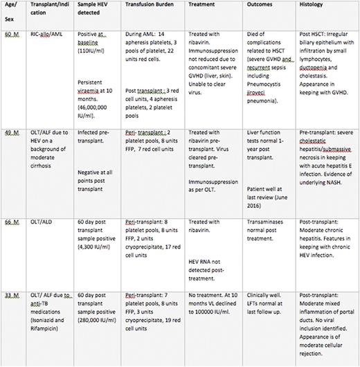
259 HSCT, 262 OLT and 349 RT patients met the inclusion criteria. Of the HSCTs there were 111 allogeneic stem cell transplants, 145 autologous transplants and 3 CD34 "top ups". The OLTs comprised 259 deceased donor, 2 live donor and 2 domino liver transplant patients. The RTs comprised 241 deceased donor, 38 live unrelated, 63 live related transplants and 1 live transplant where the relationship of the donor was unrecorded. In total 4 patients (1 HSCT and 3 OLT) were found to have HEV viraemiain our study period. This represents 0.39% of the HSCT and 1.15% of the OLT patients. All were positive with HEV genotype 3.
Virologicaland clinical data regarding these patients are presented in tables below.
Conclusion:
Chronic HEV, genotype 3 in transplant patients is associated with significant morbidity. With the increasing incidence of HEV, immunosuppressed patients with high transfusion burden have an infection risk equivalent to 7-9 years' dietary exposure.
Our results show that prevalence of HEV viraemia in OLT, RT and HSCT patients is higher than expectedbased on comparison from UK blood donors. Secondly, infection is not cleared as would be expected in the general population with 3 of the 4 patients requiring treatment to clear the virus.
The source of infection for these patients is unclear; on a population wide basis HEV is usually a dietary acquired infection, with pork products carrying the greatest risk. In the transplant patient population, the risk of iatrogenic infection via infected blood products or an infected transplanted organ is an important consideration and UK guidance has recently changed so that transplant recipients receive dietary advice and HEV-negative blood to avoid infection (SaBTO2016).
In this study 2 patients were found to be positive for HEV RNA in the baseline sample. One, the recipient of HSCT, did not acquire it through transfusion as testing of the transfused products that the patient had received were negative for the virus. It is not clear how the two OLT patients who tested positive acquired the virus. The absence of any positive results in the renal transplant recipients may reflect the lower transfusion burden in this patient group. Given the multiple routes of possible infection with HEV, a strategy if screening recipients for the virus in addition to screening blood and organ donors would be an effective way of detecting viral transmission via all routes.
Sekhar:Novartis: Research Funding.
Author notes
Asterisk with author names denotes non-ASH members.

This icon denotes a clinically relevant abstract


