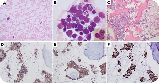
A 58-year-old man with a history of JAK2-mutated essential thrombocythemia (ET) progressing to myelofibrosis 10 years earlier presented with fatigue, melena, and cytopenia refractory to transfusion. Complete blood count revealed severe pancytopenia (hemoglobin 7.5 g/dL, white blood cells 2.3 × 10³/μL, platelets 3 × 10³/μL). Peripheral blood smears demonstrated granulocytic dysplasia and rare circulating erythroblasts (panel A, ×1000 original magnification). Bone marrow touch preparation showed 93% of cells were of erythroid lineage and 60% were proerythroblasts with basophilic cytoplasm and cytoplasmic blebs; periodic acid–Schiff stain highlighted chunky globules in erythroblasts (panel B, inset, ×1000 original magnification). Biopsy showed hypercellular (90%) bone marrow with increased sheets of blasts in a sinusoidal pattern (panel C, ×200 original magnification), positive for glycophorin A, CD71, E-cadherin, and p53 (panels D-F, ×400 original magnification), suggestive of a TP53 mutation, and negative for CD34, CD117, lysozyme, and myeloperoxidase. Fluorescence in situ hybridization showed 83% of cells with one copy loss of TP53, consistent with isochromosome 17(10q) by karyotyping (46,XY,del(13)(q12q14),i(17)(q10)[14]/58∼70,XXY,+Y,+1,+2,+3,−4,+6,+7,+8,−9,−10,−11,−12,−13,+14,+15,−16,−17,+19,+20,+21,−22,+mar[cp6]). A next-generation sequencing panel revealed mutations in ASXL1, JAK2 (variant allele frequency 97%), FLT3, ETNK1, and TP53 (variant allele frequency 16%), with the TP53 mutation newly identified. The diagnosis of acute erythroid leukemia was rendered.
Acute transformation of myeloproliferative neoplasm typically presents as AML, or rarely as B-cell acute lymphocytic leukemia. This case illustrates the extremely rare presentation of acute erythroid leukemia evolving from myeloproliferative neoplasm, supported by the persistent JAK2 mutation. The emergence of and/or expanding TP53 alterations associated with the complex karyotype likely contributes to the pathogenesis.
For additional images, visit the ASH Image Bank, a reference and teaching tool that is continually updated with new atlas and case study images. For more information, visit https://imagebank.hematology.org.


