Abstract
During the innate immune response to infection, monocyte-derived cytokines (monokines), stimulate natural killer (NK) cells to produce immunoregulatory cytokines that are important to the host's early defense. Human NK cell subsets can be distinguished by CD56 surface density expression (ie, CD56bright and CD56dim). In this report, it is shown that CD56bright NK cells produce significantly greater levels of interferon-γ, tumor necrosis factor-β, granulocyte macrophage–colony-stimulating factor, IL-10, and IL-13 protein in response to monokine stimulation than do CD56dim NK cells, which produce negligible amounts of these cytokines. Further, qualitative differences in CD56bright NK-derived cytokines are shown to be dependent on the specific monokines present. For example, the monokine IL-15 appears to be required for type 2 cytokine production by CD56bright NK cells. It is proposed that human CD56bright NK cells have a unique functional role in the innate immune response as the primary source of NK cell–derived immunoregulatory cytokines, regulated in part by differential monokine production.
Introduction
Natural killer (NK) cells are innate immune effectors that produce immunoregulatory cytokines, such as interferon (IFN)-γ and granulocyte macrophage–colony-stimulating factor GM-CSF, critical to early host defense against a variety of viral, bacterial, and parasitic pathogens.1-4 Human NK cells comprise approximately 10% of all peripheral blood lymphocytes and are characterized phenotypically by the presence of CD56 and the lack of CD3.1 There are 2 distinct subsets of human NK cells identified by cell surface density of CD56. The majority (approximately 90%) of human NK cells are CD56dim and express high levels of FcγRIII (CD16), whereas a minority (approximately 10%) are CD56bright and CD16dim/neg.5
CD56bright NK cells constitutively express the high- and intermediate-affinity IL-2 receptors and expand in vitro and in vivo in response to low (picomolar) doses of IL-2.6-8These NK cells also express the c-kit receptor tyrosine kinase whose ligand enhances IL-2–induced proliferation.9,10 In contrast, resting CD56dim NK cells express only the intermediate affinity IL-2 receptor, are c-kitneg, and proliferate weakly in response to high doses of IL-2 (1 to 10 nM) in vitro, even after induction of the high-affinity IL-2 receptor.6,7 Resting CD56dim NK cells are more cytotoxic against NK-sensitive targets than CD56bright NK cells.11 However, after activation with IL-2 or IL-12, CD56bright cells exhibit similar or enhanced cytotoxicity against NK targets compared to CD56dimcells.11-13
NK cell subsets have differential natural killer receptor (NKR) repertoires. All resting CD56bright NK cells have high expression of CD94/NKG2 C-type lectin receptors.14 A small percentage (less than 10%) expresses killer cell immunoglobulin-like receptors (KIR),15 while most (more than 85%) resting CD56dim NK cells are KIR+ and have low expression of CD94/NKG2.
CD56bright NK cells also express the adhesion molecule L-selectin (CD62L), which mediates initial interactions with vascular endothelium.16 CD56dim NK cells lack this receptor but have recently been found to express PEN5, an NK cell-restricted sulfated lactosamine epitope that partially mediates the binding of L-selectin,15 thus suggesting the potential for differential trafficking of human NK cell subsets in vivo. Therefore, CD56bright and CD56dim NK cells differ in their proliferative response to IL-2, intrinsic cytotoxic capacity, NKR repertoire, and adhesion molecule expression.
NK cells constitutively express receptors for monocyte-derived cytokines (monokines) and produce critical cytokines, such as IFN-γ, in response to monokine stimulation.17-20 In the current study we examine CD56bright and CD56dim NK cell production of multiple cytokines—including IFN-γ, tumor necrosis factor (TNF)-β, IL-10, IL-13, TNF-α, and GM-CSF—in response to stimulation with monokines. We show that CD56bright NK cells are the primary population responsible for NK cell cytokine production in response to monokines. These data support a model whereby CD56bright and CD56dim NK cells represent functionally distinct subsets of mature human NK cells.
Materials and methods
Cell culture reagents and antibodies
Human NK cells and macrophages were cultured in RPMI-1640 with 10% human serum (C-6 Diagnostics, Mequon, WI) and antibiotics. Recombinant human IL-12 was provided by Genetics Institute (Cambridge, MA); rIL-15 was a gift from Immunex (Seattle, WA); rIL-18 was a gift from BASF Bioresearch (Worcester, MA); and rIL-1β was purchased from Peprotech (Rocky Hill, NJ). PMA and ionomycin (calcium salt) were obtained from Calbiochem (La Jolla, CA). Anti-CD56-phycoerythrin (PE) (NKH1), CD16-fluorescein isothiocyanate (FITC) monoclonal antibodies, and isotype controls were purchased from Coulter (Miami, FL).
Purification of human NK cell subsets and macrophages
Human NK cells were isolated from fresh normal donor leukopacs (American Red Cross, Columbus, OH) as previously described9,18 or with RosetteSep NK cell cocktail (StemCell Technologies, Vancouver, BC) according to the manufacturer's directions. NK cells were stained with anti–CD56-PE or control PE, and subsets were purified based on CD56 cell-surface density by FACS (Elite Flow Cytometer; Coulter) as previously described.6 Cells were routinely greater than 98% pure by post-FACS cytometric analysis of CD56 and CD16, as shown in Figure 1. Human macrophages were isolated by adherence and gentle scraping (more than 85% CD14+ by flow cytometry).
Purification of human NK cell subsets.
CD56bright and CD56dim NK cell subsets were FACS-purified from fresh peripheral blood lymphocytes. Flow cytometric analysis of CD56-PE (A) and CD56-PE/CD16-FITC expression before (B) and after (C) FACS purification of a representative donor is shown, and the percentages of cells are indicated in the upper right quadrant. (A) Gating on total peripheral blood lymphocytes; 12.8% of unpurified lymphocytes were CD56+, and (B) 11.8% were positive for both CD56 and CD16. (C) Purified CD56bright and CD56dim NK cell subsets were more than 98% pure, as shown by flow cytometry analysis of CD56 and CD16. Of CD56brightcells, 51.5% expressed CD16 at low levels (mean fluorescence intensity [MFI] 49.2), whereas 98% of CD56dim cells had high expression of CD16 (MFI 213.9). NK cell subsets were gated on total viable cells (more than 98% of all collected events).
NK cell monokine and PMA–ionomycin stimulation
Purified NK cells (5 × 10⁴ cells/well) were stimulated with combinations of IL-12 (10 ng/mL), IL-15 (100 ng/mL), IL-18 (100 ng/mL), IL-1β (10 ng/mL), PMA (20 ng/mL), or ionomycin (5 μM), and cell-free culture supernatants were harvested at 72 hours. Cell culture supernatants were assayed for IFN-γ, IL-10, GM-CSF, TNF-α, IL-13, IL-5 (Endogen, Woburn, MA), and TNF-β (R&D Systems, Minneapolis, MN) protein in duplicate enzyme-linked immunosorbent assay (ELISA) wells. Results represent the mean ± SEM of 3 or more donors.
Quantitation of cytokine transcripts by real-time RT-PCR
FACS-purified CD56bright and CD56dim NK cells (1 × 10⁵) were either immediately lysed for RNA (Qiagen RNeasy lysis buffer; Qiagen, Valencia, CA) or cultured at 1 × 10⁵ cells/well with recombinant monokines. Cells were harvested at 24 hours and lysed with 300 μL RNA lysis buffer. Total cellular RNA was isolated (Qiagen RNeasy Mini-kits; Qiagen) and cDNA was generated with random hexamer primers and MMLV-RT according to the manufacturer's recommendations (Gibco Life Technologies, Rockville, MD). cDNA was then used as a template for real-time polymerase chain reaction (PCR).
Real-time quantitative reverse transcription (RT)-PCR is a novel method to accurately measure amplified target copy number through the use of a dual-labeled fluorogenic probe.21 Real-time PCR reactions for human IFN-γ, GM-CSF, and IL-10 transcripts were performed as previously described and as multiplex reactions with primer and probe sets specific for the cytokine transcript of interest and an internal control (rRNA, 18s; PE Applied Biosystems, Foster City, CA).18 cDNA from PHA-activated human lymphocytes served as positive controls for cytokine transcripts, and murine cDNA (P815 cell line) was used as a negative control. Reactions were performed using an ABI prism 7700 sequence detector (Taqman; PE Applied Biosystems), and data were analyzed with the Sequence Detector version 1.6 software to establish the PCR cycle at which the fluorescence exceeded a set threshold, CT, for each sample. Data were analyzed according to the comparative CT method, as previously described, using internal control (18s) transcript levels to normalize differences in sample loading and preparation. Results are semiquantitative and represent the n-fold difference of transcript levels in a particular sample compared to calibrator cDNA (for these experiments, cDNA samples of resting NK cells from each donor). Results are expressed as the mean ± SEM of triplicate reaction wells.
NK cell and macrophage co-cultures
Purified CD56bright and CD56dim NK cells (1.0 × 10⁵) were co-cultured with autologous macrophages (1.0 × 10⁵) as previously described22 and stimulated with 10 μg/mL lipopolysaccharide (LPS; serotype 0127 B8; Sigma, St Louis, MO) for 72 hours.
Statistical analysis
Statistical analysis was performed using the Student paired t test; P < .05 was considered significant.
Results
CD56bright NK cells produce abundant type 1 and type 2 cytokines compared to CD56dim NK cells
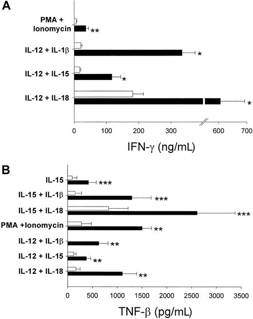
We stimulated sorted resting CD56bright and CD56dim NK cells (Figure 1) with the recombinant monokines IL-12, IL-15, IL-18, and IL-1β alone and in combination with IL-12 or IL-15. To examine the stimulation of NK cells independent of monokine receptor expression, NK cell subsets were also activated with phorbol esters (PMA) plus ionomycin. The CD56bright subset produced significantly more of the type 1 cytokines IFN-γ and TNF-β than CD56dim NK cells cultured under identical conditions after stimulation with monokines or PMA plus ionomycin (Figure2). CD56bright NK cells co-stimulated with IL-18 plus IL-12 produced the most IFN-γ protein (Figure 2A), whereas stimulation with IL-18 plus IL-15 or IL-1β plus IL-15 induced the highest levels of TNF-β protein production.
NK cell production of type 1 cytokines IFN-γ and TNF-β.
(A) IFN-γ protein was measured by ELISA in cell culture supernatants of freshly isolated CD56bright and CD56dim NK cells after culture with monokines or PMA plus ionomycin for 72 hours. CD56bright NK cells produced significantly more IFN-γ in response to all positive stimuli; data represent the mean ± SEM of 5 to 10 donors (*P < .004; **P < .03). IL-12 or IL-15 alone induced minimal IFN-γ (less than 2 ng/mL) whereas no IFN-γ was detected in cultures with IL-18, IL-1β, PMA, or ionomycin alone (data not shown; sensitivity < 25 pg/mL). (B) CD56bright NK cells also produced significantly more TNF-β protein after activation with monokines or PMA plus ionomycin (n = 3-5 donors per condition; **P < .03; ***P < .05). There was no detectable TNF-β protein with IL-12, IL-1β, IL-18, PMA, or ionomycin alone (data not shown; sensitivity less than 20 pg/mL). ■ indicates CD56dim; ▪, CD56bright.
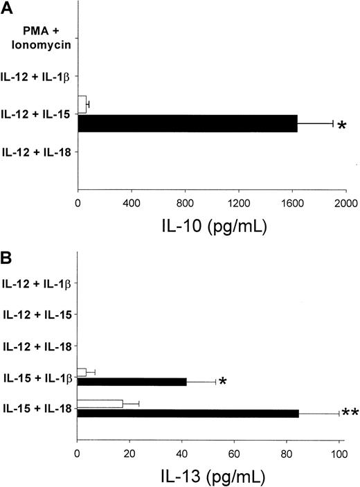
NK cell production of IL-10, a type 2 cytokine, was only detected after co-stimulation with IL-12 plus IL-15 (Figure3A), with CD56bright NK cells producing in excess of 25-fold more IL-10 protein than CD56dim cells. Interestingly, other monokine combinations, including IL-12 plus IL-18, failed to elicit any production of IL-10 from either subset. Modest amounts of IL-13, another cytokine produced by committed Th2 cells, were detected in cultures of CD56bright NK cells stimulated with IL-15 plus IL-18 or IL-1β, and this production was always significantly greater than in CD56dim NK cells (Figure 3B). We did not detect any NK cell production of IL-5 protein in response to monokine or PMA plus ionomycin stimulation (data not shown; sensitivity less than 2 pg/mL).
CD56bright NK cells produce the type 2 cytokines IL-10 and IL-13.
(A) IL-12 plus IL-15 was the only monokine combination that induced IL-10 production in human NK cell subsets. Significantly more protein was detected in CD56bright NK cells (1635.5 ± 266.3 vs 61.2 ± 21.3 pg/mL CD56bright vs CD56dim; *P < .0012; n = 8). (B) The CD56brightsubset produced significantly more IL-13 protein than CD56dim NK cells after stimulation with IL-15 plus IL-1β or IL-18 (*P < .0018; **P < .014; n = 7). No IL-13 was detected in cultures with IL-12 plus IL-1β, IL-15, or IL-18 (n = 3) or with PMA plus ionomycin (n = 2). ■ indicates CD56dim; ▪, CD56bright.
Thus, CD56bright NK cells produce high levels of 2 principal type 1 cytokines, IFN-γ and TNF-β, after stimulation with monokines or phorbol esters plus ionomycin. However, only specific combinations of monokines that included IL-15 as a co-stimulus induced IL-10 or IL-13 protein in cultures of CD56bright NK cells, suggesting that production of these type 2 cytokines by human NK cells requires specific monokine (eg, IL-15)-induced signaling.
CD56bright NK cell production of other pro-inflammatory cytokines, GM-CSF, and TNF-α
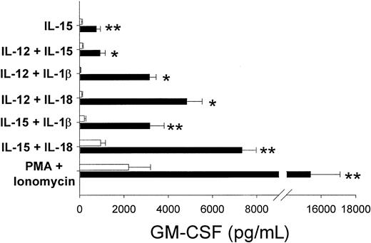
CD56bright NK cells produced significantly more of the macrophage-activating GM-CSF than CD56dim NK cells in response to all positive stimuli (Figure4). IL-15 was the only monokine that induced GM-CSF protein without a co-stimulus (sensitivity less than 15 pg/mL). The combination of IL-15 plus IL-18 was the most potent monokine stimulus for GM-CSF production, similar to TNF-β. Unlike other NK-derived cytokines, stimulation with PMA plus ionomycin induced the highest levels of GM-CSF protein from CD56bright NK cells. CD56bright NK cell production of the pro-inflammatory cytokine TNF-α, after culture with monokines or PMA plus ionomycin, was modest (less than 300 pg/mL) and somewhat variable, but it was consistently greater than in the CD56dim NK cell subset (data not shown).
GM-CSF production by human NK cell subsets.
Stimulation with monokines or PMA plus ionomycin induced significantly more GM-CSF protein in cultures of CD56bright NK cells than in CD56dim cells (*P < .007; **P < .04; n = 3-7 donors per condition). IL-15 alone induced moderate levels of GM-CSF (751.7 ± 190.4 pg/mL, CD56bright NK cells), whereas there was no detectable GM-CSF in cultures with IL-12, IL-1β, IL-18, PMA, or ionomycin alone (sensitivity less than 15 pg/mL). ■ indicates CD56dim; ▪, CD56bright.
Cytokine transcript levels in NK cell subsets
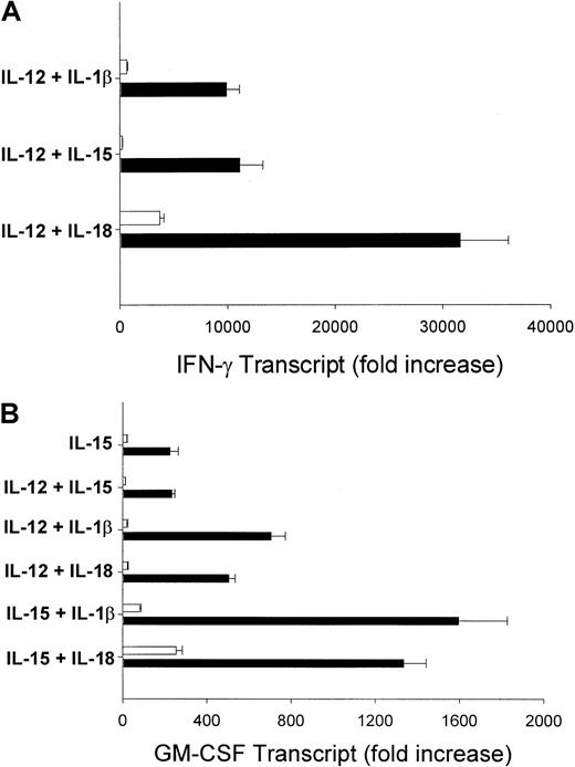
To determine whether NK cell subsets have differential baseline expression of cytokines not detectable by ELISA that might account for observed differences in protein production, we measured transcript levels of 3 primary NK-derived cytokines in resting and monokine-activated NK cell subsets by real-time quantitative RT-PCR. Resting subsets lacked any detectable expression of IL-10 (data not shown), but both CD56bright and CD56dim NK cell subsets expressed equal amounts of IFN-γ and GM-CSF transcript (data not shown; n = 3). Therefore, there is no detectable difference in the baseline production of these cytokine transcripts by resting NK cell subsets. After 24 hours of monokine stimulation, the CD56bright NK subset produced higher levels of IFN-γ and GM-CSF transcript than the CD56dim NK subset, consistent with their production of the respective proteins (Figure5A,B).
CD56bright cells produce abundant IFN-γ and GM-CSF transcript.
NK cell subsets were activated for 24 hours with the indicated monokines, and RNA was harvested. Resultant cDNA was analyzed for IFN-γ and GM-CSF transcripts by real-time PCR. Results represent fold-increase in cytokine transcript compared to resting NK cell subsets ± SEM and are representative of 3 experiments. (A) CD56bright NK produced high levels of IFN-γ transcript in response to IL-12 plus IL-1β, IL-15, and IL-18 compared to the CD56dim subset. (B) CD56bright NK cells also produced more GM-CSF transcript in response to monokine stimulation, including activation with IL-15 alone. All results were calibrated to an internal control (18s ribosomal RNA) to normalize for differences in total cDNA. ■ indicates CD56dim; ▪, CD56bright.
CD56bright NK cells produce significantly more IFN-γ than CD56dim NK cells when cultured with LPS-activated macrophages
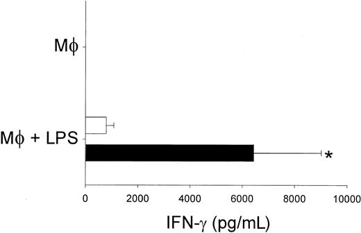
Gram-negative bacteria-derived LPS stimulate macrophages to secrete a number of monokines, including IL-1, IL-12, and IL-15.22-24 We have previously shown that the production of monokines by LPS-activated macrophages induces IFN-γ production by human NK cells in vitro.22 To determine the subset of NK cells responsible for IFN-γ production after monokine activation by LPS, purified CD56bright and CD56dim NK cells were co-cultured with LPS-stimulated autologous macrophages (Figure 6). Similar to results obtained with recombinant monokines, CD56bright NK cells produced 8-fold more IFN-γ protein than CD56dim cells (n = 7). Thus, CD56brightNK cells are the primary producers of IFN-γ in response to both recombinant and endogenous monokines.
CD56bright NK cell co-cultured with LPS-activated macrophages produce abundant IFN-γ.
Resting CD56bright and CD56dim NK cells (1 × 10⁵/well) were co-cultured with autologous macrophages (1 × 10⁵/well) and control (phosphate-buffered saline) or LPS for 72 hours, and cell culture supernatants were assayed for IFN-γ protein. CD56brightNK cells co-cultured with LPS-activated macrophages (Mφ + LPS) produced significantly more IFN-γ protein than CD56dim NK cells (6439.41 ± 2579.16 vs 791.5 ± 289.2 pg/mL;P < .05; n = 7). No IFN-γ protein was detected in co-cultures without LPS (Mφ) or in cultures of LPS-activated NK cells or macrophages (data not shown). ■ indicates CD56dim; ▪, CD56bright.
Discussion
Collectively, our results reveal that CD56bright human NK cells are the primary source of NK-derived immunoregulatory cytokines, including IFN-γ, TNF-β, IL-10, IL-13, and GM-CSF, whereas the CD56dim NK cell subset consistently produces significantly less of these cytokines in vitro. The results confirm our earlier observations of CD56bright NK IFN-γ production18 but also provide evidence for a more generalized property that can be attributed to this distinct human NK cell subset. Although the possibility exists that differences in cytokine production may be attributed to differential monokine receptor expression, density, or both, the activation of NK cell subsets with phorbol esters plus ionomycin, which is not dependent on monokine receptor activation, resulted in significantly greater production of IFN-γ, TNF-β, and GM-CSF by CD56bright NK cells. Furthermore, LPS, a bacterial component recognized by host innate immune effector cells, indirectly induced CD56bright NK cells to produce much greater IFN-γ than CD56dim NK cells when co-cultured with macrophages. Therefore, the CD56bright NK subset has a significantly higher capacity for cytokine production than the CD56dim subset.
Peritt et al25 recently reported the differentiation of human NK cells into NK1 and NK2 subsets by generating NK cell clones after 8-day in vitro culture under type 1– or type 2–inducing conditions. After stimulation with PMA plus ionomycin, NK1 cells produced IFN-γ and TNF-β but also produced IL-10, a type 2 cytokine, and NK2 cells produced IL-5 and IL-13. However, because of the clear role of NK cells as efficient producers of cytokines early in the innate immune response to infection long before clonal responses are mounted, it is unlikely that prolonged peripheral NK cell differentiation is necessary in vivo. In this report we show that freshly isolated CD56bright NK cells produce abundant type 1 and type 2 cytokines immediately after monokine stimulation, consistent with their known in vivo role. Hence, though we do not exclude the possibility for subsets of human NK cells that produce type 1 or type 2 cytokines, our data suggest that these subsets would be found within the resting CD56bright NK cell population.
Additional data presented here indicate that the qualitative and quantitative production of monokines after host infection are likely important in distinguishing the induction of type 1 and type 2 cytokines by CD56bright NK cells. IL-15 co-stimulation was requisite for CD56bright NK cell production of type 2 cytokines (eg, IL-10 and IL-13), whereas IL-12 co-stimulation was required for optimal production of the type 1 cytokine IFN-γ. Although each of these monokines had the capacity to stimulate both type 1 and type 2 responses (eg, IL-12 co-stimulation of IL-10 and IL-15 co-stimulation of optimal TNF-β production), the relative quantities of each and the presence of other monokines (eg, IL-1 or IL-18) could influence the predominant CD56bright NK cell cytokine response. Thus, the monokine milieu induced by infection may dictate CD56bright NK cell production of type 1 or type 2 cytokines, which could then, in part, influence the consequent development of a T-helper response.26 27
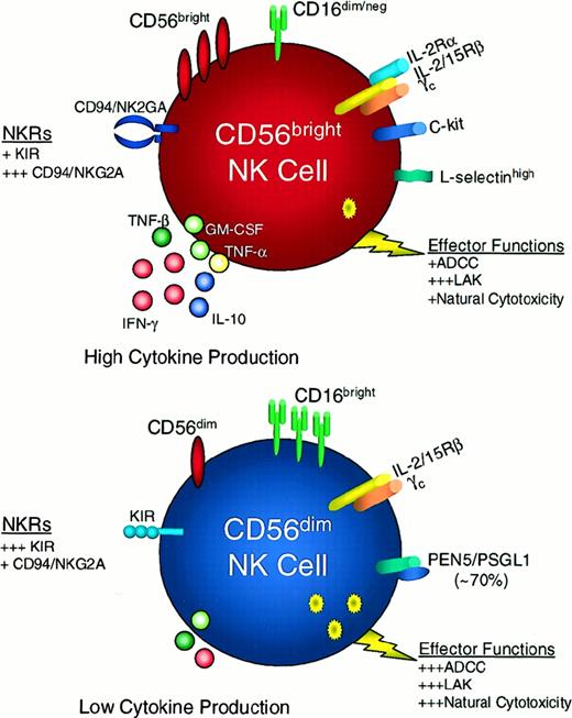
It has been hypothesized that CD56bright and CD56dim NK cells represent different stages of NK cell maturation, with CD56dim NK cells being the more differentiated cell type.11 This paradigm is based on observations that resting CD56dim NK cells are more cytotoxic than CD56bright NK cells, express high surface density expression of both KIR15 and CD16 (which mediate natural NK cytotoxicity28 and antibody-dependent cellular cytotoxicity,29 respectively), and do not readily proliferate in response to IL-2. In the current study we provide new evidence to suggest that CD56bright NK cells are the major cytokine-producing subset of human NK cells, an attribute that is associated with essential host function. Based on these data, we propose that CD56bright and CD56dim NK cells represent functionally and phenotypically distinct subsets of NK cells with unique immunoregulatory roles in vivo (Figure7).
CD56bright and CD56dim NK cell subsets exhibit differential receptor profiles and innate immune functions.
Many of the receptors and functions of CD56bright (red cell) and CD56dim (blue cell) NK cells are schematized in this figure. CD56brightCD16dim/neg NK cells produce abundant immunoregulatory cytokines (some of which are depicted here) and exhibit potent LAK activity but are less effective mediators of ADCC and natural cytotoxicity. By contrast, CD56dimCD16bright NK cells produce low levels of NK-derived cytokines and are potent mediators of ADCC, LAK activity, and natural cytotoxicity.11,12 CD56bright NK cells have a less granular morphology when compared with CD56dim NK cells.5 CD56dim NK cell exhibit much higher levels of KIR, whereas resting CD56bright NK cells have high expression of CD94/NKG2A.14,15 L-selectin is highly expressed on CD56bright NK cells, whereas CD56dim NK cells express PEN5/PSGL1 which appears to mediate interactions with L-selectin.15 16 ADCC, antibody-dependent cellular cytotoxicity; KIR, killer-immunoglobulin-like receptor; LAK, lymphokine-activated killer; NKRs, natural killer receptors.
Numerous studies from our laboratory and those of others have definitively identified IL-15 as the critical factor for the development of human and murine NK cells.30-32 Culture of CD34+Linneg human hematopoietic cells with IL-15 results in the differentiation of mature, functional CD56+ NK cells. Culturing progenitor cells with flt3 ligand (FL) or c-kit ligand (KL) can increase the NK cell precursor frequency through up-regulation of the IL-15R complex.30,33 However, in vitro-generated NK cells are consistently CD56brightCD16dim/negKIRlow/neg, making them more phenotypically similar to the minor (approximately 10%) population of CD56bright peripheral blood NK cells.33 Further, these cell also produce immunoregulatory cytokines. Until recently, evidence for the differentiation of distinct CD56bright and CD56dim NK cell populations has been lacking because there were no reports of in vitro–generated CD56dim NK cells, lending support to the theory that CD56bright NK cells may be the more immature cell type. The discovery of a novel cytokine, IL-21, by Foster et al34may shed some light on the developmental differences between CD56bright and CD56dim human NK cell subsets. IL-21 is a unique cytokine most closely related to IL-2 and IL-15, with effects on B- and T-cell proliferation and NK cell differentiation, proliferation, and cytotoxicity. The authors cultured CD34+Linneg hematopoietic progenitors with FL ± IL-21 and FL + IL-15 ± IL-21. IL-21, in combination with IL-15 and FL, induced the differentiation of CD56+CD16+ NK cells, which, by flow cytometric analysis, appear to be CD56dimCD16+. FL plus IL-21 alone did not induce NK cell differentiation, whereas stimulation with FL plus IL-15 resulted in the expected population of CD56brightCD16neg NK cells.34 The discovery of this cytokine, which can, in combination with IL-15, induce differentiation of CD56dimCD16+ NK cells, supports a hypothesis whereby human CD56bright and CD56dim NK cells are terminally differentiated cell types that develop within the bone marrow under the influence of differential growth factors, such as IL-15 and IL-21. Further studies are required to investigate definitively the developmental relation between CD56bright and CD56dim NK cells. It will be interesting to determine the role of IL-21/IL-21R in the development of human NK cell subsets and NK receptors (eg, CD16 and KIR).
As we continue to develop immunotherapeutic strategies that target human NK cells, such as the selective expansion of CD56bright NK cells with low-dose IL-2 treatment of malignancies8,35 and human immunodeficiency virus,36 it will be important to understand the functional differences between these human NK cell subsets. Knowledge of the distinct functional attributes of CD56bright (eg, immunoregulatory cytokine production) and CD56dim (eg, cytotoxicity and antibody-dependent cellular cytotoxicity) human NK cell subsets and the factors involved in their development and expansion may enable us to design strategies that preferentially activate that subset with the greatest therapeutic potential for a particular disease.
We thank A. Oberyszyn for cell sorting and J. Bush and A. Ponnappan for technical assistance.
Supported by National Institutes of Health grants CA-68458, CA-65670, and P30CA-16058. M.A. Cooper is a Howard Hughes Medical Institute Medical Student Research Training Fellow. T.A.F. is the recipient of the Medical Scientist Training Program and the Bennett Fellowships from The Ohio State University College of Medicine and Public Health.
M.A. Cooper and T.A.F. contributed equally to this work.
The publication costs of this article were defrayed in part by page charge payment. Therefore, and solely to indicate this fact, this article is hereby marked “advertisement” in accordance with 18 U.S.C. section 1734.
References
Author notes
Michael A. Caligiuri, Department of Internal Medicine, Division of Hematology/Oncology, The Ohio State University, 458A Starling-Loving Hall, 320 West 10th Ave, Columbus, OH 43210; e-mail: caligiuri-1@medctr.osu.edu.

![Fig. 1. Purification of human NK cell subsets. / CD56bright and CD56dim NK cell subsets were FACS-purified from fresh peripheral blood lymphocytes. Flow cytometric analysis of CD56-PE (A) and CD56-PE/CD16-FITC expression before (B) and after (C) FACS purification of a representative donor is shown, and the percentages of cells are indicated in the upper right quadrant. (A) Gating on total peripheral blood lymphocytes; 12.8% of unpurified lymphocytes were CD56+, and (B) 11.8% were positive for both CD56 and CD16. (C) Purified CD56bright and CD56dim NK cell subsets were more than 98% pure, as shown by flow cytometry analysis of CD56 and CD16. Of CD56brightcells, 51.5% expressed CD16 at low levels (mean fluorescence intensity [MFI] 49.2), whereas 98% of CD56dim cells had high expression of CD16 (MFI 213.9). NK cell subsets were gated on total viable cells (more than 98% of all collected events).](https://ash.silverchair-cdn.com/ash/content_public/journal/blood/97/10/10.1182_blood.v97.10.3146/6/m_h81011074001.jpeg?Expires=1765934418&Signature=Wam1Op4y0Gx4jJ5gf-K1CxXWkSX07gNrFCO-NKSP7f2U1912Rb36829UptOuHbidQfi2v6PzsXWA6E9l4rUBexCFcuvrhN84BC9lEolawcEU2aiQ4WMrg7yrgEpnHgwwRrBCL0zG8OAJAF471D3~sgmnpSHy6l8zoPIWx9ve7hJPTbEn5uPSlkxNZgj3zD-FWks6DJeBOawYLbRK6gqoGYPw2UNv5IQmfikNli8CrlwufInr37lL~X0vQTjv4vtcORh4mA~KSRZXqYk7Ionbi7~7B~BDC4fekL3tqbAfNLKrFkbHHjnyMRrnLQp-AjhZPdYvtvm-MPgDSpfjopOS4Q__&Key-Pair-Id=APKAIE5G5CRDK6RD3PGA)
