Activating mutations of c-kit at codon 816 (Asp816) have been implicated in a variety of malignancies, including acute myeloid leukemia (AML). The mutant c-Kit receptor confers cytokine-independent survival of leukemia cells and induces tumorigenicity. Changes in the signal transduction pathways responsible for Asp816 mutant c-Kit–mediated biologic effects are largely undefined. The results of this study show that Asp816 mutant c-Kit induces constitutive activation of signal transducer and activator of transcription 3 (STAT3) and STAT1, and up-regulates STAT3 downstream targets, Bcl-xL and c-myc. The phosphatidylinositol-3-kinase (PI-3K)/Akt pathway, but not the Ras-mediated mitogen-activated protein (MAP) kinase pathway, is also constitutively activated by Asp816 mutant c-Kit. Suppression of STAT3 activation by a dominant negative molecule in MO7e leukemia cells transduced with mutant c-kit inhibits stem cell factor (SCF)-independent survival and proliferation, accompanied by the down-regulation of Bcl-xL and c-myc. However, activated STAT3 does not appear to be the sole mediator that is responsible for the phenotypic changes induced by Asp816 mutant c-Kit, because expression of constitutively activated STAT3 in MO7e cells does not completely reconstitute cytokine independence. Activation of other signaling components by mutant c-Kit, such as those in the PI-3K/Akt pathway, is demonstrated and may also be needed for the mutant c-Kit–mediated biologic effects. The investigation of altered signal transduction pathways and the resulting functional consequences mediated by Asp816 mutant c-Kit should provide important information for the characterization of subsets of leukemia and potential molecular pathways for therapeutic targeting.
Introduction
Signal transducer and activator of transcription (STAT) proteins are latent transcription factors that are activated by phosphorylation of a conserved tyrosine residue at their C-terminus, typically on stimulation of surface receptors by extracellular ligands. Tyrosine phosphorylated STAT molecules in the form of homodimers or heterodimers subsequently translocate to the nucleus and bind specific DNA elements, leading to transcriptional activation.1,2STAT signaling is involved in control of diverse biologic processes regulated by extracellular ligands, including cell proliferation, differentiation, development, and survival. Accumulating evidence has also demonstrated that abnormal activation of STAT signaling plays a critical role in oncogenesis. For example, activation of STAT3 or STAT5 induced by certain oncogenes is required for cellular transformation.3-5 Activated STAT3 by itself is able to transform fibroblasts from mouse and rat, indicating that STAT3 can serve as an oncoprotein.6 Constitutive activation of STAT proteins is found in a wide variety of malignancies.7
The proto-oncogene c-kit encodes a cellular receptor, c-Kit, which is classified as a type III receptor tyrosine kinase (RTK).8,9 This subfamily of receptors is structurally characterized by the presence of 5 immunoglobulin (Ig)-like motifs in the extracellular domain and a cytoplasmic kinase domain interrupted by a sequence that separates the kinase domain into an adenosine triphosphate (ATP)-binding region and a phosphotransferase region.8-10 The ligand for c-Kit is stem cell factor (SCF), also known as kit ligand, mast cell growth factor, or Steel factor.11-14 Binding of SCF to c-Kit promotes dimerization and autophosphorylation of the receptor at specific tyrosine residues. The phosphorylated receptor then serves as a docking site for the assembly of multi-subunit complexes that are further activated and transmit a series of biochemical signals leading to a variety of cellular responses.10,15 Several pathways have been implicated in SCF/c-Kit–mediated signal transduction, including the Ras-Raf-MAP (mitogen-activated protein) kinase cascade, phosphatidylinositol-3-kinase (PI-3K), Src family kinases, and STATs.15
Activating mutations in the phosphotransferase region of the kinase domain of c-kit at codon 814 (Asp814) in mouse and codon 816 (Asp816) in humans have been identified in hematopoietic cell lines.16-18 These mutations promote receptor autophosphorylation without the requirement of SCF stimulation. In murine experimental systems, Asp814mutant c-kit can convert cytokine-dependent cell lines to cytokine independence with associated tumorigenicity.17-20 Asp816 mutations of c-kit have been found in some malignancies, including acute myeloid leukemia (AML),21-23mastocytosis,24,25 and germ cell tumors26 However, the molecular mechanisms, especially the changes in signal transduction pathways, responsible for the functional effects mediated by Asp816(human)/814(mouse) mutations of c-kit are not known.
During the process of investigating genes involved in the inhibition of apoptosis, we have identified a point mutation of c-kit at codon 816 (D816H) (single-letter amino acid codes) from a revertant of the cytokine-dependent human AML cell line, MO7e. Another Asp816 mutation of c-kit (D816N) was subsequently identified in childhood AML. Functional studies reveal that these Asp816 c-kit mutants confer MO7e cells with cytokine-independent survival and a growth advantage, as well as tumorigenicity and resistance to apoptosis in response to chemotherapeutic drugs and ionizing radiation.23 In the present study, we report that activation of STAT3 and its downstream targets, Bcl-xL and c-myc, is a consequence of Asp816 mutant c-Kit signaling in leukemia cells. Functional studies reveal that STAT3 activation is required but not sufficient for mutant c-Kit–mediated cytokine-independent survival and proliferation.
Materials and methods
Reagents
Unless otherwise specified, all reagents for cell culture were purchased from Gibco-BRL (Grand Island, NY). Recombinant human SCF (rhSCF) was purchased from Peprotech (Princeton, NJ). Antibodies against c-Kit, STAT1, STAT3, STAT5, Bcl-xL, and extracellular signal-related kinase (ERK-1) were purchased from Santa Cruz Biotechnology (Santa Cruz, CA). Antibodies against tyrosine-phosphorylated STAT3 and STAT5, and phospho–ERK-1/ERK-2 MAP kinase were purchased from New England Biolabs (Beverly, MA). Anti-FLAG antibody (M2) was purchased from Sigma (St Louis, MO). All oligonucleotide primers were synthesized and provided by Gibco-BRL.
Cell lines
MO7e is a human AML cell line27 whose viability and proliferation depend on the addition of exogenous cytokines, including SCF. Parental MO7e cells and the cells transfected with wild-type or constitutively activated STAT3 were maintained in RPMI 1640 media with 10% fetal calf serum (FCS; Biowhittaker, Walkersville, MD) and antibiotics, supplemented with 0.5% SCF-containing supernatant from a Chinese hamster ovary (CHO) cell line transduced with the human SCF complementary DNA (cDNA; provided by Genetics Institute, Cambridge, MA). G50, a SCF-independent revertant of MO7e cells bearing the D816H c-kit mutation, and MO7e cells transduced with D816H or D816N mutant c-kit were routinely maintained in RPMI media without SCF addition, unless otherwise indicated. The human embryonic kidney cell line, 293, and the murine amphotropic retroviral packaging cell line, PA317, were obtained from the American Type Culture Collection and maintained in DMEM media containing 10% FCS and antibiotics.
Plasmids and cDNAs
The complete cDNA open reading frame of wild-type c-kit from MO7e cells was obtained by reverse transcription–polymerase chain reaction (RT-PCR) and cloned into a retroviral plasmid vector containing the neomycin resistance gene, pLNCX (from Dr A. Miller, Fred Hutchinson Cancer Research, Seattle, WA). DNA sequencing showed that the cloned cDNA encodes a GNNK− isoform of c-Kit, which is characterized by the absence of 4 amino acids in the juxtamembrane region of the extracellular domain due to the alternative messenger RNA (mRNA) splicing.28,29 D816H or D816N mutant c-kitplasmids were subsequently constructed by replacement of a 440-bp fragment from the wild-type cDNA with a region containing the corresponding point mutations. Wild-type STAT3 cDNA30tagged with a FLAG epitope and STAT1 cDNA31 were cloned into a mammalian expression vector containing the hygromycin resistance gene, pcDNA3.1/Hygro(+) (Invitrogen, Carlsbad, CA). The cDNAs for dominant-negative STAT3 (Phe substitution at Tyr705, STAT3-YF),32 constitutively activated STAT3 (Cys substitutions at Ala662 and Asn664, STAT3-C),6 and dominant-negative STAT1 (Phe substitution at Tyr701, STAT1-YF)32 were made by PCR-mediated mutagenesis and cloned into pcDNA3.1/Hygro(+). Wild-type STAT3 and STAT3-C cDNA inserts containing FLAG epitope were also cloned into the pLNCX vector.
Gene transduction by retrovirus vector
Packaging cell line, PA317, was transfected with pLNCX retroviral plasmid containing different cDNA inserts by DOSPER reagents (Roche Molecular Biochemicals, Indianapolis, IN) following the manufacturer's instructions. Supernatants were collected from PA317 cells 48 hours after transfection and passed through 0.45 μm filters. Parental MO7e cells were incubated with individual retroviral supernatant for 5 hours in the presence of SCF and 2 μg/mL polybrene (Sigma). Cells were then centrifuged and resuspended in regular media containing SCF. At 48 hours after infection, 0.8 mg/mL G418 (Gibco-BRL) was added to the cell culture for selecting cells with neomycin resistance.
Colony assay
MO7e cells stably transduced with D816H mutant c-kitcDNA were subsequently transfected with pcDNA3.1/Hygro(+) vector containing different STAT cDNAs by electroporation using the Gene Pulser apparatus with capacitance extender plus (Biorad, Hercules, CA). Cells (6 × 10⁶) were washed twice with cold phosphate-buffered saline (PBS) and resuspended in 0.8 mL RPMI medium without serum and antibiotics. Cell suspension was mixed with 20 μg plasmid DNA and transferred into a 0.4-cm cuvette. Electroporation was carried out using a combination of 270 V in voltage and 960 μF in capacitance. After electroporation cells were resuspended in 8 mL methylcellulose medium containing 10% FCS without SCF and aliquoted into 3 35-mm tissue culture dishes. Forty-eight hours later, culture dishes were overlaid with 1.5 mL RPMI medium supplemented with hygromycin (0.5 mg/mL, Invitrogen). Fresh hygromycin-containing medium was replaced every 3 days. Three weeks after transfection, colonies that were visually recognizable (consisting of > 100 cells by microscope) were counted both with and without a microscope, the latter used to confirm the identity of colonies as defined. The data presented for each experimental group were the colony numbers that were added from all 3 dishes.
Electrophoretic mobility shift assay
Cytosolic extracts were prepared as described by Vignais and colleagues33 with modifications. Cells (1 × 10⁷) were resuspended in 100 μL hypotonic buffer (20 mM Hepes/pH 7.9,1 mM EDTA,1 mM EGTA,1 mM DTT, and 0.2% NP-40) with protease inhibitors (Protease Inhibitor Cocktail Tablets, Roche Molecular Biochemicals). After incubation on ice for 15 minutes, cells were lysed by passage through a 28.5-gauge needle. Insoluble debris was removed by a brief centrifugation at 13 000g. Supernatant was recovered and NaCl added to a final concentration of 0.1 M. After centrifugation at 13 000g for 30 minutes, the resulting fractions (cytosolic extracts) with glycerol added to a final concentration of 10%, were used for electrophoretic mobility shift assay (EMSA). Radiolabeled high-affinity SIEm6734 probe was obtained by incubating Klenow enzyme (High Prime, Roche Molecular Biochemicals) and α-[32P]dCTP (NEN Life Science Products, Boston, MA) with the annealed oligonucleotides 5′-GTCGACATTTCCCGTAAATC-3′ and 5′-TCGACGATTTACGGGAAATG-3′. Cytosolic extract (20 μg) was incubated with 1 μg poly (dI-dC)-poly (dI-dC) (Amersham Pharmacia Biotech, Piscataway, NJ) in binding buffer (50 mM NaCl, 6 mM Hepes, 1 mM EDTA, 1 mM DTT, and 6% glycerol) for 20 minutes on ice before adding 30 000 cpm radiolabeled probe. For supershift assays, nuclear extracts were preincubated with indicated antibody for 40 minutes on ice. Competition was performed with 50 × excess of unlabeled probe. After binding for 20 minutes at room temperature, protein-DNA complexes were separated by electrophoresis in 5% polyacrylamide gels containing 0.5 × TBE buffer with 2.5% glycerol. Gels were dried and subsequently analyzed by autoradiography.
Immunoblotting
Total cellular lysates were prepared from cells lysed with modified RIPA2 buffer (20 mM Tris/pH 7.4, 137 mM NaCl, 10% glycerol, 0.1% sodium dodecyl sulfate (SDS), 1% Triton X-100, 2 mM EDTA), supplemented with protease inhibitors and 1 mM DTT. Lysates were centrifuged at 13 000g at 4°C for 20 minutes to pellet cellular debris and the supernatants were collected. Protein (30 μg) from each sample was loaded onto SDS–polyacrylamide gel (SDS-PAGE; acrylamide percentage varying from 8% to 12%) for electrophoresis. Immunoblotting was conducted according to the Research Applications provided by Santa Cruz Biotechnology. Immunoblots were visualized using enhanced chemiluminescence (ECL) reagents (Amersham Pharmacia Biotech) following the manufacturer's instructions.
Northern blot analysis
Total RNA was isolated from cells by using TRIzol reagent (Gibco-BRL) according to the manufacturer's instructions. Samples of RNA (2 μg) were electrophoresed through 1% agarose gels containing 0.8 M formaldehyde. RNA was transferred to nylon membrane (Micron Separations, Westboro, MA) and hybridized with 32P-labeled probes. The c-myc probe is a human 1.8-kb cDNA insert in pSP65.35 A glyceraldehyde phosphate dehydrogenase (GAPDH) gene probe derived from mouse36 was used for verifying sample loading.
Akt kinase assay
In vitro Akt kinase assays were carried out by using the Akt Kinase Assay Kit (Cell Signaling Technology, Beverly, MA) according to the manufacturer's instructions. Essentially, Akt protein from MO7e cells was immunoprecipitated with immobilized Akt antibody. The immunoprecipitated Akt protein was resuspended in 40 μL kinase buffer with 1 μg glycogen synthase kinase 3 (GSK-3) fusion protein (substrate for Akt) and 200 μM ATP. The kinase reaction was carried out for 30 minutes at 30°C and terminated with the addition of 20 μL 2 × SDS sample buffer. After a brief centrifugation, the supernatant (20 μL) was loaded onto a 12% SDS-PAGE gel for electrophoresis. The proteins were transferred to the nitrocellulose membrane (Hybond-C Extra, Amersham Pharmacia Biotech) and probed with either anti–phospho–GSK-3 or Akt antibody. Immunoblots were visualized using ECL reagents.
Proliferation and apoptosis analysis
To evaluate DNA synthesis, MO7e cells (5 × 10⁴/200 μL) were placed in 96-well culture plates in triplicate. After 24 hours for MO7e cells and 48 hours for 293 cells, 1 μCi [3H]thymidine was added to each well and incubated for another 4 hours. Cells were harvested and the amount of incorporated radioactivity was quantitated by liquid-scintillation analysis (Packard Instrument, Meriden, CT). In some situations, cell proliferation was also determined by cell counts.
To determine apoptotic cell death, MO7e cells (5 × 10⁴/200 μL) were placed in 96-well culture plates in triplicate in the presence or absence of different concentrations of rhSCF. Twenty-four hours later, the percentage of apoptotic cells was determined by acridine orange staining as previously described.36
Results
Constitutive activation of STAT3 and up-regulation of STAT3 downstream targets are associated with Asp816mutant c-Kit
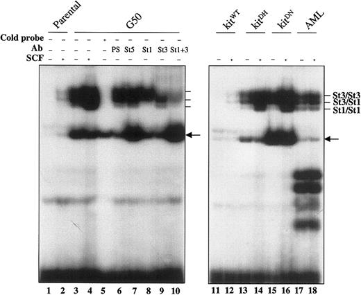
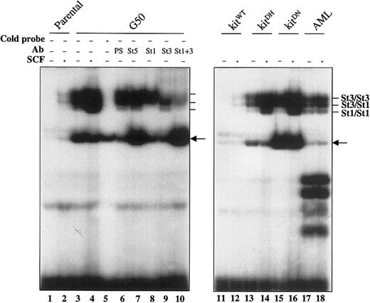
To determine whether activation of STATs might play a role in mutant c-Kit–mediated biologic effects, we first examined STAT3 and STAT1 activation status by EMSA using a DNA probe derived from the c-fos SIE (sis-inducible element)34 in cells expressing the mutant receptor. Constitutive DNA binding activity of STAT3 homodimers, STAT3/STAT1 heterodimers, and, to a lesser extent, STAT1 homodimers, was present in the cellular extracts from the G50 revertant cell line and from cells transduced with D816H or D816N mutant c-kit (Figure 1, lanes 3, 13, and 15). Constitutive DNA binding activity of STAT3 and STAT1 was also observed in the AML sample from which the D816N mutation of c-kit was identified (Figure 1, lane 17). Although SCF only weakly induced STAT DNA binding activity in MO7e cells expressing wild-type c-Kit, superinduction of activation of STAT3 and STAT1 by SCF was observed in cells expressing the mutant c-Kit receptor (Figure 1, lanes 4, 14, and 16). Similar amounts of c-Kit between wild-type and mutant c-Kit expressing MO7e cells have been demonstrated,23 indicating that the differential activation of STATs in these cells is not due to the difference of receptor expression. SIE-binding proteins were confirmed to be composed of specific STAT dimers, because the protein-DNA complexes disappeared or were significantly diminished following coincubation of cellular extracts with specific antibodies against either STAT3 or STAT1 or both (Figure 1, lanes 6-10).
Constitutive DNA binding activity of STAT3 and STAT1 in leukemia cells expressing Asp816 mutant c-Kit.
Parental MO7e cells and cells transduced with the wild-type c-kit cDNA (kitWT) were cultured without SCF for 5 hours. The G50 clone (a cytokine-independent revertant from MO7e cells containing the D816H c-kit mutation), MO7e cells transduced with the mutant c-kit cDNAs (kitDHand kitDN) and the AML cells bearing the D816N mutant c-kit were cultured in the absence of SCF. These cells were then stimulated with rhSCF (40 ng/mL) for 5 minutes. Cytosolic extracts were prepared from the cells before and after SCF stimulation and subjected to EMSA (see “Materials and methods”). In the left panel, some samples were preincubated with a 50-fold excess of unlabeled (cold) probe, rabbit preimmune serum (PS), or defined STAT (St) antibodies. Protein-DNA complexes corresponding to the specific STAT dimers are indicated. A nonspecific band, which could not be completed by the addition of cold probe and was preferentially present in MO7e cells bearing the mutant c-kit, is also indicated by an arrow in both left and right panels.
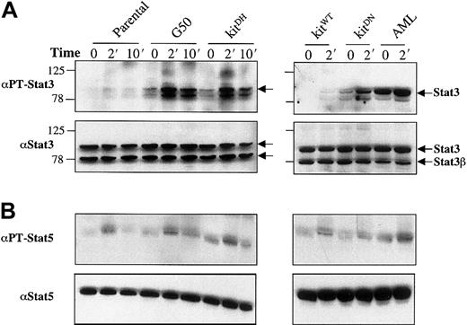
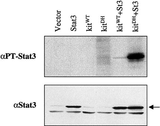
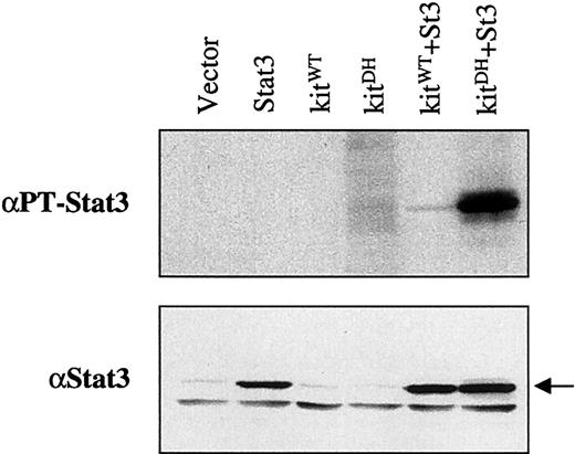
Consistent with EMSA results, Western blot analysis demonstrated that the constitutive tyrosine phosphorylation of STAT3 could be further superinduced by SCF in leukemia cells expressing Asp816mutant c-kit (Figure 2A, upper panels). The antibody reactive with tyrosine-phosphorylated STAT3 also recognized a protein (∼85 kd) that showed patterns of activation similar to that of STAT3 (Figure 2A, upper panels). This protein was not reactive with an antibody against the internal domain of STAT3 (Figure 2A, lower panels), nor to antibodies against STAT1 or STAT5 (data not shown), indicating that the observed band does not represent those STAT proteins. Similar patterns of STAT5 tyrosine phosphorylation were observed in leukemia cells bearing the wild-type and mutant c-Kit receptor in the presence or absence of SCF stimulation (Figure 2B). To determine that the activation of STAT3 was dependent on the expression of Asp816 mutant c-Kit, we transfected both D816H mutant c-kit and STAT3 cDNAs into the human embryonic kidney cell line, 293, which does not express any endogenous c-Kit receptor. Figure3 shows that STAT3 activation occurs in this cell line as a result of cotransfection with both cDNAs, thus demonstrating the dependence of STAT3 activation on D816H c-Kit activity.
Constitutive tyrosine phosphorylation of STAT3 in leukemia cells expressing Asp816 mutant c-Kit.
Total cellular lysates (20 μg) from indicated cells unstimulated or stimulated with SCF (40 ng/mL) for the times shown were analyzed by Western blotting. (A) The upper panels show the antibody against tyrosine- phosphorylated STAT3 reacting to 2 cellular-phosphorylated proteins. The top band was confirmed to be STAT3 by using the antibody against the internal domain of the protein, which recognizes both STAT3 and STAT3β, as shown in the lower panels using the stripped blots from the upper panels. The lower band observed as part of the doublet recognized by the antibody against tyrosine-phophorylated STAT3, could not be definitively identified as STAT3, 1, or 5. Size markers are shown in kilodaltons. (B) Blots in the upper panels were probed with the antibody against tyrosine- phosphorylated STAT5. Blots from the upper panels were stripped and reprobed with the antibody against STAT5 to illustrate the total STAT5 protein, as shown in the lower panels.
Constitutive tyrosine phosphorylation of STAT3 in 239 cells cotransfected with D816H mutant c-kit and STAT3.
Each plasmid alone (1.5 μg) or in combinations as indicated was transfected into 293 cells mediated by DOSPER reagents. Forty-eight hours later, cellular lysates were obtained from the cells and analyzed by Western blotting. The blot in the upper panel was probed with the antibody against tyrosine-phosphorylated STAT3. The blot from upper panel was stripped and reprobed with the antibody against the internal domain of STAT3, as shown in the lower panel.
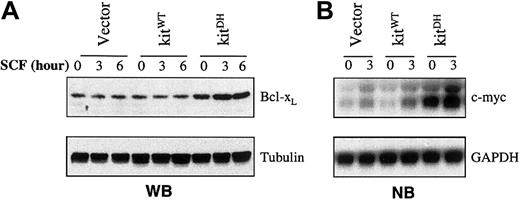
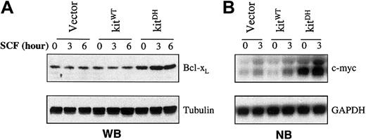
A number of genes that function to protect against apoptosis or promote cell cycle progression, such as Bcl-xL and c-myc, are activated by STAT3.6 37 To determine whether activation of Bcl-xL or c-myc was associated with Asp816 mutant c-Kit, MO7e cells stably transduced with either wild-type or D816H mutant c-kit cDNAs were analyzed. Western blot analysis showed that Bcl-xL in MO7e cells transduced with the empty vector or wild-type c-kit did not appear to be regulated by SCF stimulation (Figure 4A), consistent with the observations that the protein levels did not notably decline in these cells following SCF withdrawal (data not shown). However, a significantly higher level of Bcl-xL expression, which could be further induced by SCF, was observed in MO7e cells transduced with D816H mutant c-kit (Figure 4A). A significantly higher level of c-myc RNA expression was also observed in MO7e cells expressing the mutant c-Kit receptor (Figure 4B).
Up-regulation of Bcl-xL and c-myc in MO7e cells transduced with D816H mutant c-kit.
MO7e cells transduced with empty pLNCX vector or wild-type c-kit were SCF-starved for 5 hours, and then stimulated, together with the cells transduced with D816H mutant c-kit, with rhSCF (40 ng/mL) for the times indicated. (A) Western blot (WB) analysis of the total cellular extracts from the cells. The blot in the upper panel was probed with anti–Bcl-xL antibody. The blot was stripped and reprobed with antitubulin antibody to verify the protein loading, as shown in the lower panel. (B) Northern blot (NB) analysis of total RNA from the indicated cell types. The blot in the upper panel was hybridized with a c-myc–specific probe as described in “Materials and methods.” The blot was stripped and rehybridized with a GAPDH-specific probe to verify RNA loading, as shown in the lower panel.
These results demonstrate that constitutive activation of STAT3 and up-regulation of STAT3 downstream targets are associated with Asp816 mutant c-Kit, suggesting that the activated components may play an important role in mutant c-Kit–mediated biologic effects in leukemia cells.
STAT3 activation is required for SCF-independent survival and proliferation mediated by Asp816 mutant c-Kit
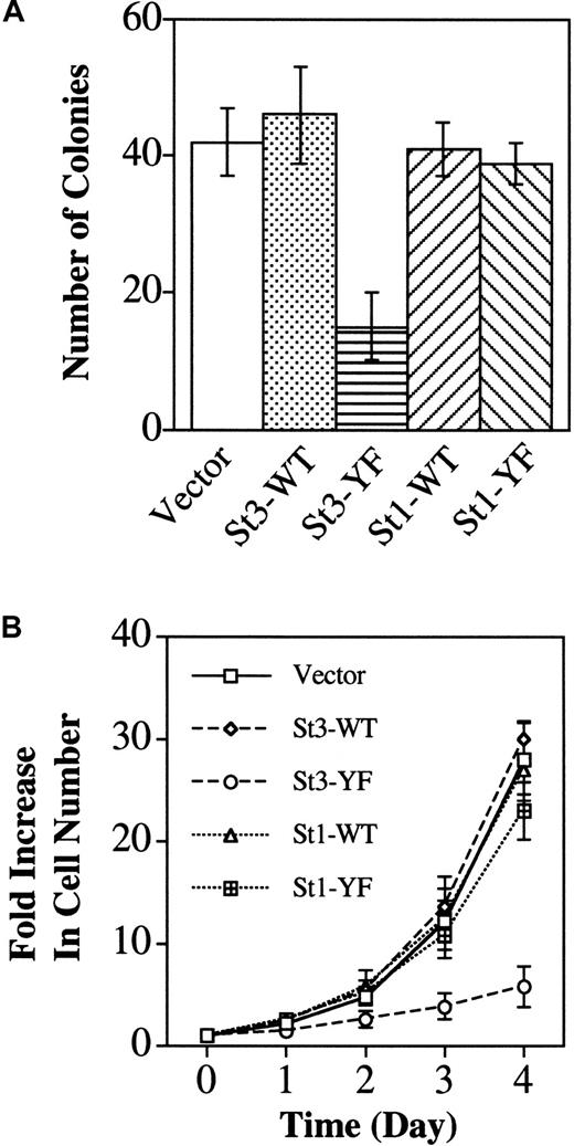
To determine whether activation of STAT3 or STAT1 is required for the SCF-independent survival and proliferation mediated by Asp816 mutant c-Kit, MO7e cells, stably transduced with D816H mutant c-kit cDNA, were subsequently transfected with plasmids encoding either the dominant-negative STAT3 or STAT1, as well as various control plasmids. Cytokine-independent survival of D816H mutant bearing MO7e cells was evaluated by colony formation assay, in which the cells were cultivated in the absence of SCF in methylcellulose-based medium containing the appropriate antibiotics. Although D816H c-Kit–expressing cells showed similar colony formation when they were transfected with the empty vector, wild-type STAT3 or STAT1 and dominant-negative STAT1, significantly fewer colonies were observed in cells transfected with dominant-negative STAT3 (Figure5A). Furthermore, the size of most colonies from cells transfected with dominant-negative STAT3 was remarkably smaller than that observed from cells transfected with other constructs (data not shown), suggesting that the surviving cells had a slower growth rate. To quantitate their proliferative status, individual clones were isolated and expanded in liquid culture in the absence of SCF, followed by counting cell numbers during a period of 4 days. Three clones each from cells transfected with different STAT cDNAs or control vector were evaluated. As shown in Figure 5B, although cells transfected with wild-type STAT3/1 and dominant-negative STAT1 gave rise to a similar growth rate, proliferation was significantly slower in cells transfected with dominant-negative STAT3 (Figure 5B). These results indicate that activation of STAT3, but not STAT1, is required for mutant c-Kit–mediated SCF-independent survival and proliferation in MO7e leukemia cells.
Expression of dominant-negative STAT3 inhibits SCF-independent survival and proliferation mediated by D816H mutant c-Kit in MO7e cells.
(A) D816H mutant c-kit–transduced MO7e cells were subsequently transfected with the empty pcDNA3.1/Hygro(+) vector, vector containing the wild-type (WT) STAT3 (St3) or STAT1, and vector containing dominant-negative (YF) STAT3 or STAT1. SCF-independent survival was evaluated by counting the numbers of colonies grown in methylcellulose culture 3 weeks following transfection (see “Materials and methods”). The results are presented as mean ± SE from 3 independent experiments. (B) Clones were isolated from colonies in methylcellulose and cultured in suspension in the absence of SCF. SCF-independent proliferation was evaluated from 3 clones in each experimental group by counting the cell numbers over a period of 4 days. The results are presented as mean ± SE from 3 independent experiments.
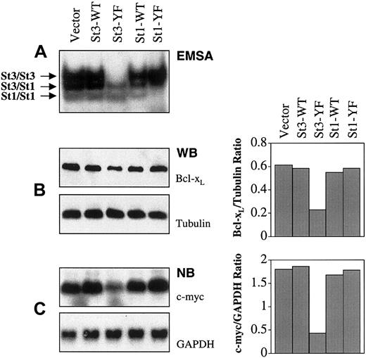
Cloned cells stably transfected with STAT cDNAs or control vector were further expanded in liquid culture. Cytosolic abstracts from 3 clones each were isolated and incubated with 32P-labeled SIE probe, followed by EMSA. Activation of STAT3 and STAT1 by D816H mutant c-Kit was significantly suppressed in cells transfected with the corresponding dominant-negative STAT cDNAs, although there was still considerable DNA binding activity from STAT3/1 heterodimers in dominant-negative STAT3–transfected cells (Figure6A). The protein level of Bcl-xL and the mRNA level of c-myc were also examined in clones transfected with different STAT cDNAs (Figure 6B,C). Significant reduction of Bcl-xL and c-myc was evident only in D816H mutant c-Kit–expressing MO7e cells that were transfected with dominant-negative STAT3 (Figure 6B,C). Clones within the same experimental group showed similar patterns of EMSA binding as well as expression of Bcl-xL and c-myc when compared to tubulin and GAPDH, respectively. The results from representative clones are shown in Figure 6. These results demonstrate that STAT3 activation plays an essential role in mutant c-Kit–induced up-regulation of Bcl-xL and c-myc, which is likely to be important for mutant c-Kit–mediated biologic effects.
Down-regulation of Bcl-xL and c-myc by expression of dominant- negative STAT3 in MO7e cells bearing D816H mutant c-Kit.
(A) Cytosolic extracts from representative clones in each experimental group were incubated with 32P-labeled SIEm67 probe, followed by EMSA. (B) Total cellular lysates were analyzed by Western blotting (WB). In the left panel, the upper part was the blot probed with anti–Bcl-xL antibody. Blot from the upper part was stripped and reprobed with antitubulin antibody to verify the protein loading, as shown in the lower part. Density of the bands was determined by densitometry analysis and the Bcl-xL–tubulin ratios are shown in the right panel. (C) Total RNA was analyzed by Northern blotting (NB). In the left panel, the upper part was the blot hybridized with a c-myc–specific probe. Blot from the upper part was stripped and rehybridized with GAPDH-specific probe to verify RNA loading, as shown in the lower part. Density of the bands was determined by densitometry analysis and the c-myc/GAPDH ratios are shown in the right panel. These analyses were performed on 3 different clones transfected with each of the STAT (St) cDNAs or vector alone. Within each group of transfectants, similar patterns of EMSA binding as well as Bcl-xL and c-myc expression were observed. Results of representative clones are depicted.
Constitutively activated STAT3 promotes survival of MO7e leukemia cells
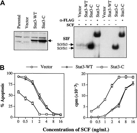
To further investigate the role of STAT3 activation in SCF-independent survival and proliferation in MO7e leukemia cells, a cDNA plasmid encoding constitutively activated STAT3 (STAT3-C)6 was constructed. Functional analysis of STAT3-C shows that it induces cellular transformation in 293 cells, whereas wild-type STAT3 does not (manuscript in preparation), consistent with the original observations that STAT3-C transforms rat and mouse fibroblasts.6 Parental MO7e cells were transduced with the retroviral particles containing the wild-type STAT3 or STAT3-C tagged with the FLAG epitope. Exogenous STAT3 expression and DNA binding activity from the cytosolic extracts were examined in stable cell lines. Western blot analysis using an anti-FLAG antibody revealed that both wild-type STAT3 and STAT3-C proteins were expressed in the corresponding cell populations (Figure7A, left panel). However, a greater amount of STAT3-SIE complex, which could be supershifted with anti-FLAG antibody, was observed only in the cytosolic extracts from STAT3-C–transduced MO7e cells (Figure 7A, right panel). Significant DNA binding activity of STAT3-C was still present in the transduced cells even after SCF withdrawal for 12 hours (Figure 7A, right panel). The extent of apoptotic cell death from these cells was determined in the presence or absence of various concentrations of SCF. Although cells transduced with wild-type STAT3 showed a similar percentage of apoptosis compared to cells transduced with the empty vector, significantly less apoptotic cell death was observed in cells transduced with STAT3-C on complete withdrawal of SCF or in the presence of lower SCF concentrations (Figure 7B, left panel). Furthermore, STAT3-C–transduced MO7e cells retained long-term viability in culture at a low concentration (1 ng/mL) of SCF, at which both the empty vector and wild-type STAT3-transduced cells were not able to survive (data not shown). DNA synthesis was also determined by3H-thymidine incorporation in different transduced cells using the same conditions as described above. Cells transduced with STAT3-C consistently showed more active DNA synthesis than those transduced with the wild-type STAT3 or empty vector in the presence or absence of different concentrations of SCF (Figure 7B, right panel). These results demonstrate that activated STAT3 is able to mediate antiapoptotic and proliferative signals in MO7e cells. However, activated STAT3 by itself is not sufficient to confer SCF-independent survival and proliferation, suggesting that the activation of other signaling pathways by Asp816 mutant c-Kit may be required, together with activated STAT3, for the complete reversal of cytokine dependence of MO7e cells.
Constitutively activated STAT3 promotes survival and proliferation of MO7e cells.
(A) Left panel: MO7e cells were transduced with either empty pLNCX vector or vector containing the wild-type STAT3-FLAG or STAT3-C–FLAG. After stable lines of cells were obtained by selection in G418 (0.8 mg/mL), total cellular lysates were prepared and subjected to Western blot analysis with an anti-FLAG antibody (arrow). Right panel: Cytosolic extracts from MO7e cells, in the presence or absence of SCF, were incubated with 32P-labeled SIEm67 probe, followed by EMSA. Some samples were preincubated with an antibody against FLAG before incubation with labeled probe. (B) MO7e cells transduced with the empty vector, wild-type STAT3, or STAT3-C were cultured in the presence or absence of different concentrations of rhSCF for 24 hours. Apoptosis (left panel) and DNA synthesis (right panel) were determined by acridine orange staining and 3H-thymidine incorporation, respectively. The results are presented as a mean ± SE from 3 independent experiments.
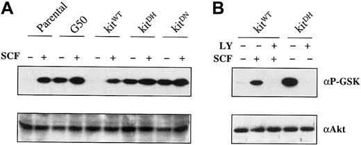
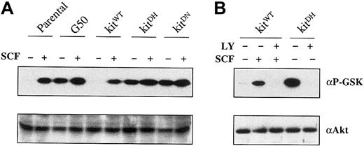
To determine whether other major signaling pathways that have been associated with the regulation of apoptosis might be constitutively activated by mutant c-Kit, we examined PI-3K/Akt activation in MO7e cells. Akt protein was immunoprecipitated from cells expressing either wild-type or mutant c-Kit in the presence or absence of SCF, followed by in vitro kinase assays using recombinant GSK-3, the Akt downstream target, as substrate. Although Akt kinase activity could be induced by SCF stimulation in wild-type c-Kit–expressing cells, constitutive activation of Akt, which could be moderately increased by SCF stimulation, was observed in cells expressing Asp816 mutant c-Kit (Figure 8A). In addition, Akt kinase activity could be suppressed by a PI-3K inhibitor, LY294002 (Figure 8B), indicating that PI-3K is responsible for the Akt activation in MO7e cells. These results suggest that constitutive activation of PI-3K/Akt pathway by Asp816 mutant c-Kit may contribute, together with activated STAT3, to cytokine-independent survival and proliferation in MO7e cells expressing the mutant receptor. The induction of apoptosis in MO7e cells expressing either wild-type or mutant c-Kit by 20 μM LY294002 further supports a contribution of PI-3K activation to mutant c-Kit–mediated SCF-independent survival (manuscript in preparation).
Constitutive activation of PI-3K/Akt pathway in MO7e cells expressing Asp816 mutant c-Kit.
(A) Different cells as indicated were incubated in 0.5% FCS in the absence of SCF for 5 hours and then stimulated with rhSCF (40 ng/mL) for 5 minutes. Akt protein was immunoprecipitated and the in vitro Akt kinase assay using GSK-3 fusion protein as substrate was carried out as described in “Materials and methods,” followed by Western blot analysis. The blot in the upper panel was probed with antiphospho–GSK-3 antibody. This blot was subsequently stripped and reprobed with anti-Akt antibody, as shown in the lower panel. (B) MO7e cells transduced with wild-type or D816H mutant c-kit were incubated in 0.5% FCS in the absence of SCF for 5 hours. LY294002 (20 μM) was added to some samples as indicated at the fourth hour of the incubation, followed by the experimental procedures as described in panel A.
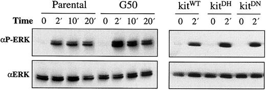
D814H c-kit, the mouse mutant corresponding to the human D816H c-kit, induces constitutive activation of MAP kinases in a mouse erythroleukemia cell line.18 We examined MAP kinase activation by Western blot analysis using an antibody recognizing phosphorylated ERK-1 and ERK-2 (p44 and p42) in MO7e cells bearing the mutant c-Kit receptor. Basal levels of activation were comparable between wild-type and mutant c-Kit–expressing cells, but mutant c-Kit–expressing cells appeared to have moderately higher levels of MAP kinase phosphorylation than wild-type c-Kit–expressing cells after SCF stimulation (Figure 9). The absence of constitutive activation of MAP kinases in MO7e cells expressing Asp816 mutant c-Kit suggests that MAP kinase activation does not play a major role in mutant c-Kit–mediated biologic effects in these leukemia cells.
Similar activation patterns of MAP kinase in MO7e cells with or without Asp816 mutations of c-kit.
Total cellular lysates (20 μg) from cells unstimulated or stimulated with SCF (40 ng/mL) for the times indicated were examined by Western blot analysis. The upper panels show the MAP kinase phosphorylation recognized by the antibody against phosphorylated ERK-1 and ERK-2. The same blots were stripped and reprobed with the antibody reactive with both ERK-1 and ERK-2, as shown in the lower panels.
Discussion
Mutations of c-kit at codon 816 have been identified in various malignancies, including AML,21,22mastocytosis,24,38 and germ cell tumors.26The biologic consequences of these activating mutations, demonstrated mostly by the equivalent mouse c-kit mutations at codon 814, include converting cytokine-dependent cell lines to cytokine independence and inducing tumorigenicity.17-20 39 However, the molecular mechanisms, particularly the changes in the signal transduction pathways, which are responsible for these effects mediated by Asp816(human)/814(mouse) mutations of c-kit,are largely undefined. Data presented in this report demonstrate that constitutive activation of STAT3 and the up-regulation of STAT3 downstream targets are associated with Asp816 mutations of c-kit in leukemia cells. Functional studies using dominant-negative or constitutively activated STAT3 show that STAT3 activation is required for cytokine independent survival and proliferation mediated by Asp816 mutant c-Kit.
Multiple pathways have been implicated in SCF/c-Kit–mediated signal transduction, including the Ras-Raf-MAP kinase cascade, PI-3K/Akt, Src family kinases, and JAK/STATs.15 STAT140 and STAT541 have been shown to be activated by SCF in hematopoietic cell lines. STAT3 was reported to be serine, but not tyrosine-phosphorylated by SCF stimulation.42 In contrast to these findings, other studies show that SCF/c-Kit interaction has no measurable effect on STAT family members in other cell lines.43,44 By evaluating either tyrosine phosphorylation or DNA binding activity, we demonstrate that STAT3 and STAT1 are only weakly activated by SCF at its optimal concentration in wild-type c-Kit–expressing MO7e cells. However, when cells are transduced with D816H or D816N mutant c-kit, a significant constitutive activation of STAT3 and STAT1 to levels higher than that in SCF-stimulated cells expressing wild-type c-Kit is observed. Furthermore, activation of these STAT proteins is superinduced by SCF in cells expressing mutant c-Kit. In contrast, the pattern of Ras-mediated MAP kinase activation without SCF stimulation in this system is comparable in cells that express either wild-type or mutant c-Kit. The different observations between these results and others18 regarding MAP kinase activation by mutant c-Kit may reflect the differences of species or leukemia cell lines studied.
Constitutive activation of the selective signaling components by Asp816 mutant c-Kit demonstrated in our studies suggests that mutant c-Kit does not simply replace the normal interaction between SCF and the wild-type receptor, but instead has distinct signaling characteristics. This interpretation is supported by 2 other lines of evidence from studies on the mouse Asp814 c-Kit mutants. First, although wild-type c-Kit in the presence of SCF forms dimerized receptors in the extracellular domain, D814Y mutant dimerizes in the cytoplasmic region in the absence of SCF.45 Second, D814V mutant has been shown to possess altered sites of receptor autophosphorylation and shifted specificity for peptide substrates.46 Taken together, our findings and those from others45 46 show that Asp816 mutant c-Kit has certain changes in receptor binding or catalytic properties or both, which selectively activate signal transduction pathways leading to profound phenotypic changes that are distinguishable from the effects of the wild-type receptor.
How STAT3 and STAT1 are activated by Asp816 mutant c-Kit is currently unknown. Studies of the platelet-derived growth factor receptor, another type III RTK, have shown that STAT3 and STAT1 can be activated either directly by the receptor47 or in conjunction with other tyrosine kinases, including JAK47and Src48 kinases. Although STAT1 is reported to associate with c-Kit,40 the physical interactions of c-Kit with other kinases, such as JAK249 and the Src family member, Lyn,50 have also been demonstrated. Future experiments are being directed toward elucidating whether STATs are directly activated by the mutant c-Kit receptor or indirectly activated by other tyrosine kinases.
STAT3 has been implicated in protection from apoptosis51-55and promotion of cell growth.51,54-58 Constitutive activation of STAT3 occurs in a variety of malignancies, including AML.59-63 Our experiments demonstrate that expression of dominant-negative STAT3 in MO7e cells bearing D816H mutant c-Kit significantly inhibits colony formation in methylcellulose, suggesting that STAT3 activation by mutant c-Kit is required for SCF-independent survival in these cells. However, some small colonies were formed from cells transfected with dominant STAT3. We speculate that this could be due to the difference in the extent of STAT3 inhibition by a dominant-negative molecule within the cell population. If STAT3 activation was completely suppressed in a given cell, that cell might not be able to survive, and thus, fewer colonies would be formed. However, if STAT3 activation was incompletely blocked, a cell might survive and proliferate, though with a lower proliferation rate. To support this speculation, our experiments show that surviving clones transfected with dominant-negative STAT3 have little DNA binding activity for STAT3 homodimers, but retain some activity for STAT3/STAT1 heterodimers (Figure 6A). Although a system for inducible expression of dominant-negative STAT3 could also be used in the future to further address the role of inhibition of STAT3 activation in cytokine-independent survival and proliferation mediated by Asp816 mutant c-Kit, these results demonstrate that STAT3 is required for cytokine-independent survival and proliferation.
Accumulating evidence suggests that STAT3 inhibits apoptosis and promotes proliferation by regulating the expression of antiapoptosis suppressors, such as Bcl-xL,6,53 and cell cycle modulators, such as c-myc6,37 and cyclins.6 56 Our results show that higher levels of Bcl-xL and c-myc are present in Asp816 mutant c-Kit–expressing MO7e cells compared with that observed in wild-type c-Kit–expressing cells, consistent with the differential activation status of STAT3. Furthermore, expression of Bcl-xL and c-myc is down-regulated by blockade of STAT3 activation in cells expressing mutant c-Kit. These findings imply that Bcl-xL and c-myc are regulated by mutant c-Kit–mediated STAT3 activation and that the up-regulation of these genes may also be involved in SCF-independent survival and proliferation.
To address whether activated STAT3 by itself could restore Asp816 mutant c-Kit–induced phenotypic changes, we have expressed a constitutively activated STAT3 molecule6 in parental MO7e cells. In contrast to the results that STAT3 activation is able to reconstitute mutant c-Kit–induced cellular transformation in 293 cells (manuscript in preparation), activated STAT3 by itself is not sufficient to confer SCF-independent survival of MO7e cells. Thus, other signaling pathway(s) that are constitutively activated by mutant c-Kit are likely to be required, together with STAT3 activation, for the complete reversal of cytokine dependence in MO7e cells.
This possibility is highlighted by our demonstration that the PI-3K/Akt pathway is also constitutively activated in MO7e cells expressing Asp816 mutant c-Kit and that the blockade of PI-3K activity by a specific inhibitor induces apoptosis in these cells. The PI-3K/Akt pathway has been shown to play a major role in multiple biologic responses mediated by SCF/c-Kit interaction,64-66including cell survival and proliferation.67,68 In terms of the cooperation between STATs and PI-3K signaling pathway in cellular functions, recent studies have shown that STAT3 acts as an adapter to couple the activation of the interferon receptor by its ligand to the PI-3K pathway,69 and that STAT5 and PI-3K cooperate in the interleukin 3–dependent survival of a bone marrow–derived cell line.70 Our laboratory is currently coexpressing constitutively activated STAT3 and the components of the PI-3K pathway in leukemia cells to further evaluate the role of activation of multiple signaling pathways in Asp816 mutant c-Kit–mediated biologic effects.
Activating receptor mutations have been implicated in the pathogenesis of a variety of different forms of cancer, including leukemia.21,22,26,71-74 In the case of AML, although mutations resulting from an internal tandem duplication in the juxtamembrane domain of the Flt3 gene have been reported in a frequency of nearly 20% in AML,71 a recent study has shown that there is also a high frequency of Asp816mutations of c-kit in AML-M2 with t(8;21) and AML-M4Eo with inv(16): 6 of 15 patients screened had either D816V or D816Y mutation of c-kit.22 The characterization of altered signal transduction pathways and the resulting functional consequences mediated by mutant receptors should provide essential information for the molecular identification of subsets of leukemia and solid tumors as well as the development of nongenotoxic therapies designed to specifically block the function of abnormal receptors and their critical downstream signaling pathways. The results that we have presented demonstrate a mechanism by which a mutant receptor is preferentially linked to STAT3 activation, and, thereby, contributes to leukemia cell survival. These studies may also provide a model system to screen for small molecules that specifically inhibit the aberrant activation of STAT3 by the mutant c-Kit receptor in leukemia cells.
We thank Paul Jubinsky, Hélène Paradis, and Eric Raabe for their helpful discussion and advice, and the Leukemia Research Foundation.
The publication costs of this article were defrayed in part by page charge payment. Therefore, and solely to indicate this fact, this article is hereby marked “advertisement” in accordance with 18 U.S.C. section 1734.
References
Author notes
Robert J. Arceci, Johns Hopkins Oncology Center, Division of Pediatric Oncology, Bunting-Blaustein Cancer Research Bldg, 1650 Orleans St, 2M51, Baltimore, MD 21231; e-mail: arcecro@jhmi.edu.

