Erythropoietin and its receptor are required for definitive erythropoiesis and maturation of erythroid progenitor cells. Mice lacking the erythropoietin receptor exhibit severe anemia and die at about embryonic day 13.5. This phenotype can be rescued by the human erythropoietin receptor transgene. Animals expressing only the human erythropoietin receptor survived through adulthood with normal hematologic parameters and appeared to respond appropriately to induced anemic stress. In addition to restoration of erythropoiesis during development, the cardiac defect associated with embryos lacking the erythropoietin receptor was corrected and the increased apoptosis in fetal liver, heart, and brain in the erythropoietin receptor null phenotype was markedly reduced. These studies indicate that no species barrier exists between mouse and human erythropoietin receptor and that the human erythropoietin receptor transgene is able to provide specific expression in hematopoietic and other selected tissues to rescue erythropoiesis and other organ defects observed in the erythropoietin receptor null mouse.
Introduction
Absence of erythropoietin or the erythropoietin receptor (EpoR) results in interruption of definitive erythropoiesis in the fetal liver,1,2 defective cardiac development,3 and eventual death at about embryonic day 13.5 (E13.5). Unlike interleukin-3 and granulocyte macrophage colony-stimulating factor, erythropoietin is cross-reactive between species. We hypothesize that the human EpoR (hEpoR) is equivalent to the mouse EpoR (mEpoR) and that it can rescue the developmental defects in liver and heart of the EpoR null mouse (mEpoR−/−) and restore normal hematopoietic development. An 80-kb hEpoR transgene recapitulates EpoR expression in hematopoietic tissue with high levels in yolk sac and fetal liver, and then adult spleen and bone marrow following the site of hematopoiesis.4 This transgene includes 6 kb of 5′ flanking sequence containing the proximal promoter that provides much of the transcription activity in erythroid cells.5-7 We show that the hEpoR transgene restores effective erythropoiesis in the EpoR null mouse and normalizes cardiac as well as brain development. These results illustrate that the mouse and human EpoRs are nearly equivalent and provide evidence for erythropoietin multiorgan function.
Study design
The rescue mEpoR−/−, hEpoR+ genotype was obtained by mating mEpoR+/−2 and hEpoR transgenic mice containing an 80-kb hEpoR gene fragment,4and intercrossing mEpoR+/−, hEpoR+, and mEpoR+/− mice. Genotyping by polymerase chain reaction (PCR) analyses of tail DNA used null mEpoR primers2 and the hEpoR primers4 as described previously. For embryos, yolk sac DNA was used. For TdT-mediated d-UTP nick end labeling (TUNEL) analysis,8 embryo sections were treated with proteinase K and analyzed using digoxigenin-labeled dUTP with terminal deoxynucleotidyl transferase (Roche Molecular Biochemicals, Indianapolis, IN), followed by incubation with antidigoxigenin antibody conjugated with alkaline phosphatase (Dako Corporation, Carpinteria, CA). New fuchsin substrate reaction was used to visualize positive staining, and sections were counterstained with hematoxylin.
Adult progenitor cell colony assays of bone marrow were carried out in triplicate cultures in Methocult GF M3434 or Methocult M3334 (Stemcell Technologies, Vancouver, BC, Canada) and monitored after 2 days for erythroid colony-forming unit (CFU-E) and after 12 days for erythroid burst-forming unit (BFU-E). Quantitative reverse transcriptase PCR was used for expression analysis of total RNA isolated from bone marrow, spleen, brain, heart, lung, kidney, and skeletal muscle, as described previously.9 Gene-specific primers and fluorescent labeled Taqman probes (7700 Sequence Detector; PE Applied Biosystems, Foster City, CA) spanning exon junctions were as follows: for hEpoR, forward primer, 5′-GCT CCC TTT GTC TCC TGC T-3′; reverse primer, 5′-CTC CCA GAA ACA CAC CAA GTC CT-3′; probe, 5′-AGC GGC CTT GCT GGC GG-3′. Primers and probe for mEpoR were described previously.9 To induce anemia, mice were injected with phenylhydrazine (Sigma, St Louis, MO) at a dose of 0.04 mg/g of body weight for 5 days.4 Mice were killed on day 7. Blood samples were collected for hematology and erythropoietin detection. Serum erythropoietin concentrations were determined by enzyme-linked immunosorbent assay using anti–human erythropoietin antibody (R&D Systems, Minneapolis, MN). Tissues were harvested and analyzed for EpoR expression.
Results and discussion
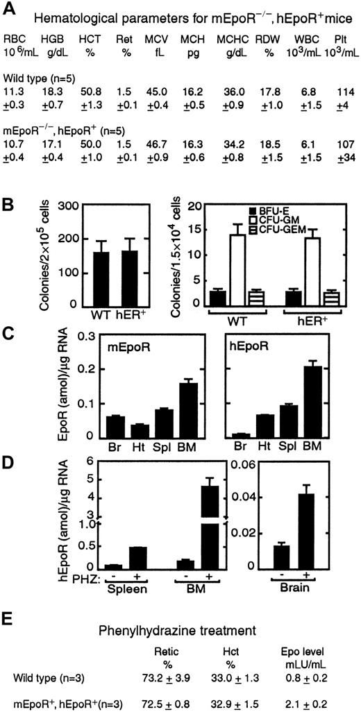
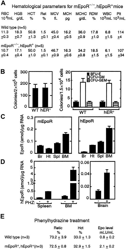
The hEpoR transgene rescued the mEpoR−/− genotype, normalized hematopoiesis, and increased survival through adulthood with no gross malformations. Although some initial difficulty in mating these mice was encountered, males and females were both fertile. The hEpoR transcript was processed to its mature form in hematopoietic cells.10 Appropriate hematopoietic expression suggests that regulatory elements in the transgene are sufficient to mimic endogenous EpoR expression, which may be useful in constructing vectors to drive gene expression, particularly in early erythroid progenitor cells where EpoR expression is maximal.11 The hEpoR-rescued mice exhibited normal hematologic parameters (Figure1A). Hematopoietic colony culture showed that BFU-E and CFU-E progenitors, and CFU-granulocyte, macrophage (CFU-GM) and CFU-granulocyte, erythroid, macrophage, and megakaryocyte (CFU-GEMM), were also analogous to those in normal controls (Figure 1B). Although some specificity for erythropoietin/EpoR has been observed for other species,12the rescue of the EpoR−/− genotype by the hEpoR transgene indicates that there is no apparent species barrier between mouse and human with regard to erythropoietin stimulation of its receptor. Only minimal differences in erythropoietin levels following anemic stress were detected (Figure 1E). These results contrast with the report that murine erythropoietin is 10-fold less active than human erythropoietin on human cells.13
Hematopoiesis in the mEpoR−/−, hEpoR+ mouse.
(A) Hematologic parameters for the mEpoR−/−, hEpoR+ (hEpoR+) genotype were comparable to those of wild-type (WT) controls. (B) CFU-E colonies (left) and BFU-E, CFU-GM, and CFU-GEMM colonies (right) were determined for bone marrow hematopoietic progenitor cells (n = 5). (C) EpoR expression was determined for spleen (Spl), bone marrow (BM), brain (Br), and heart (Ht) for wild-type controls (mEpoR) (n = 3) and mEpoR−/−, hEpoR+ (hEpoR) mice (n = 3), amol indicates attomoles. (D) Phenylhydrazine-induced (PHZ) anemia in mEpoR−/−, hEpoR+ mice (n = 3) increased hEpoR expression in hematopoietic tissues and brain (n = 3 for control). (E) Results of stimulation of erythropoiesis in phenylhydrazine-treated mice were similar in mEpoR−/−, hEpoR+ mice and controls as indicated by reticulocyte counts and hematocrit. Only differences in circulating erythropoietin levels (about 2-fold higher in mEpoR−/−, hEpoR+ mice compared with controls) were detected. Serum erythropoietin levels in untreated mice were not detected in this assay because of the relatively low affinity of the antibody for murine erythropoietin.
Quantitative analysis showed high hEpoR expression in adult hematopoietic tissues (spleen and bone marrow) comparable to mEpoR in normal controls (Figure 1C). Significant levels of hEpoR transcripts were also observed in the heart. Although transgene expression in adult brain was low, this was considerably higher than in other nonhematopoietic tissue such as skeletal muscle (data not shown). Like endogenous EpoR, the hEpoR transgene responded to anemic stress. Animals subjected to phenylhydrazine treatment to induce anemia exhibited an increase in hEpoR expression of 5-fold in spleen and of 24-fold in bone marrow (Figure 1D), with comparable erythropoietic activity and increased reticulocyte count (Figure 1E). Although no increase in hEpoR expression was observed in the heart, a 3-fold induction of hEpoR expression was observed in the brain, comparable to increases we had observed previously in normal mice.4Recovery from anemia in these mice was similar to that in treated controls. During development, endogenous EpoR and the hEpoR transgene are expressed in embryonic heart and brain in addition to hematopoietic tissue.3 4 These data suggest that the regulatory elements in the hEpoR transgene provide appropriate temporal and tissue-specific expression, including the response to anemic stress.
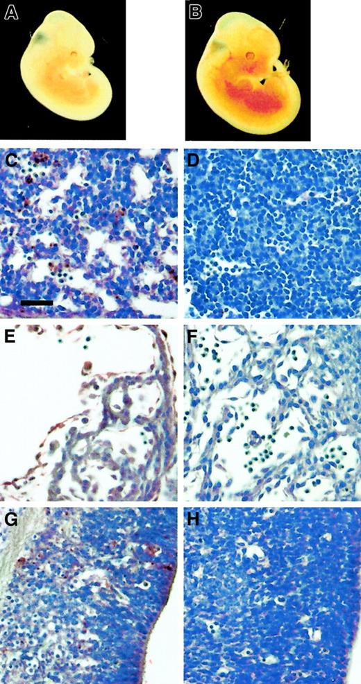
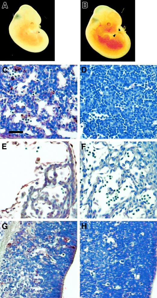
The mEpoR−/− mice exhibited a pale yolk sac and embryo (Figure 2A) with increased apoptosis (TUNEL-positive cells) in the fetal liver (Figure 2C) by E12.5. In contrast, circulating erythroid cells were readily observed in the yolk sac blood vessels and in fetal liver of the mEpoR−/−, hEpoR+ embryo (Figure 2B), with little apoptosis in the fetal liver (Figure 2D). Expression of hEpoR in adult heart and brain led us to examine these tissues during development. By E11.5, ventricular hypoplasia was noted in mEpoR−/− mice and in erythropoietin null mice, indicating a defect in proliferation and expansion of the myocardium.3 We identified extensive apoptosis in the myocardium and endocardium of the mEpoR−/− embryonic heart (Figure 2E). In comparison, expression of the hEpoR transgene normalized the proliferation of endocardium and myocardium (Figure 2F), confirming a role for EpoR in heart development. Erythropoietin stimulation of cardiomyocytes,14 of satellite cells or myoblasts from skeletal muscle,9 of endothelium,15,16and of neovascularization in vivo17 provides additional support for a broader role for erythropoietin in stimulating the proliferation of progenitor cells before terminal differentiation.
Analyses of mEpoR−/−, hEpoR+ embryos.
(A,B) The mEpoR−/− pale embryo (A) at E12.5 contrasts with the erythroid-rich mEpoR−/−, hEpoR+embryo (B). (C-H) The increased apoptotic activity (TUNEL-positive cells) in the mEpoR−/− fetal liver (C), heart (E), and brain (G) is rescued by the hEpoR transgene, with reduced TUNEL-positive cells in the corresponding fetal liver (D), heart (F), and brain (H). The bar in (C) indicates 0.05 mm.
During development, we readily observed TUNEL-positive cells in the neuroepithelial tissues of the EpoR−/− mouse (Figure2G), accompanied by hypoplasia in the region of the fourth ventricle. This increase in apoptosis in the developing brain was also rescued by the hEpoR transgene, and both the mEpoR−/−, hEpoR+ and normal controls had few apoptotic cells in corresponding sites in the nervous system (Figure 2H). These results suggest a neuroprotective and/or proliferative effect of erythropoietin, as observed in culture of NT2 cells treated with hypoxia18 and in primary embryonic neural cultures with enhanced dopamine neuron generation.19 In adults, erythropoietin is produced on both sides of the blood–brain barrier and is hypoxia inducible,20 and erythropoietin administration has been shown to be neuroprotective in animals when challenged with ischemia or other injury to the brain.21-23 In addition to induction of EpoR in the brain by anemic stress (Figure 1D), hypoxia induces EpoR expression and increases erythropoietin sensitivity in neuronal cell cultures.18 The expression of EpoR in heart and brain and the associated apoptosis in the mEpoR−/− mouse during development provide evidence for a multiorgan response to erythropoietin.
To date, the only species-distinct activity for EpoR is interaction with the spleen focus forming virus (SFFV) gp55-P viral envelope protein, which is specific for mouse but not human EpoR.24Other viruses or pathogens may target the hEpoR, such as gene-transfer vectors incorporating human erythropoietin on their surface.25 Like SFFV, these interactions may exhibit preferential binding or interactions in a species-dependent manner. The rescue of the mEpoR−/− mouse by the human transgene provides a model system to examine interactions specific to the hEpoR.
The publication costs of this article were defrayed in part by page charge payment. Therefore, and solely to indicate this fact, this article is hereby marked “advertisement” in accordance with 18 U.S.C. section 1734.
References
Author notes
Constance Tom Noguchi, Laboratory of Chemical Biology, National Institute of Diabetes and Digestive and Kidney Disorders, National Institutes of Health, Bethesda, MD 20892-1822; e-mail: cnoguchi@helix.nih.gov.


