Abstract
Heme-oxygenases (HOs) catalyze the conversion of heme into carbon monoxide and biliverdin. HO-1 is induced during hypoxia, ischemia/reperfusion, and inflammation, providing cytoprotection and inhibiting leukocyte migration to inflammatory sites. Although in vitro studies have suggested an additional role for HO-1 in angiogenesis, the relevance of this in vivo remains unknown. We investigated the involvement of HO-1 in angiogenesis in vitro and in vivo. Vascular endothelial growth factor (VEGF) induced prolonged HO-1 expression and activity in human endothelial cells and HO-1 inhibition abrogated VEGF-driven angiogenesis. Two murine models of angiogenesis were used: (1) angiogenesis initiated by addition of VEGF to Matrigel and (2) a lipopolysaccharide (LPS)–induced model of inflammatory angiogenesis in which angiogenesis is secondary to leukocyte invasion. Pharmacologic inhibition of HO-1 induced marked leukocytic infiltration that enhanced VEGF-induced angiogenesis. However, in the presence of an anti-CD18 monoclonal antibody (mAb) to block leukocyte migration, VEGF-induced angiogenesis was significantly inhibited by HO-1 antagonists. Furthermore, in the LPS-induced model of inflammatory angiogenesis, induction of HO-1 with cobalt protoporphyrin significantly inhibited leukocyte invasion into LPS-conditioned Matrigel and thus prevented the subsequent angiogenesis. We therefore propose that during chronic inflammation HO-1 has 2 roles: first, an anti-inflammatory action inhibiting leukocyte infiltration; and second, promotion of VEGF-driven noninflammatory angiogenesis that facilitates tissue repair.
Introduction
An increasing body of evidence indicates that cells from the innate immune system such as monocytes/macrophages are important regulators of angiogenesis.1,2 Granulocytes, whose role is frequently underestimated, also appear to play a primary role in the initial phases of the angiogenic process.3,4 Angiogenesis is closely associated with inflammation in several diseases including rheumatoid arthritis, atherosclerosis, carcinoma, and hematologic malignancies.5,6 Moreover, several angiogenic growth factors are not endothelial cell (EC) specific. In particular, vascular endothelial growth factor (VEGF), recognized to be fundamental to the process of angiogenesis,7 may also activate and recruit monocytes8 and has been directly implicated in the inflammatory angiogenesis associated with diseases such as atherosclerosis and rheumatoid arthritis.9-12
Recent studies have shown that heme oxygenase-1 (HO-1) may exert anti-inflammatory effects including prolongation of cardiac xenograft graft survival13 and inhibition of leukocyte transendothelial migration during complement-dependent inflammation14 and in response to low-density lipoprotein (LDL) oxidation.15 Heme oxygenases are rate-limiting enzymes that catalyze the conversion of heme into carbon monoxide (CO) and biliverdin. Human HO exists in 2 main isoforms, HO-1 (inducible) and HO-2 (constitutive). HO-2 is present in high concentration in the brain, testis, and vascular EC, while HO-1 is more widely distributed.16 HO-1 is rapidly induced during hypoxia, ischemia/reperfusion, hyperthermia, and endotoxic shock, providing cytoprotection during the resolution of stress-induced inflammatory injury.17 The importance of this is demonstrated by the severe and persistent endothelial damage observed in human HO-1 deficiency18 and in gene-targeted mice deficient in HO-1.19 Furthermore, induction of HO-1 may directly regulate EC activation, preventing adhesion molecule expression and chronic graft rejection.20-22 In vitro, HO-1 protects ECs from hydrogen peroxide–mediated cell death23 and from tumor necrosis factor α (TNFα) cytotoxicity.24 In addition to its role in vascular cytoprotection, it has been suggested that HO-1 may play a role in angiogenesis as induction of HO-1 increases EC VEGF synthesis25 and overexpression of HO-1 induces proliferation and formation of capillary-like structures.26 However, the importance of HO-1 in VEGF-driven angiogenesis in vivo is currently unknown.
In the present study, we have investigated both in vitro and in vivo the involvement of HO-1 in VEGF-dependent angiogenesis and specifically the effect of HO-1 activation on inflammatory angiogenesis in vivo.
Materials and methods
Reagents
The HO-1 inhibitors tin (IV) protoporphyrin IX dichloride (SnPP) and zinc (II) protoporphyrin IX (ZnPP) and the HO-1 activator cobalt (III) protoporphyrin IX chloride (CoPP) were purchased from Porphyrin Products (Logan, UT). Metalloporphyrins were dissolved in 0.2 M NaOH and the pH adjusted to 7.4. Acidic fibroblast growth factor (aFGF) and VEGF were from PeproTech EC (London, United Kingdom). The rat antimouse β2 integrin (CD18) monoclonal antibody (mAb) M18/2 (immunoglobulin G 2a [IgG2a]) was obtained from the American Type Culture Collection (Manassas, VA). Lipopolysaccharide (LPS) from Escherichia coli (0111:B4) was purchased from Sigma-Aldrich (St Louis, MO) and stock solutions were prepared by suspending 10 mg in 2 mL 20 mM ethylenediaminetetraacetic acid (EDTA), sonicating until clarified, and freezing at –20°C. LPS working dilutions were prepared in 10 mM HEPES (N-2-hydroxyethylpiperazine-N′-2-ethanesulfonic acid; Gibco Laboratories, Grand Island, NY). Matrigel basement membrane matrix was obtained from Becton Dickinson Labware (Bedford, MA).
Endothelial cells
Western blotting analysis
HMECs or HUVECs, treated for up to 48 hours with VEGF or aFGF, were lysed (4 mM EDTA, 50 mM Tris (tris[hydroxymethyl]aminomethane)–HCL, pH7.4 in 150 mM NaCl with 25 mM sodium deoxycholic acid, 200 μM sodium orthovanadate, 10 mM sodium pyrophosphate, 100 mM sodium fluoride, 1% Triton X-100, 1 mM phenylmethysulfonyl fluoride, and 5% protease inhibitor cocktail) and the protein content determined using the Bio-Rad Dc protein assay (Bio-Rad, Hercules, CA). Cell lysates were subjected to sodium dodecyl sulfate–polyacrylamide gel electrophoresis (SDS-PAGE) on 12.5% gels and separated proteins transferred to Immobilon-P transfer membranes (Millipore Corporation, Bedford, MA). HO-1 was detected with polyclonal anti–HO-1 (Stressgen Biotechnologies, Victoria, BC, Canada) and actin was detected as a loading control with mAb C-2 (Santa Cruz Biotechnology, Santa Cruz, CA). The blots were developed with an enhanced chemiluminescence substrate (Amersham Pharmacia Biotech UK, Little Chalfont, United Kingdom).
Heme oxygenase activity assay
HMECs were harvested by scraping, centrifuged at 2000g for 10 minutes, and stored at –80°C until required. Following 3 freeze/thaw cycles the protein concentration was determined using the Bio-Rad Dc protein assay. A chromatographic assay was then performed30 in which the samples were added to a reagent mix containing 0.2 mM MgCl2, 100 mM phosphate-buffered saline (PBS) pH7.4, 0.5 mg/mL of liver cytosol, 1.1 mM glucose-6-phosphate, 0.84 U/mL glucose-6-phosphate dehydrogenase, 28 μM hemin, and 40 mM nicotinamide adenine dinucleotide phosphate (NADPH). Following vortexing, the reagents were left to react at 37°C for 1 hour. The reaction was terminated with chloroform and the samples vortexed and centrifuged at 900g for 5 minutes. After vortexing and further centrifugation at 1100g the extracted bilirubin was quantified by calculating the difference in absorbance at 464 nm and 530 nm (extinction coefficient for bilirubin 40 mM–1cm–1). HO-1 activity was expressed as pmol of bilirubin formed per mg of protein per hour.
Endothelial cell proliferation and tube formation assays
Cell proliferation was measured as previously described.31 HUVECs (5 × 10³ cells/well) were cultured in M199/10% fetal bovine serum (FBS) in the absence of growth factor for 24 hours in 96-well plates. VEGF (25 ng/mL) was added in the absence or presence of SnPP at a range of concentrations and ECs were cultured for 72 hours with 3H-labeled thymidine added 16 hours prior to the end of the assay. ECs were harvested and proliferation quantified with an automated Betaplate 96-well harvester (Wallac Oy, Turku, Finland). EC viability was assessed by trypan blue exclusion and found to be more than 90%. EC tube formation was assessed by adaptation of a previously described assay.32 Segments (5 mm × 5 mm) of C57BL/6 murine abdominal wall muscle were placed in Matrigel in 24-well plates. Dulbecco modified Eagle medium (DMEM) supplemented with 10% FBS, heparin (10 U/mL), and VEGF in the absence and presence of SnPP (20 μM) was added and changed every 2 days. After 9 days EC tube length was measured from the edge of the embedded tissue using Optimas 6.1 Software (Optimas UK, West Malling, United Kingdom). In individual experiments each treatment was assessed in triplicate and the length of 10 EC tubes for each tissue segment was measured. The endothelial nature of the tubes was confirmed by staining with fluorescein isothiocyanate (FITC) anti–von Willebrand factor (anti-VWF; Serotec, Oxford, United Kingdom) and the lectin Griffonia simplicifolia isolectin B4 (Vector Laboratories, Peterborough, United Kingdom). In all experiments appropriate carrier controls were included.
In vivo murine angiogenesis assay
Angiogenesis was quantified by the Matrigel plug assay as previously described.33 Matrigel (8.13 mg/mL), in liquid form at 4°C, was mixed with heparin (64 U/mL, Sigma) and the experimental substances and injected (0.25 mL) into the abdominal subcutaneous tissue of mice, along the peritoneal midline. To evaluate the effect of HO-1 blockade or activation, SnPP (20 μM), ZnPP (20 μM), or CoPP (25 μM) was added to Matrigel. CoPP was also injected intraperitoneally (5 mg/kg) at day 0. To inhibit β2 integrins, mAb M18/2 or isotype-matched control mAbs were added to Matrigel at a final concentration of 20 μg/mL and in addition 100 μg of mAb M18/2 or isotype-matched control mAb was injected intraperitoneally on days 0, 3, and 5. At day 6, mice were killed and gels were processed for light microscopy and immunohistochemistry, as described.34 Rabbit anti-VWF Ab, as well as control rabbit and mice IgG (Sigma), were used as primary antibodies for indirect immunofluorescence. FITC-conjugated antirabbit IgG and antirat IgG were used as secondary antibodies (all from Sigma). Direct immunofluorescence staining was performed with antimyeloperoxidase (anti-MPO) phycoerythrin (PE)–conjugated monoclonal antibodies (Cedarlane, Hornby, ON, Canada). The total Matrigel area and the area occupied by vessels were planimetrically assessed from cross-sections of Matrigel plugs observed by light microscopy as previously described.33 Results were expressed as percentage ± SE of the vessel area with respect to the total Matrigel area.
Statistics
All data are expressed as mean ± SD. Nonparametric statistical analysis was performed by the Kruskal-Wallis test for analysis of variance (ANOVA).
Results
VEGF, but not aFGF, induces HO-1 expression
The expression of HO-1 by ECs following treatment with VEGF and aFGF was assessed by Western blotting. An increase in HO-1 expression following exposure to VEGF was seen at 24 hours and was maximal at 48 hours in HMECs (Figure 1A). Similar results were obtained with HUVECs (Figure 1B) and bovine aortic ECs (not shown). In contrast aFGF, at a concentration sufficient to induce EC proliferation, was incapable of inducing a significant change in HO-1 expression after 48 hours (Figure 1C). Endothelial expression of HO-1 after treatment with VEGF was coupled with an increase in HO-1 activity (Figure 1D).
Expression and activity of HO-1 induced by VEGF in human ECs. Lysates were prepared from HMECs and HUVECs treated with VEGF (25 ng/mL) or aFGF (100 ng/mL) for up to 48 hours and proteins were separated by SDS-PAGE and transblotted on to nitrocellulose membranes. Immunoblots were probed with a polyclonal Ab against HO-1 and mAb C-2 against actin as a loading control. (A) HMECs and (B) HUVECs. Lane 1, unstimulated ECs; 2, VEGF 24 hours; 3, VEGF 48 hours. (C) HMECs. Lane 1, unstimulated ECs; 2, aFGF 24 hours; 3, aFGF 48 hours. (D) Heme oxygenase activity was measured in lysates of HMECs cultured in the presence or absence of VEGF (25 ng/mL) for 24 hours. Data presented as mean ± SEM.
Expression and activity of HO-1 induced by VEGF in human ECs. Lysates were prepared from HMECs and HUVECs treated with VEGF (25 ng/mL) or aFGF (100 ng/mL) for up to 48 hours and proteins were separated by SDS-PAGE and transblotted on to nitrocellulose membranes. Immunoblots were probed with a polyclonal Ab against HO-1 and mAb C-2 against actin as a loading control. (A) HMECs and (B) HUVECs. Lane 1, unstimulated ECs; 2, VEGF 24 hours; 3, VEGF 48 hours. (C) HMECs. Lane 1, unstimulated ECs; 2, aFGF 24 hours; 3, aFGF 48 hours. (D) Heme oxygenase activity was measured in lysates of HMECs cultured in the presence or absence of VEGF (25 ng/mL) for 24 hours. Data presented as mean ± SEM.
Blockade of HO-1 inhibits VEGF-induced in vitro angiogenesis
The role of HO-1 induction in VEGF-driven angiogenesis was first assessed in vitro. As seen in Figure 2A, the induction of HO-1 with CoPP alone resulted in EC proliferation comparable to that seen with VEGF. The presence of the HO-1 inhibitors SnPP or ZnPP (not shown) significantly inhibited VEGF-induced EC proliferation in a dose-dependent manner. To investigate this further, murine abdominal muscle was implanted in Matrigel and capillary sprouting and neovessel development in response to VEGF was analyzed. As seen in Figure 2B-C, inhibition of HO-1 with both SnPP and ZnPP (not shown) completely inhibited the development of VEGF-induced neovessels.
Role of HO-1 in VEGF-induced angiogenesis in vitro. (A) HUVECs were cultured in M199 with 10% FBS for 24 hours prior to the addition of VEGF (25 ng/mL) and CoPP (12.5 and 25 μM) both for 72 hours. In test wells, ECs were preincubated with SnPP at the concentrations shown for 60 minutes prior to the addition of VEGF. 3H-thymidine (0.037 MBq/mL [1 μCi/mL]) was added 16 hours before the end of the assay and the plates were read on an automated plate reader. Bars show the mean + SD uptake of 3H-thymidine (n = 4). (B-F) Murine abdominal muscle was implanted in Matrigel and cultured in DMEM supplemented with 10% FBS and heparin (10 U/mL). VEGF (25 ng/mL) was added to the medium in the absence and presence of SnPP (20 μM). (B) After 9 days EC tube length was measured by image analysis. Data presented as mean + SD tube length (n = 3). Panels C-F show representative micrographs of the effect of SnPP on neovessels formed in the presence of VEGF. Panels C and E show VEGF alone and panels D and F show VEGF + SnPP. Original magnification: low power × 20 (C-D) and high power × 40 (E-F).
Role of HO-1 in VEGF-induced angiogenesis in vitro. (A) HUVECs were cultured in M199 with 10% FBS for 24 hours prior to the addition of VEGF (25 ng/mL) and CoPP (12.5 and 25 μM) both for 72 hours. In test wells, ECs were preincubated with SnPP at the concentrations shown for 60 minutes prior to the addition of VEGF. 3H-thymidine (0.037 MBq/mL [1 μCi/mL]) was added 16 hours before the end of the assay and the plates were read on an automated plate reader. Bars show the mean + SD uptake of 3H-thymidine (n = 4). (B-F) Murine abdominal muscle was implanted in Matrigel and cultured in DMEM supplemented with 10% FBS and heparin (10 U/mL). VEGF (25 ng/mL) was added to the medium in the absence and presence of SnPP (20 μM). (B) After 9 days EC tube length was measured by image analysis. Data presented as mean + SD tube length (n = 3). Panels C-F show representative micrographs of the effect of SnPP on neovessels formed in the presence of VEGF. Panels C and E show VEGF alone and panels D and F show VEGF + SnPP. Original magnification: low power × 20 (C-D) and high power × 40 (E-F).
HO-1 blockade promotes VEGF-induced inflammation and angiogenesis
The murine Matrigel model was used to investigate the role of HO-1 in VEGF-induced angiogenesis in vivo. Surprisingly, HO-1 inactivation by SnPP (20 μM) increased the angiogenic effect induced by VEGF (Figure 3A). In contrast, no such effect was observed when SnPP was added to Matrigel plugs containing aFGF (Figure 3A). Further experiments with ZnPP (20 μM) confirmed these results (Figure 3B). Histologic examination of the implants containing VEGF plus SnPP or ZnPP showed the formation of enlarged hemorrhagic vessels when compared with the neovessels seen in plugs containing VEGF alone (Figure 4A-B). In addition, the presence of a dense inflammatory infiltrate was noted within implants treated with VEGF + SnPP or ZnPP (Figure 4C) and was confirmed by immunofluorescence staining for myeloperoxidase (Figure 4D). Quantification demonstrated a significant leukocytic infiltrate in plugs treated with a combination of VEGF and SnPP or ZnPP when compared with VEGF alone (P < .05) and a smaller but significant infiltrate was also seen with SnPP or ZnPP alone (P < .05; Figure 3C). Interestingly, in a recent study of wound healing, heme-induced influx of leukocytes was also significantly elevated following pharmacologic inhibition of HO-1.35
Role of HO-1 during in vivo angiogenesis. (A) Quantitative evaluation of neovessels infiltrating Matrigel after injection of aFGF (50 ng/mL), VEGF (40 ng/mL), or vehicle alone in the absence or presence of SnPP (20 μM). The results are expressed as percentage ± SE of the vessel area to the total Matrigel area. (B) Effect of SnPP (20 μM) and ZnPP (20 μM) on angiogenesis induced by VEGF (40 ng/mL). (C) Quantitative evaluation of inflammatory cells infiltrating Matrigel that were counted in sections stained with hematoxylin and eosin. The results are expressed as the mean ± SE of cells/field (× 400). Six mice were used per condition in each experiment.
Role of HO-1 during in vivo angiogenesis. (A) Quantitative evaluation of neovessels infiltrating Matrigel after injection of aFGF (50 ng/mL), VEGF (40 ng/mL), or vehicle alone in the absence or presence of SnPP (20 μM). The results are expressed as percentage ± SE of the vessel area to the total Matrigel area. (B) Effect of SnPP (20 μM) and ZnPP (20 μM) on angiogenesis induced by VEGF (40 ng/mL). (C) Quantitative evaluation of inflammatory cells infiltrating Matrigel that were counted in sections stained with hematoxylin and eosin. The results are expressed as the mean ± SE of cells/field (× 400). Six mice were used per condition in each experiment.
Histologic and immunohistochemical analysis of Matrigel implants. Representative hematoxylin and eosin–stained sections of Matrigel containing VEGF (A) or VEGF plus SnPP (B) implanted in mice and excised 6 days after injection. A dense inflammatory infiltrate around vessels was observed in plugs containing VEGF plus SnPP when compared with VEGF alone (original magnification × 160). (C-D) Double immunofluorescence staining for VWF (C) and MPO (D) showing the presence of leukocytes around vessels in plugs with VEGF plus SnPP (original magnification × 250). (E-F) Representative hematoxylin and eosin–stained sections of Matrigel containing LPS (E) or LPS in the presence of CoPP, which prevented leukocyte-dependent angiogenesis (F) (original magnification × 160).
Histologic and immunohistochemical analysis of Matrigel implants. Representative hematoxylin and eosin–stained sections of Matrigel containing VEGF (A) or VEGF plus SnPP (B) implanted in mice and excised 6 days after injection. A dense inflammatory infiltrate around vessels was observed in plugs containing VEGF plus SnPP when compared with VEGF alone (original magnification × 160). (C-D) Double immunofluorescence staining for VWF (C) and MPO (D) showing the presence of leukocytes around vessels in plugs with VEGF plus SnPP (original magnification × 250). (E-F) Representative hematoxylin and eosin–stained sections of Matrigel containing LPS (E) or LPS in the presence of CoPP, which prevented leukocyte-dependent angiogenesis (F) (original magnification × 160).
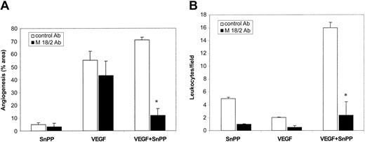
In order to investigate further the influence of the migrating leukocytes on angiogenesis the inhibitory anti-β2 mAb M18/2 was used. Treatment of mice with M18/2 did not affect VEGF-induced angiogenesis per se but abrogated the previously observed enhancement of VEGF-induced angiogenesis in the presence of SnPP (P < .05; Figure 5A). This was related to a specific suppression of leukocytic infiltration into Matrigel plugs by M18/2, as an isotype-matched control mAb did not affect the proinflammatory effects of SnPP (Figure 5B) and no effect of mAb M18/2 was seen on VEGF-induced angiogenesis (Figure 5A). Furthermore, in the absence of leukocyte infiltration, SnPP completely inhibited VEGF-induced angiogenesis (Figure 5A) supporting a role for HO-1 in angiogenesis directly driven by VEGF.
Effect of anti–β2-integrinAb M18/2 on angiogenesis and leukocyte infiltration. M18/2 or a isotype-matched control mAb was added to Matrigel and injected intraperitoneally every other day, as described in “Materials and methods.” (A) Quantitative evaluation of neovessels infiltrating Matrigel after treatment with SnPP, VEGF, or VEGF plus SnPP in presence of M18/2 or a control mAb. The results are expressed as percentage ± SE of the vessel area with respect to the total Matrigel area. (B) Quantitative evaluation of inflammatory cells infiltrating Matrigel counted in hematoxylin and eosin–stained sections. The results are expressed as the mean ± SE of cells/field (× 400). Four mice were used per condition in each experiment. ANOVA with Dunnett multicomparison test was performed between control Ab and M18/2 (*P < .05).
Effect of anti–β2-integrinAb M18/2 on angiogenesis and leukocyte infiltration. M18/2 or a isotype-matched control mAb was added to Matrigel and injected intraperitoneally every other day, as described in “Materials and methods.” (A) Quantitative evaluation of neovessels infiltrating Matrigel after treatment with SnPP, VEGF, or VEGF plus SnPP in presence of M18/2 or a control mAb. The results are expressed as percentage ± SE of the vessel area with respect to the total Matrigel area. (B) Quantitative evaluation of inflammatory cells infiltrating Matrigel counted in hematoxylin and eosin–stained sections. The results are expressed as the mean ± SE of cells/field (× 400). Four mice were used per condition in each experiment. ANOVA with Dunnett multicomparison test was performed between control Ab and M18/2 (*P < .05).
HO-1 activation inhibits leukocyte recruitment and inflammation-related angiogenesis
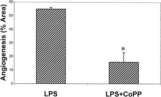
To explore these findings further, lipopolysaccharide was added to Matrigel plugs as a representative model of inflammatory angiogenesis in vivo. This model is characterized by an intense leukocytic infiltration and an associated angiogenic response.36 As expected, the inclusion of LPS alone induced leukocytic infiltration into the Matrigel plug with significant angiogenesis (Figure 4E and 6). To investigate the effect of HO-1 activation in this model of angiogenesis mice were treated with the CoPP. As seen in Figure 4F and Figure 6, addition of CoPP significantly inhibited leukocyte-induced angiogenesis, suggesting that HO-1 may act to suppress inflammatory angiogenesis in vivo.
Effect of HO-1 induction on inflammatory angiogenesis in vivo. Matrigel containing LPS (10 ng/mL) plus CoPP (25 μM) was injected subcutaneously into C57BL/6 mice. After 6 days plugs were explanted and fixed in formalin and paraffin embedded. Quantification of neovascularization was performed on hematoxylin and eosin–stained sections as described in “Materials and methods.” The results are expressed as percentage ± SE of the vessel area with respect to the total Matrigel area. Nine mice were used per condition in each experiment. ANOVA with Dunnett multicomparison test was performed between LPS and LPS plus CoPP (*P < .05).
Effect of HO-1 induction on inflammatory angiogenesis in vivo. Matrigel containing LPS (10 ng/mL) plus CoPP (25 μM) was injected subcutaneously into C57BL/6 mice. After 6 days plugs were explanted and fixed in formalin and paraffin embedded. Quantification of neovascularization was performed on hematoxylin and eosin–stained sections as described in “Materials and methods.” The results are expressed as percentage ± SE of the vessel area with respect to the total Matrigel area. Nine mice were used per condition in each experiment. ANOVA with Dunnett multicomparison test was performed between LPS and LPS plus CoPP (*P < .05).
Discussion
Previous in vitro studies have reported a role for HO-1 in EC survival, proliferation, and tube formation, suggesting that HO-1 induction may promote angiogenesis and that this is mediated, at least in part, by CO.25,26 A recent study has also reported HO-1 induction in the chicken chorioallantoic membrane following treatment with VEGF.37 We found that VEGF, a potent angiogenic factor, induced HO-1 expression and increased HO-1 activity in human ECs. The potential involvement of endothelial HO-1 in VEGF-driven angiogenesis was demonstrated by the ability of the HO-1 antagonists SnPP and ZnPP to significantly inhibit EC proliferation and tube formation in vitro. Furthermore, the results presented herein demonstrate for the first time that HO-1 plays an important role in VEGF-driven angiogenesis in vivo and acts as a key regulator in the control of the anti-inflammatory versus proinflammatory actions of VEGF.
VEGF is known to act on both leukocytes and ECs. However, in vivo models have suggested that VEGF is principally an inducer of noninflammatory angiogenesis.38 This may reflect a predominant influence of VEGF on ECs rather than leukocytes. In our in vivo experiments inhibition of HO-1 with SnPP shifted VEGF-driven angiogenesis from noninflammatory to proinflammatory, as evidenced by the presence of a dense leukocytic infiltrate. This suggests that induction of HO-1 by VEGF may act to both promote endothelial proliferation and to limit proinflammatory leukocytic infiltration. This hypothesis was confirmed in vivo by the effect of the inhibitory anti-β2 integrin mAb M18/2. M18/2 did not affect VEGF-induced angiogenesis per se but completely abrogated the VEGF-induced leukocytic infiltration observed in the presence of HO-1 inhibitors. Moreover, when leukocyte infiltration was inhibited by M18/2, the HO-1 antagonists ZnPP and SnPP inhibited VEGF-induced angiogenesis confirming the role of HO-1 in this process. Furthermore, experiments performed with aFGF, which did not induce HO-1, showed that blockade of HO-1 did not influence aFGF-induced angiogenesis, thus demonstrating that the HO-1 inhibitors themselves do not affect angiogenesis. These data support previous studies showing that VEGF and FGF induce angiogenesis through distinct pathways.39
One of the products of HO-1 activation, CO, is an endogenously produced vasorelaxant molecule that activates soluble guanylate cyclase leading to an increase in intracellular cyclic guanosine monophosphate (cGMP).40 Recent work has demonstrated that treatment with CO prevents arteriosclerosis in a model of aortic transplantation in which CO exerts a marked inhibitory effect on graft leukocyte infiltration and activation and reduces vascular smooth muscle cell proliferation via generation of cGMP and activation of p38 mitogen-activated protein kinase (MAPK).41 Activation of guanylate cyclase is also induced by nitric oxide (NO), the major secondary mediator of VEGF effects.42 It is therefore possible that HO-1 and NO synthase exert a synergistic effect during VEGF-induced angiogenesis. Moreover, HO-1 may play a role in different angiogenic settings, such as during hypoxia, where NO production and endothelial NO synthase expression are suppressed.43 Indeed, a role for HO-1 in angiogenesis during hypoxia has been suggested by the observation that HO-1 activation enhances VEGF production and that VEGF induced by hypoxia was down-regulated by HO-1 inhibitors but not by inhibitors of NO synthase.44
HO-1 breaks down heme to equimolar amounts of CO, biliverdin, and free iron. Biliverdin is rapidly reduced to bilirubin by the action of biliverdin reductase. Although less is known about the anti-inflammatory and cytoprotective effects of bilirubin, these have been clearly demonstrated.45,46 Hence, the relative contribution of bilirubin and CO in mediating the effects of VEGF-induced HO-1 in angiogenesis merits further investigation.
An anti-inflammatory effect of HO-1 has been demonstrated in vitro and in vivo. The induction of HO-1 decreases EC expression of the cellular adhesion molecule ICAM-122 and inhibits monocyte chemotaxis.15 Pretreatment with HO-1 agonists attenuates tissue damage in a variety of models including carrageenan-induced pleuritis, endotoxic shock, and oxidant-induced lung injury.14,47,48 Moreover, increased leukocyte adhesion to the vessel wall and spontaneous perivascular infiltration of leukocytes into the liver, lungs, and kidneys was observed in HO-1–deficient mice.19 To investigate the role of HO-1 in inflammatory angiogenesis we used LPS-conditioned Matrigel. In this model,36 neutrophils invade and begin to degrade the gel creating clefts and this is followed by migration of macrophages. Growth factors, such as VEGF and basic FGF (bFGF), released by neutrophils and macrophages subsequently induce EC migration and tube formation. Thus, this model of angiogenesis is directly dependent upon leukocyte migration. Treatment of mice with the HO-1 inducer CoPP significantly inhibited LPS-induced leukocyte infiltration and the subsequent angiogenic response. This suggests that HO-1–mediated inhibition of leukocyte recruitment represents an important mechanism for the control of angiogenesis associated with inflammation.
The data presented herein, and in other recent studies, suggests that many anti-inflammatory and cytoprotective molecules act through a mechanism dependent upon the expression of HO-1 and that the products of HO-1 facilitate the protective effects.46 This is supported by the demonstration that HO-1 expression, or treatment with CO, reproduces these beneficial actions. Thus, HO-1 and CO have been shown to mediate the protective effects of interleukin-10 in LPS-induced septic shock in mice.49 Further examples of the pivotal role of HO-1 include its importance in the antiproliferative effects of rapamycin,50 the anti-inflammatory actions of 15-Deoxy-12,14-prostaglandin J2,51 and the cytoprotective effects of heat-shock preconditioning.52
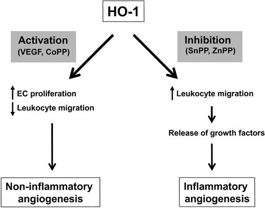
VEGF may act both as a potent proangiogenic factor and as a leukocyte chemoattractant. We propose that HO-1 induced by VEGF acts to promote angiogenesis while inhibiting the local recruitment of leukocytes (Figure 7). Thus, in the setting of inflammatory diseases associated with VEGF-driven angiogenesis, such as rheumatoid arthritis, the action of VEGF-induced HO-1 may be to maximize angiogenesis associated with the resolution of tissue injury while inhibiting leukocyte adhesion and transmigration. However, HO-1 induction may be impaired during inflammation, as recently reported in human chronic graft rejection.53 Thus, therapeutic induction of HO-1, or delivery of CO or bilirubin, may be beneficial in the treatment of chronic inflammatory diseases, not only through their anti-inflammatory actions but also via a proangiogenic effect enhancing tissue repair.
Proposed mechanism for the actions of HO-1 in vivo. HO-1 activation by VEGF favors endothelial cell proliferation while inhibiting leukocyte migration and activity, thus resulting in a predominantly noninflammatory angiogenesis. However, when HO-1 is inhibited, there is increased leukocyte migration with subsequent local release of growth factors and induction of inflammatory angiogenesis.
Proposed mechanism for the actions of HO-1 in vivo. HO-1 activation by VEGF favors endothelial cell proliferation while inhibiting leukocyte migration and activity, thus resulting in a predominantly noninflammatory angiogenesis. However, when HO-1 is inhibited, there is increased leukocyte migration with subsequent local release of growth factors and induction of inflammatory angiogenesis.
Prepublished online as Blood First Edition Paper, October 2, 2003; DOI 10.1182/blood-2003-06-1974.
Supported by grants from Arthritis Research Campaign grant M0620, British Heart Foundation Programme grant RG/98/0003, and the Italian Ministry of University and Research (MIUR) ex 60%, and by the Special Project “Oncology,” Compagnia San Paolo, Torino, Italy. J.C.M. is an Arthritis Research Campaign Senior Fellow.
The publication costs of this article were defrayed in part by page charge payment. Therefore, and solely to indicate this fact, this article is hereby marked “advertisement” in accordance with 18 U.S.C. section 1734.


![Figure 2. Role of HO-1 in VEGF-induced angiogenesis in vitro. (A) HUVECs were cultured in M199 with 10% FBS for 24 hours prior to the addition of VEGF (25 ng/mL) and CoPP (12.5 and 25 μM) both for 72 hours. In test wells, ECs were preincubated with SnPP at the concentrations shown for 60 minutes prior to the addition of VEGF. 3H-thymidine (0.037 MBq/mL [1 μCi/mL]) was added 16 hours before the end of the assay and the plates were read on an automated plate reader. Bars show the mean + SD uptake of 3H-thymidine (n = 4). (B-F) Murine abdominal muscle was implanted in Matrigel and cultured in DMEM supplemented with 10% FBS and heparin (10 U/mL). VEGF (25 ng/mL) was added to the medium in the absence and presence of SnPP (20 μM). (B) After 9 days EC tube length was measured by image analysis. Data presented as mean + SD tube length (n = 3). Panels C-F show representative micrographs of the effect of SnPP on neovessels formed in the presence of VEGF. Panels C and E show VEGF alone and panels D and F show VEGF + SnPP. Original magnification: low power × 20 (C-D) and high power × 40 (E-F).](https://ash.silverchair-cdn.com/ash/content_public/journal/blood/103/3/10.1182_blood-2003-06-1974/6/m_zh80030456170002.jpeg?Expires=1769082910&Signature=l9yGK53HNQ62XbQ1R00vsPn-LLay5Mu6LcCtHWX6~xJDcxcRsbRFf4YL0WSjcwo8qE2tNq6How3EP9i2RwxkEk-IyJlXV67xA3Uy6uOLr5YQbcN3U0Fi8vmnnSSMXyuj~ULTs4rrxj~x7lblxOwXdprUGco9PSMTTjkH7r5yPxpLUw-ZS70vHqkBVSmkDwFlo4UrKEib-uipYhwUmpxNKfquAP9TJoc-~qPwxVEFjKfuzEtRE~qBuxPKxGM~oNifctP3SRM2SSxDGmg7KidlzGausxlblDFiUGQVieMSEbw0GArS4JZpv6mkOAZD2i1NVQg~AcY7r-3Ob2J7f9MOtA__&Key-Pair-Id=APKAIE5G5CRDK6RD3PGA)


This feature is available to Subscribers Only
Sign In or Create an Account Close Modal