Abstract
The transcription factor NF-κB plays a central role in regulating inflammation and apoptosis, making it a compelling target for drug development. We identified a small molecule inhibitor (ML120B) that specifically inhibits IKKβ, an Ikappa-B kinase that regulates NF-κB. IKKβ and NF-κB are required in vivo for prevention of TNFα-mediated apoptosis. ML120B sensitized mouse bone marrow progenitors and granulocytes, but not mature B cells to TNFα killing in vitro, and induced apoptosis in vivo in the bone marrow and spleen within 6 hours of a single oral dose. In vivo inhibition of IKKβ with ML120B resulted in depletion of thymocytes and B cells in all stages of development in the bone marrow but did not deplete granulocytes. TNF receptor–deficient mouse thymocytes and B cells were resistant to ML120B-induced depletion in vivo. Surprisingly, surviving bone marrow granulocytes expressed TNFR1 and TNFR2 after dosing in vivo with ML120B. Our results show that inhibition of IKKβ with a small molecule in vivo leads to rapid TNF-dependent depletion of T and B cells. This observation has several implications for potential use of IKKβ inhibitors for the treatment of inflammatory disease and cancer.
Introduction
NF-κB plays a central role in inflammation and apoptosis, making it a primary target for inhibition for treatment of inflammatory disease and cancer.1,2 NF-κB consists of subunits that associate in dimers.3,4 The commonly described dimer forms are p50 (NF-κB1) with RelA (p65) and p52 (NF-κB2) with RelB. IκBα holds the p50/RelA complex in the cytoplasm in an inactive form. NF-κB nuclear translocation is regulated by upstream signaling events initiated primarily by proinflammatory stimuli, resulting in activation of IKK.5 IKK contains 3 related subunits: α, β, and γ. IKKβ is the subunit responsible for phosphorylating IκBα, resulting in its ubiquitination and subsequent proteasomal degradation.6,8 Released NF-κB translocates to the nucleus to initiate transcription of response genes, which include proinflammatory and antiapoptotic genes.9 In addition to phosphorylating IκBα, IKKβ also phosphorylates the transcription factor FOXO3a, which is also degraded by the proteasome.10 FOXO3a that is not phosphorylated by IKKβ can translocate to the nucleus where it upregulates expression of genes that control cell cycle progression11,13 and induce apoptosis.14,15
NF-κB activation has been associated with several human immune modulated diseases including arthritis, asthma, diabetes, stroke, inflammatory bowel disease, and atherosclerosis.16 In addition, IKKβ and NF-κB have been shown to play a direct role in regulating the development of insulin resistance17,18 and in severe muscle wasting.19 Recently, it has been demonstrated that IKKβ and NF-κB play a key role in inflammation-associated cancer and tumor progression.20,24 These data suggest that inhibition of IKKβ in vivo with a specific small molecule may be a potential therapeutic approach for several human diseases.
In addition to controlling inflammatory response and apoptosis, IKKβ and NF-κB are required for normal lymphocyte development. Mice deficient in the p65 (RelA) subunit of NF-κB or IKKβ die during fetal development through a TNF-dependent mechanism.6,25,26 Generation of p65- or IKKβ-deficient mice on a TNFα- or TNFR-deficient background rescues mice from embryonic lethality.27,29 Studies evaluating the requirement for NF-κB in hematopoiesis using transplantation of fetal liver stem cells from p65- or IKKβ-deficient mice into lethally irradiated hosts have revealed a specific requirement for these molecules in development of T cells, B cells, and common lymphoid progenitors but not myeloid cells or stem cells and a role in regulating the production of granulocytes.30,32
We identified ML120B, a specific small chemical inhibitor of IKKβ that protects against cartilage and bone destruction in adjuvant and collagen-induced models of arthritis.33 Here we demonstrate that IKKβ inhibition using a specific chemical inhibitor results in TNFR-dependent thymocyte depletion and B-cell progenitor death in vitro and in vivo.
Materials and methods
IKK complex enzyme assay
The IKK complex was purified as previously described.34,35 Inhibition of the IKK complex by ML120B was evaluated at 50 μM ATP using biotinylated GST–tagged IκBα (5-55) as a substrate and the reaction was detected using Europium-labeled (Perkin Elmer, Wellesley, MA) anti–phospho-IκBα monoclonal antibody 12C2 (Molecular Probes, Eugene, OR). ATP competitive inhibition was performed by evaluating phosphorylation of biotinylated GST–tagged IκBα and varying concentrations of ATP.
Mice
All animal procedures were done in accordance with the guidelines of the Institutional Animal Care and Use Committee. In vivo and in vitro assays were carried out with female C57BL/6 mice (Charles River Laboratories, Bedford, MA). TNFR1-deficient (C57BL/6-Tnfrsf1atm1Imx/J), TNFR2-deficient (B6.129S2-Tnfrs1btm1Mwm/J), and TNFR1/TNFR2 double knockout mice (B6;129S-Tnfrsf1a tm1Imx Tnfrsf1b tm1Imx/J) were obtained from The Jackson Laboratory (Bar Harbor, ME). Transgenic mice expressing luciferase driven by NF-κB response elements36 were obtained and licensed from MiceTech (Oslo, Norway).
Pharmacokinetic studies
Mice were administered a single oral dose of ML120B (300 mg/kg) and blood was collected at 1, 4, 8, and 24 hours after dosing (n = 3 mice per time point). ML120B plasma concentrations were determined by a HP-1100 LC (Hewlett-Packard, Houston, TX), a CTC leap auto sampler (LEAP Technologies, Carrboro, NC), and a SCIEX API 3000 detector (Applied Biosystems, Concord, ON, Canada) according to standard analytic methods. A peak area ratio versus concentration calibration curve was used to quantify the sample concentrations. Separation was performed on a YMC-ODS-AQ 2.0 × 3.0 mm high-pressure liquid chromatography (HPLC) column (Waters, Milford, MA) and the analytes were eluted at a flow rate of 0.4 mL/min. Mobile phase A consisted of 0.1% (vol/vol) formic acid in water, and the mobile phase B consisted of 0.1% (vol/vol) formic acid in acetonitrile. ML120B was detected by positive ion spray in the multiple-reaction monitoring (MRM) mode. The parent-daughter mass transition monitored was 519.30:279.96 mass-charge ratio (m/z). The calibration curve was linear in the concentration range of 1.0 to 2500 nM.
Flow cytometry and in vivo imaging
Bone marrow single-cell suspensions were incubated with fluorescein isothiocyanate (FITC)–, phycoerythrin (PE)–, or allophycocyanin (APC)–conjugated antibodies after red blood cell lysis. Stained cells were detected on a BD LSR flow cytometer (Becton Dickinson, San Jose, CA). PE-conjugated anti-IgM (R6-60.2), anti–GR-1 (RB6-8C5), anti-TNFR type II/p75 (TR75-89), APC-conjugated anti-Mac1 (M1/70), anti-B220 (RA-60.2), anti-TNFR type I/p55 (55R-286), FITC-conjugated anti-CD43 (S7), and anti–hamster IgG were from Pharmingen (San Diego, CA). NF-κB–dependent luminescence in transgenic mice expressing luciferase was performed as described previously36 using an IVIS imaging system and Living Image software version 2.5 (Xenogen, Alameda, CA).
B-cell isolation
Bone marrow single-cell suspensions from C57BL/6 female mice were incubated with Mac1-PE (M1/70), CD3-PE (145-2C11), GR-1–PE (RB6-8C5), and Ter199-PE (Ter-110; Pharmingen). PE-positive cells were removed by Macs separation columns (Miltenyi Biotech, Auburn, CA) after incubating with Macs anti-PE–conjugated beads (Miltenyi Biotech).
Western blot analysis
Antibodies against IκBα (C-21), HSC70 (K-19), and FOXO3a (sc-11351) were from Santa Cruz Biotech (Santa Cruz, CA). Phospho-specific antibody against IκBα (Ser32/36; polyclonal) was from Cell Signaling (Beverly, MA). Horseradish peroxidase (HRP)–conjugated secondary antibodies for anti–mouse IgG and anti–rat IgG were from Amersham Biosciences (Piscataway, NJ). Whole bone marrow cell lysates were prepared from 1 × 10⁷ cells in cell lysing buffer (Cell Signaling) with protease inhibitor cocktail (Roche Applied Science, Indianapolis, IN). Lysates were resolved on 4% to 12% NuPage gel (Invitrogen, Carlsbad, CA) and transferred onto nitrocellulose paper (Invitrogen). The blots were blocked with 0.5% nonfat milk in TBS with 0.1% Tween 20 and then incubated with primary antibodies. Bound HRP-conjugated secondary antibodies were detected by enhanced chemiluminescence (ECL) Western blotting detection reagents (Amersham) on light-sensitive film.
Histologic analysis
For histologic analysis, tissues were fixed with formalin and sections were stained hematoxylin and eosin (H&E). Microscopic evaluation was performed using a Nikon Eclipse E800 microscope and objectives as listed in the figure legends (Nikon, Melville, NY). Images were captured with a SPOT RT color model 2.2.1 camera and analyzed using SPOT software version 3.5 (Diagnostic Instruments, Sterling Heights, MI).
Detection of apoptosis
Bone marrow cells were flushed from femurs of C57BL/6 mice that had been administered ML120B at either 100 mg/kg or 300 mg/kg and vehicle only were fixed in 3.7% formaldehyde. Apoptosis was detected by using apoptosis detection kits (R&D Systems, Minneapolis, MN) following the manufacturer's protocol.
In vitro colony-forming assays
Bone marrow single-cell suspensions were plated in methylcellulose semisolid media in Iscove modified Dulbecco media (IMDM) supplemented with 10 ng/mL human IL-7 (2 × 10⁵ cells/mL) for B-cell colony growth or with 10 ng/mL human IL-6, 10 ng/mL murine IL-3, and 50 ng/mL murine SCF with 4 × 10⁴ cells/mL for myeloid colony growth (Stem Cell Technologies, Vancouver, BC, Canada). Colonies were scored on day 7. In experiments where cells were cultured in liquid suspension prior to plating in colony-forming assays, cells were plated at 2 × 10⁶ cells in 1 mL of IMDM with 10% FCS and the presence or absence of 100 ng/mL mouse or human TNFα (Pharmingen) and cultured for 4 hours prior to washing and plating.
Results
ML120B is a specific inhibitor of IKKβ and inhibits cytokine production and IκBα phosphorylation in bone marrow cells in response to LPS
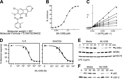
We identified a specific small molecule inhibitor of IKKβ, ML120B. ML120B inhibits IKKβ with a 50% inhibitory concentration (IC50) of 0.06 μM (Figure 1A-B) and is an ATP competitive inhibitor (Figure 1C). We evaluated ML120B for selectivity in a screen of 30 tyrosine and serine-threonine kinases and found ML120B is greater than 1000 times more potent in inhibiting IKKβ than other inhibiting kinases (data not shown). IKKα was not inhibited by ML120B at a concentration of 100 μM. Specific chemical and pharmacodynamic properties of ML120B will be published elsewhere (Julie Fields Liu, M.H., Prakash Raman, Jeremy D. Little, François Soucy, Robert S. Murray, Yingchun Ye, Julian Adams, G.H., Alfredo Castro, Hormoz Mazdiyasni, Paul Fleming, Dominic Reynolds, T.O., B.J., Y.X., H.Y., D.W., and A.B., manuscript in preparation). In order to determine potency of ML120B on primary cells, we cultured mouse bone marrow cells in media containing lipopolysaccharide (LPS) in the presence of an increasing concentration of ML120B, and after 4 hours supernatant was evaluated for quantity of IL-6 and RANTES. ML120B inhibited IL-6 and RANTES production in response to LPS in a dose-dependent manner, with a half-maximal inhibition (IC50) at 1 μM ML120B (Figure 1D). Phosphorylation and degradation of IκBα and nuclear translocation of p65 in mouse bone marrow in response to LPS was also evaluated. LPS induced phosphorylation of IκBα and nuclear translocation of p65 within 15 minutes and detectable degradation of IκBα within 60 minutes (Figure 1E-F). It is of interest to note that p65 translocation occurred in this setting prior to detection of IκBα degradation. Data from a number of laboratories suggest that mechanisms other than degradation of IκBα are involved in regulating trafficking of NF-κB between the nucleus and cytoplasm.4 ML120B inhibited IκBα phosphorylation and degradation and nuclear translocation of p65. Therefore, the presence of ML120B in mouse bone marrow cultures inhibited both cytokine production IκBα phosphorylation and degradation.
ML120B is a reversible competitive inhibitor of IKKβ and inhibits cytokine production and IκBα phosphorylation and degradation in stimulated bone marrow cells. (A) Molecular structure of ML120B. (B) Inhibition of the IKK complex by ML120B at 50 μM ATP, using biotinylated GST–tagged IκBα (5-55) as a substrate. The data represent the average of triplicate measurements, with error bars indicating SD. (C) ML120B is an ATP competitive inhibitor of the IKK complex. Phosphorylation of biotinylated GST–tagged IκBα (5-55) was measured at fixed inhibitor concentrations of 167 μM (▪), 111 μM (▴), 75 μM (♦), 50 μM (•), 33 μM (□), 22 μM (▵), and 0 μM (⋄). (D) Bone marrow was isolated from a C57BL/6 female mouse and red blood cells were lysed and plated into a 96-well plate (4 × 10⁵ cells/well). Cells were preincubated with ML120B for 1 hour and then stimulated with LPS (1 μg/mL). Supernatants were collected after 4 hours and IL-6 and RANTES levels were evaluated. (E) Western blot analysis of bone marrow preincubated with ML120B (30 μM) or media with equivalent DMSO (0.5%) for 1 hour and then stimulated with LPS (1 μg/mL) for 5, 15, 30, and 60 minutes. (F) Nuclear translocation of p65. Bone marrow cells were stimulated as described in panel D and nuclear extracts were analyzed by immunoblotting. Error bars indicate SD.
ML120B is a reversible competitive inhibitor of IKKβ and inhibits cytokine production and IκBα phosphorylation and degradation in stimulated bone marrow cells. (A) Molecular structure of ML120B. (B) Inhibition of the IKK complex by ML120B at 50 μM ATP, using biotinylated GST–tagged IκBα (5-55) as a substrate. The data represent the average of triplicate measurements, with error bars indicating SD. (C) ML120B is an ATP competitive inhibitor of the IKK complex. Phosphorylation of biotinylated GST–tagged IκBα (5-55) was measured at fixed inhibitor concentrations of 167 μM (▪), 111 μM (▴), 75 μM (♦), 50 μM (•), 33 μM (□), 22 μM (▵), and 0 μM (⋄). (D) Bone marrow was isolated from a C57BL/6 female mouse and red blood cells were lysed and plated into a 96-well plate (4 × 10⁵ cells/well). Cells were preincubated with ML120B for 1 hour and then stimulated with LPS (1 μg/mL). Supernatants were collected after 4 hours and IL-6 and RANTES levels were evaluated. (E) Western blot analysis of bone marrow preincubated with ML120B (30 μM) or media with equivalent DMSO (0.5%) for 1 hour and then stimulated with LPS (1 μg/mL) for 5, 15, 30, and 60 minutes. (F) Nuclear translocation of p65. Bone marrow cells were stimulated as described in panel D and nuclear extracts were analyzed by immunoblotting. Error bars indicate SD.
ML120B inhibits NF-κB activation and rapidly induces apoptosis in lymphoid organs in vivo
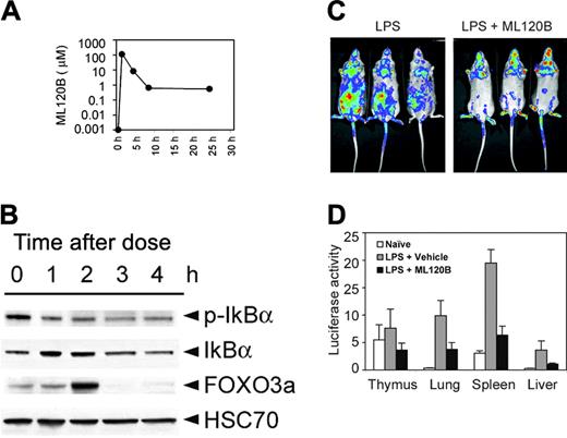
In order to evaluate pharmacokinetics, mice were administered a single oral dose of ML120B (300 mg/kg). Plasma was collected prior to dosing and 1, 4, 8, and 24 hours after dosing for pharmacokinetic analysis (Figure 2A). Maximal exposure (Cmax) of ML120B at 300 mg/kg was 113 μM, which occurred at 1 hour after dosing. Based on the pharmacokinetic analysis, a predicted effective level would be maintained for 4 to 6 hours in vivo after a single oral dose of 300 mg/kg. The 2 known targets for IKKβ phosphorylation and subsequent ubiquitination and proteasomal-mediated degradation are IκBα and transcription factor FOXO3a. We purified bone marrow B cells and evaluated IκBα and FOXO3a at various times after a single oral dose of ML120B (Figure 2B). Within one hour after a single oral dose, phosphorylation of IκBα decreased. Total IκBα and FOXO3a increased for 2 hours before returning to baseline levels. Similarly, FOXO3a levels accumulated at 2 hours and declined by 3 hours after a single oral dose. We evaluated ML120B in vivo by analyzing transgenic mice that express luciferase under the control of NF-κB response elements.34 Treating mice with LPS results in an increase in luminescence (not shown). A single oral dose of ML120B 1 hour prior to treating with LPS resulted in a decrease of luminescence (Figure 2C). Analysis of individual organs (thymus, lung, spleen, liver) from mice treated with LPS and a single oral dose of vehicle alone showed increased luminescence when compared with naive mice (Figure 2D). A single oral dose of ML120B 1 hour prior to treating mice with LPS effectively decreased luminescence. These data indicate ML120B is effective in inhibiting IKKβ-dependent NF-κB activation in vivo.
NF-κB is known to inhibit TNF-mediated apoptosis in vivo. We therefore evaluated spleen and thymus sections and bone marrow cells for evidence of apoptosis 6 hours after a single oral dose of ML120B. Areas of apoptotic cells were clearly visible by histology in sections of both spleen and bone marrow treated with ML120B when compared with those treated with vehicle alone (Figure 3A,D). Analysis of viable pre–B-cell content in the bone marrow (B220+/IgM–/annexin V–) demonstrated a decrease at 100 and 300 mg/kg without a significant change in total marrow cellularity (Figure 3B). A similar decrease of viable B cells in the spleen (B220+ annexin V–) was also observed (Figure 3D). Cytospins of bone marrow cells showed a greater than 2-fold increase of apoptotic cells detected by labeling fragmented DNA (Figure 3C). Apoptotic cells were evident in the spleen and thymus within 6 hours of a single oral dose of ML120B by staining for nuclear fragmentation (Figure 3D-E). There was a dose-dependent increase in apoptotic cells within the follicles and a disruption of splenic structure as determined by B220 staining within 18 hours. Similarly, ML120B induced a significant increase in apoptotic thymocytes within 6 hours (Figure 3E). Interestingly, significant changes in luciferase levels were not observed in NF-κB transgenic mice (Figure 2D), suggesting that apoptosis in the thymus may be sensitive to small changes in NF-κB activity. The data suggest that ML120B induces a rapid apoptotic response in primary and secondary lymphoid organs.
ML120B inhibits NF-κB activation in vivo. (A) Pharmacokinetic analysis of IKKβ inhibitor ML120B. Mice were given a single oral dose (300 mg/kg) and plasma concentration was determined at 1, 4, 8, and 24 hours after dosing (n = 3 mice per time point). (B) Western blot analysis of IκBα, phosphorylated IκBα (p-IκBα), and FOXO3a in purified bone marrow B cells 1, 2, 3, and 4 hours after a single oral dose of ML120B. Marrow was isolated at indicated times and B cells were purified by negative selection by removing non–B cells with antibodies to Mac1, CD3, Gr-1, and Ter199-PE and passing cells through a magnetic bead separation column. (C) Image of luciferase activity in transgenic mice. Mice were given a single oral dose of vehicle or ML120B (300 mg/kg) and 1 hour later injected with LPS and photographed 4 hours later after injection of D-luciferin. (D) Naive mice, mice treated with vehicle and LPS, or mice treated with ML120B and LPS were evaluated. Individual organs were removed and photon counts were collected (n = 3 per group).
ML120B inhibits NF-κB activation in vivo. (A) Pharmacokinetic analysis of IKKβ inhibitor ML120B. Mice were given a single oral dose (300 mg/kg) and plasma concentration was determined at 1, 4, 8, and 24 hours after dosing (n = 3 mice per time point). (B) Western blot analysis of IκBα, phosphorylated IκBα (p-IκBα), and FOXO3a in purified bone marrow B cells 1, 2, 3, and 4 hours after a single oral dose of ML120B. Marrow was isolated at indicated times and B cells were purified by negative selection by removing non–B cells with antibodies to Mac1, CD3, Gr-1, and Ter199-PE and passing cells through a magnetic bead separation column. (C) Image of luciferase activity in transgenic mice. Mice were given a single oral dose of vehicle or ML120B (300 mg/kg) and 1 hour later injected with LPS and photographed 4 hours later after injection of D-luciferin. (D) Naive mice, mice treated with vehicle and LPS, or mice treated with ML120B and LPS were evaluated. Individual organs were removed and photon counts were collected (n = 3 per group).
IKKβ inhibition by ML120B induces apoptosis in bone marrow and spleen in vivo. (A) Histologic evaluation and nuclear fragmentation detection in situ with C57BL/6 bone marrow cytospins 6 hours after a single dose of vehicle alone or ML120B (300 mg/kg). Arrow indicates area of apoptotic foci. (B) Total cell count and percent viable pre–B cells (B220+/IgM–/annexin V–) in the bone marrow 6 hours after a single oral dose of ML120B. Mice were given a single oral dose as indicated (n = 4/group) and 6 hours later cell counts per femur (× 106) and flow cytometry were performed. Percent B220+/IgM–/annexin V– cells were calculated. (C) Nuclear fragmentation detection in situ with C57BL/6 bone marrow cytospins 6 hours after single oral dose with ML120B or vehicle control as indicated. One hundred to 200 cells were scored from cytospins from bone marrow of individual mice (n = 4/group). Histology and detection of nuclear fragmentation in spleens (D) and thymi (E) 6 hours after a single oral dose of ML120B. Arrow indicates area of apoptotic foci. B220 staining of spleen sections was performed 18 hours after a single oral dose, and percent viable B cells (B220+/annexin V–) in the spleen (n = 4 per group) 6 hours after a single dose are shown. For quantitation of cells positive for nuclear fragmentation, 3 to 4 spleens and 4 thymi were scored per dose of ML120B, 3 to 4 fields within 3 follicles were scored per spleen, and 3 fields were scored per thymus in the cortex region. Spleen and thymus data show average number of positive cells per field. Error bars indicate SD. Original magnifications: (A, C) 400 × (objective 40 ×/0.75 numeric aperture [NA]); (D, H&E and apoptotic) 200 × (objective 20 ×/0.50 NA); (D, B220) 40 × (objective 4 ×/0.13 NA); and (E) 100 × (objective 10 ×/0.30 NA).
IKKβ inhibition by ML120B induces apoptosis in bone marrow and spleen in vivo. (A) Histologic evaluation and nuclear fragmentation detection in situ with C57BL/6 bone marrow cytospins 6 hours after a single dose of vehicle alone or ML120B (300 mg/kg). Arrow indicates area of apoptotic foci. (B) Total cell count and percent viable pre–B cells (B220+/IgM–/annexin V–) in the bone marrow 6 hours after a single oral dose of ML120B. Mice were given a single oral dose as indicated (n = 4/group) and 6 hours later cell counts per femur (× 106) and flow cytometry were performed. Percent B220+/IgM–/annexin V– cells were calculated. (C) Nuclear fragmentation detection in situ with C57BL/6 bone marrow cytospins 6 hours after single oral dose with ML120B or vehicle control as indicated. One hundred to 200 cells were scored from cytospins from bone marrow of individual mice (n = 4/group). Histology and detection of nuclear fragmentation in spleens (D) and thymi (E) 6 hours after a single oral dose of ML120B. Arrow indicates area of apoptotic foci. B220 staining of spleen sections was performed 18 hours after a single oral dose, and percent viable B cells (B220+/annexin V–) in the spleen (n = 4 per group) 6 hours after a single dose are shown. For quantitation of cells positive for nuclear fragmentation, 3 to 4 spleens and 4 thymi were scored per dose of ML120B, 3 to 4 fields within 3 follicles were scored per spleen, and 3 fields were scored per thymus in the cortex region. Spleen and thymus data show average number of positive cells per field. Error bars indicate SD. Original magnifications: (A, C) 400 × (objective 40 ×/0.75 numeric aperture [NA]); (D, H&E and apoptotic) 200 × (objective 20 ×/0.50 NA); (D, B220) 40 × (objective 4 ×/0.13 NA); and (E) 100 × (objective 10 ×/0.30 NA).
Inhibition of IKKβ in vivo with ML120B results in depletion of bone marrow B cells and thymocytes
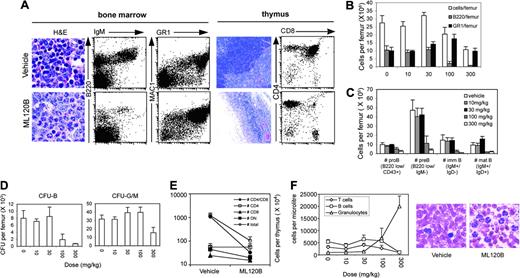
In order to determine consequences of prolonged inhibition of IKKβ, mice were administered ML120B twice daily by oral gavage for 4 consecutive days. On day 5, bone marrow and thymus were evaluated for cellular content. ML120B-treated mice showed dramatic depletion of lymphocytes in the bone marrow with maintenance of polymorphonuclear leukocytes when analyzed histologically (Figure 4A). Flow cytometric analysis showed a reduction in B220+ B cells and retention of granulocytes. Evaluation of cellular content in bone marrow from femurs showed a decrease in cellularity in all mice administered 100 and 300 mg/kg ML120B (Figure 4B). This effect was primarily due to a decrease in the number of B220+ B cells per femur, whereas the GR-1+ granulocyte numbers did not decline. We analyzed different stages of B-cell development in the bone marrow by cell surface expression of specific markers. All stages of B-cell development (pro-B, pre-B, immature, and mature) were depleted in the marrow at the higher doses of ML120B. We also evaluated progenitor (colony-forming unit [CFU]) content in the marrow (Figure 4D). While the B-cell progenitor (CFU-B) content declined after oral dosing of ML120B at 100 mg/kg, myeloid progenitor content remained unchanged. Similarly, thymocytes were depleted, primarily due to a reduction of CD4+CD8+ cells, whereas more mature CD4+ and CD8+ numbers were not significantly affected (Figure 4A,E). In order to determine consequences of prolonged exposure of ML120B on peripheral hematopoietic cells, we dosed mice for 7 days and analyzed peripheral blood content (Figure 4F). Interestingly, peripheral blood B-cell and T-cell numbers were not affected at a dose of 100 mg/kg, in contrast to the depletion of B cells observed in the bone marrow (Figure 4B) and the depletion of the thymus at the same dose (Figure 4E), suggesting either the stage of development or the local environment may play a role in sensitivity to ML120B-induced depletion. Granulocyte numbers increased dramatically at the higher doses. The granulocytosis is similar to what has been shown in bone marrow transplantation studies using donor cells from IKKβ- or p65-deficient mice.29,31 These results demonstrate ML120B induced a significant reduction in all stages of B-cell development in the bone marrow and a depletion of developing CD4+CD8+ thymocytes, whereas myeloid progenitors and granulocyte numbers were resistant to depletion in vivo.
ML120B sensitizes bone marrow cells and thymocytes to TNFα killing
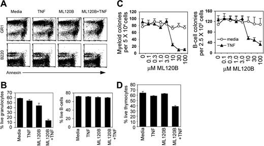
IKKβ is required in many cell types to prevent apoptosis in response to TNFα. To study the mechanism of depletion in the bone marrow we evaluated bone marrow B cells, granulocytes, and colony-forming units (CFUs) as well as thymocyte survival in vitro in the presence of ML120B and TNFα. Bone marrow and thymocytes were cultured in the presence or absence of ML120B in combination with TNFα and evaluated for induction of apoptosis by flow cytometry or plated in semisolid media to evaluate surviving myeloid and B-cell CFU content. ML120B and TNFα alone had little effect on granulocyte (GR-1+/annexin–) and thymocyte cell survival (Figure 5A-B,D), however, combined ML120B and TNFα significantly depleted the live granulocyte and thymocyte content in culture. Similarly, myeloid CFU survival was not affected by increasing concentrations of ML120B but was severely depleted in combination with TNFα, with a 10-fold shift in sensitivity (ML120B did not deplete myeloid CFU content at 100 μM but did at 10 μM in combination with TNFα; Figure 5C). This is in contrast to the IC50 of ML120B (1 μM) for inhibition of LPS-induced cytokine production and may suggest variable contribution of IKKβ activity to cytokine production and inhibition of apoptosis. In contrast to granulocyte survival, B-cell survival (B220+/annexin–) was not affected by TNFα or ML120B alone or in combination (Figure 5A-B). However, B-cell CFUs were depleted by ML120B in combination with TNFα (Figure 5C), which suggested that early B-cell progenitors but not pre-B or mature B cells in the bone marrow are sensitized to TNFα killing during IKKβ inhibition.
IKKβ inhibition by ML120B depletes all stages of B cells in the marrow and thymocytes but maintains granulocytes in vivo. (A) Histologic and flow cytometric analysis of bone marrow and thymus after twice daily oral dosing of ML120B. Representative bone marrow and thymus sections and flow cytometry after 4 days of dosing (100 mg/kg) with ML120B or vehicle alone. (B) Total cells, B220+ (B cells), GR-1+ (granulocytes), and B-cell subsets (C) per femur after oral dosing twice per day at indicated doses. All stages of B-cell development analyzed were depleted at 100 mg/kg and 300 mg/kg. (D) Bones were flushed and red blood cells (RBCs) were lysed and plated in semisolid media with IL-7 for B-cell colony growth or IL-6, IL-3, and SCF for myeloid colony growth. Colony-forming units (CFUs) were scored on day 7 and total number per individual femur was calculated (n = 4 per group). B-cell progenitors (CFU-Bs) are depleted at 100 mg/kg and 300 mg/kg. Myeloid progenitors (CFU-G/Ms) are unaffected at 100 mg/kg. (E) Thymocyte CD4, CD8, CD4/CD8 double positive (DP), and CD4/CD8 double negative (DN) numbers after 4 days of oral dosing at 100 mg/kg. (F) Numbers of T cells (CD4 and CD8+), B cells (B220+), and granulocytes (GR-1+) in peripheral blood after 7 days of ML120B at the indicated doses. Representative blood smears are shown from blood obtained in vehicle and a 300 mg/kg ML120B. Error bars indicate SD. Original magnifications: (A, bone marrow) 200 × (objective 20 ×/0.50 NA); (A, thymus) 40 × (objective 4 ×/0.13 NA); and (F) 400 × (objective 40 ×/0.75 NA).
IKKβ inhibition by ML120B depletes all stages of B cells in the marrow and thymocytes but maintains granulocytes in vivo. (A) Histologic and flow cytometric analysis of bone marrow and thymus after twice daily oral dosing of ML120B. Representative bone marrow and thymus sections and flow cytometry after 4 days of dosing (100 mg/kg) with ML120B or vehicle alone. (B) Total cells, B220+ (B cells), GR-1+ (granulocytes), and B-cell subsets (C) per femur after oral dosing twice per day at indicated doses. All stages of B-cell development analyzed were depleted at 100 mg/kg and 300 mg/kg. (D) Bones were flushed and red blood cells (RBCs) were lysed and plated in semisolid media with IL-7 for B-cell colony growth or IL-6, IL-3, and SCF for myeloid colony growth. Colony-forming units (CFUs) were scored on day 7 and total number per individual femur was calculated (n = 4 per group). B-cell progenitors (CFU-Bs) are depleted at 100 mg/kg and 300 mg/kg. Myeloid progenitors (CFU-G/Ms) are unaffected at 100 mg/kg. (E) Thymocyte CD4, CD8, CD4/CD8 double positive (DP), and CD4/CD8 double negative (DN) numbers after 4 days of oral dosing at 100 mg/kg. (F) Numbers of T cells (CD4 and CD8+), B cells (B220+), and granulocytes (GR-1+) in peripheral blood after 7 days of ML120B at the indicated doses. Representative blood smears are shown from blood obtained in vehicle and a 300 mg/kg ML120B. Error bars indicate SD. Original magnifications: (A, bone marrow) 200 × (objective 20 ×/0.50 NA); (A, thymus) 40 × (objective 4 ×/0.13 NA); and (F) 400 × (objective 40 ×/0.75 NA).
ML120B sensitizes bone marrow granulocytes and B-cell progenitors to killing by TNFα. (A) Bone marrow cells were isolated and incubated in media, ML120B alone (30 μM), murine TNFα alone (100 ng/mL), or ML120B + TNFα for 4 hours and then analyzed by flow cytometry for annexin with granulocyte marker GR-1 or B-cell marker B220. (B) Percent live granulocytes (GR-1+ annexin–) and live B cells (B220+annexin–) after 4 hours of culture. Values are averages of triplicates; error bars indicate SD. (C) Bone marrow cells were cultured in increasing concentrations of ML120B in the presence or absence of murine TNFα (100 ng/mL) for 4 hours then washed and plated in culture conditions for myeloid colony growth or B-cell colony growth. Colonies were scored 7 days after plating. All cultures contained 0.5% DMSO. (D) Percent live thymocytes (annexin–) after 6 hours of culture. Values are averages of triplicates; error bars indicate SD. One representative experiment of 3 with similar results is shown.
ML120B sensitizes bone marrow granulocytes and B-cell progenitors to killing by TNFα. (A) Bone marrow cells were isolated and incubated in media, ML120B alone (30 μM), murine TNFα alone (100 ng/mL), or ML120B + TNFα for 4 hours and then analyzed by flow cytometry for annexin with granulocyte marker GR-1 or B-cell marker B220. (B) Percent live granulocytes (GR-1+ annexin–) and live B cells (B220+annexin–) after 4 hours of culture. Values are averages of triplicates; error bars indicate SD. (C) Bone marrow cells were cultured in increasing concentrations of ML120B in the presence or absence of murine TNFα (100 ng/mL) for 4 hours then washed and plated in culture conditions for myeloid colony growth or B-cell colony growth. Colonies were scored 7 days after plating. All cultures contained 0.5% DMSO. (D) Percent live thymocytes (annexin–) after 6 hours of culture. Values are averages of triplicates; error bars indicate SD. One representative experiment of 3 with similar results is shown.
ML120B-induced bone marrow B-cell and thymocyte depletion in vivo is TNFR dependent
Multiple studies have demonstrated that TNFα mediates apoptosis in IKKβ- and NF-κB–deficient mice, resulting in embryonic lethality and lymphocyte deficiency in adoptive transfer experiments.27,29 Introducing a TNFα- or TNFR gene–deficient background reverses these events. In order to determine if TNF mediates thymocyte and B-cell depletion after IKKβ inhibition in vivo, we administered vehicle alone or ML120B to TNFR1- and TNFR2-deficient mice, TNFR1– and TNFR2–double-deficient mice, or wild-type mice at 100 mg/kg twice daily for 3 consecutive days. Mice were evaluated on day 4. Pre–B cells were depleted in control mice and TNFR2-deficient mice as evaluated by histology and flow cytometry (Figure 6A-B). However, marrow in TNFR1- and TNFR1/TNFR2-deficient mice appeared normal by histology and B220+IgM– pre–B-cell numbers were not significantly altered. Similarly, thymocyte depletion was rescued in TNFR1/TNFR2-deficient mice but rescue was not complete, suggesting other non-TNF–mediated mechanisms may be involved. In order to understand the mechanism of granulocyte survival in vivo, we next evaluated expression of TNFR1 and TNFR2 in the marrow of normal mice after dosing with vehicle alone or with ML120B. In vehicle-treated mice, greater than 90% of granulocytes express TNFR1 and this was not altered by treatment with ML120B (Figure 6C-D). Surprisingly, approximately 50% of granulocytes express TNFR2 in vehicle-treated control mice, whereas greater than 80% of granulocytes expressed TNFR2 after treatment with ML120B. In order to determine if myeloid CFUs present in the bone marrow after treatment were resistant to TNFα+ ML120B–induced killing in vitro, cells were removed from bone marrow, plated in media or TNFα+ ML120B for 4 hours, then washed and plated in myeloid CFU assay (Figure 6E). TNFα+ ML120B treatment for 4 hours in vitro–depleted bone marrow from both vehicle-treated mice and ML120B-treated mice, indicating that myeloid cells in the marrow did not develop a resistance to ML120B-induced apoptosis in vivo.
ML120B-induced B-cell depletion in vivo is TNFR dependent. (A) Representative bone marrow histology and (B) percent cellular decrease in bone marrow and thymus from wild-type control mice (wt), TNFR1-deficient mice, TNFR2-deficient mice, and TNFR1/TNFR2 double knockout mice (R1/R2–/–) after twice daily oral doses for 3 days (analysis on day 4), with vehicle alone or with ML120B (100 mg/kg). Percent decrease in pre–B cells (B220+/IgM–) and decrease in thymocyte numbers in ML120B-treated animals when compared with vehicle controls (n = 4 per group). (A) Original magnification, 400 × (objective 40 ×/0.75 NA). (C) TNFR1 and TNFR2 expression in bone marrow granulocytes (GR-1+). (D) The percent of GR-1+ cells expressing TNFR2 but not TNFR1 increases after ML120B dosing. (E) Myeloid progenitors (CFUs) surviving 3 days of ML120B dosing are not resistant to ML120B + TNFα killing in vitro. Bone marrow cells from individual mice (n = 4) were cultured in media or ML120B (30 μM) in the presence of murine TNFα (100 ng/mL) for 4 hours then washed and plated in culture conditions for myeloid colony growth. Colonies were scored 7 days after plating. All cultures contained 0.5% DMSO.
ML120B-induced B-cell depletion in vivo is TNFR dependent. (A) Representative bone marrow histology and (B) percent cellular decrease in bone marrow and thymus from wild-type control mice (wt), TNFR1-deficient mice, TNFR2-deficient mice, and TNFR1/TNFR2 double knockout mice (R1/R2–/–) after twice daily oral doses for 3 days (analysis on day 4), with vehicle alone or with ML120B (100 mg/kg). Percent decrease in pre–B cells (B220+/IgM–) and decrease in thymocyte numbers in ML120B-treated animals when compared with vehicle controls (n = 4 per group). (A) Original magnification, 400 × (objective 40 ×/0.75 NA). (C) TNFR1 and TNFR2 expression in bone marrow granulocytes (GR-1+). (D) The percent of GR-1+ cells expressing TNFR2 but not TNFR1 increases after ML120B dosing. (E) Myeloid progenitors (CFUs) surviving 3 days of ML120B dosing are not resistant to ML120B + TNFα killing in vitro. Bone marrow cells from individual mice (n = 4) were cultured in media or ML120B (30 μM) in the presence of murine TNFα (100 ng/mL) for 4 hours then washed and plated in culture conditions for myeloid colony growth. Colonies were scored 7 days after plating. All cultures contained 0.5% DMSO.
Discussion
NF-κB is an important regulator in inflammatory response and in protecting cells from apoptosis through diverse signal cascades. Because of these properties, therapeutic strategies for inhibiting NF-κB are being sought for treating inflammatory diseases such as arthritis and for treating cancers such as B-cell malignancies.37,39 IKKβ is a key kinase involved in linking multiple signals to NF-κB activation. Both NF-κB and IKKβ are required for normal lymphocyte development.40
IKKβ is required to protect T cells from apoptosis induced by TNF, and IKKβ-deficient thymocytes fail to develop in radiation chimeras unless in a TNF receptor–deficient mouse background.29 Both IKKα and IKKβ are required for peripheral B-cell maturation, maintenance, and function.41,44 Analysis of hematopoiesis in bone marrow of radiation chimeras using IKKα- and IKKβ-deficient donors revealed that IKKβ deficiency results in complete absence of B cells in the bone marrow,42 presumably due to defects in B-cell progenitors, whereas IKKα chimeras have normal numbers of pro– and pre–B cells in the marrow but have deficiencies in mature B cells.41
We developed ML120B, a specific small molecule inhibitor of IKKβ, and evaluated its impact on hematopoiesis in normal adult mice. ML120B inhibited cytokine production and IκBα phosphorylation in stimulated bone marrow cells in vitro and killed B cells, thymocytes, and myeloid progenitors in the presence of TNFα in a dose-dependent manner. Interestingly, under the same conditions, GR-1+ bone marrow granulocytes but not B220+ B cells were killed in response to ML120B and TNFα. TNFα induces apoptosis through binding of TNF receptors, inducing activation of a caspase cascade and activating pathways that lead to disruption of mitochondrial homeostasis.45 NF-κB is thought to prevent TNFα-induced apoptosis through nuclear translocation and subsequent upregulation of expression of genes involved in inhibiting caspase activation and in maintaining mitochondrial stability.46,48 Analysis of the expression of both TNFα receptors in bone marrow revealed that most GR-1+ granulocytes express TNFR1 and approximately 50% express TNFR2, consistent with the TNFα sensitivity to killing in the presence of ML120B in vitro. In contrast to granulocytes, most B cells in the bone marrow do not express either TNF receptor at levels sufficient enough to be detected by flow cytometry. This is consistent with our observation that B220+ bone marrow B cells do not become annexin+ in the presence of ML120B and TNFα in vitro, but B-cell progenitors are depleted under the same conditions. The role of TNF receptor expression in normal B-cell development in the marrow is unknown; TNFR1/TNFR2 double knockout mice do not display significant defects in lymphocyte development.49
Multiple dosing of ML120B in vivo lead to extensive depletion of thymocytes and all stages of B-cell development in the bone marrow but spared granulocyte production and induced granulocytosis in the peripheral blood. B-cell progenitors in the marrow were more sensitive to ML120B treatment in vivo than myeloid progenitors. This is similar to what has been previously shown in experiments with genetically deficient mice. This phenotype is observed in experiments where IKKβ - or p65-deficient fetal liver cells were transplanted into lethally irradiated hosts.29,31 Although the phenotype observed in p65 and IKKβ knockout experiments is similar to what we observed by inhibition with ML120B in adult animals, it is difficult to draw direct comparisons. Genetically deficient mice and transplantation studies require de novo hematopoiesis from stem cells during development or during reconstitution, respectively, whereas a chemical inhibitor affects ongoing hematopoiesis in the adult, and to some extent the phenotype is dependent on the pharmacokinetics of the molecule. ML120B has a maximal concentration in the serum of mice 1 hour after a single oral dose, and by 6 hours the concentration is below the dose required to kill B-cell progenitors in vitro. In vivo ML120B alters both IκBα and FOXO3a in B cells within 2 hours of a single oral dose, and bone marrow showed evidence of apoptosis within 6 hours. These data suggest that the depletion of B cells in the bone marrow in vivo occurs rapidly in response to ML120B. However, it is possible that at least some of the lymphocyte depletion observed in the thymus and bone marrow after long-term dosing is due to the effects of ML120B on the common lymphoid progenitor.
We evaluated the in vivo role of TNF in B-cell decline by dosing TNFR-deficient mice and found that depletion was attenuated significantly. Previous studies demonstrated TNFR1 deficiency rescued T-cell development in IKKβ -deficient mice,29 but the effect on B-cell development was not evaluated. Bone marrow produces approximately 2 × 10⁷ B cells per day, and the differentiation process requires approximately 3 days.50 B-cell development in the bone marrow is altered at the B-cell progenitor stage by ML120B. Surprisingly, the majority of granulocytes in the bone marrow following ML120B treatment expressed both TNFR1 and TNFR2. In vitro experiments showed that this was not due to an acquired resistance to killing by combined IKKβ inhibition and TNFα-induced apoptosis. It is possible that the bone marrow in vivo has factors that are responsible for inhibiting TNF-mediated apoptosis in granulocytes and this is removed when cells are placed in vitro.
Our results show that inhibition of IKKβ with a small molecule leads to rapid TNF-dependent depletion of lymphocytes in vivo. This observation has several implications for proposed use of IKKβ inhibitors for the treatment of inflammatory disease and cancer. Rheumatoid arthritis has been a widely accepted potential therapeutic target for inhibitors of NF-κB.1,2,37 Inhibition of NF-κB and IKKβ is efficacious in in vivo models of arthritis,51,54 and B-cell depletion has a beneficial effect on rheumatoid arthritis in clinical trials.55 Similarly, it has been proposed that NF-κB inhibition may be therapeutic in treating some B-cell malignancies.38,56 The potential applications of IKKβ inhibitors extend beyond arthritis and B-cell malignancies and understanding potential therapeutic uses and limitations are in early stages. Further understanding of the role of TNF and TNFR family members in regulating apoptotic response in vivo while inhibiting NF-κB will help guide therapeutic approaches and outcomes.
Prepublished online as Blood First Edition Paper, January 26, 2006; DOI 10.1182/blood-2005-09-3852.
All of the authors have declared a financial interest in a company, Millennium Pharmaceuticals Inc, whose potential product was studied in the present work.
The publication costs of this article were defrayed in part by page charge payment. Therefore, and solely to indicate this fact, this article is hereby marked “advertisement” in accordance with 18 U.S.C. section 1734.
Kumiko Nagashima is now working at Merck, 33 Avenue Louis Pasteur, Boston, MA.


![Figure 3. IKKβ inhibition by ML120B induces apoptosis in bone marrow and spleen in vivo. (A) Histologic evaluation and nuclear fragmentation detection in situ with C57BL/6 bone marrow cytospins 6 hours after a single dose of vehicle alone or ML120B (300 mg/kg). Arrow indicates area of apoptotic foci. (B) Total cell count and percent viable pre–B cells (B220+/IgM–/annexin V–) in the bone marrow 6 hours after a single oral dose of ML120B. Mice were given a single oral dose as indicated (n = 4/group) and 6 hours later cell counts per femur (× 106) and flow cytometry were performed. Percent B220+/IgM–/annexin V– cells were calculated. (C) Nuclear fragmentation detection in situ with C57BL/6 bone marrow cytospins 6 hours after single oral dose with ML120B or vehicle control as indicated. One hundred to 200 cells were scored from cytospins from bone marrow of individual mice (n = 4/group). Histology and detection of nuclear fragmentation in spleens (D) and thymi (E) 6 hours after a single oral dose of ML120B. Arrow indicates area of apoptotic foci. B220 staining of spleen sections was performed 18 hours after a single oral dose, and percent viable B cells (B220+/annexin V–) in the spleen (n = 4 per group) 6 hours after a single dose are shown. For quantitation of cells positive for nuclear fragmentation, 3 to 4 spleens and 4 thymi were scored per dose of ML120B, 3 to 4 fields within 3 follicles were scored per spleen, and 3 fields were scored per thymus in the cortex region. Spleen and thymus data show average number of positive cells per field. Error bars indicate SD. Original magnifications: (A, C) 400 × (objective 40 ×/0.75 numeric aperture [NA]); (D, H&E and apoptotic) 200 × (objective 20 ×/0.50 NA); (D, B220) 40 × (objective 4 ×/0.13 NA); and (E) 100 × (objective 10 ×/0.30 NA).](https://ash.silverchair-cdn.com/ash/content_public/journal/blood/107/11/10.1182_blood-2005-09-3852/4/m_zh80110696130003.jpeg?Expires=1769176663&Signature=ncuxJLjc5RP2vnD0rHHCAfj9AH1aTZAvmknfMKKOyF49u8cg92dsrY1Er~7uXsKzx41I5Z0UGhtUJm~Uk7KRddgqjhVzQ--nRjhPiiv-jp3AjqREe30dD8Ele6DqQDKJQOCEAW2vqo4NsLx4pRQidBWWUMc1WFu3Bv7U3Um0XN5xax1ZcBebjAoK83KloiHaMc0sqJObGDZOoppxyiadtAGSPcOOT7JpJ9dUY6htvhU2OEsFp1r32ExSObTipv9K1WM9liLC4pVwSLAvJUJJFXdQM4dEu4yhKCe4hh-Z3627qLJ7zMOqqe2n~d~Ls7b4Zi3b84qzcIYiWTA5e38Auw__&Key-Pair-Id=APKAIE5G5CRDK6RD3PGA)


This feature is available to Subscribers Only
Sign In or Create an Account Close Modal