Abstract
PAX5 is a transcription factor acting both as an activator and a repressor, known to be frequently altered (30% of the cases) in B Cell Precursor Leukemia (BCP-ALL). PAX5 aberrations include point mutations, deletions, amplifications and translocations with several partner genes. All the originated fusion genes retain the PAX5 DNA binding domain but acquire the regulatory regions of the partner gene, showing a dominant negative mechanism on endogenous PAX5. However, the function of PAX5 fusion proteins and their common mechanisms of leukemogenesis are poorly understood.
We have established an in vitro model in which wild-type murine pre-BI cells were transduced with the most recurrent PAX5 fusion gene, PAX5/ETV6. Gene expression profiling of transduced PAX5/ETV6 pre-BI cells showed a significantly altered transcription profile compared to empty vector control. Interestingly, pathway analyses revealed that many of the genes implicated in the B-cell receptor (BCR) assembly and signaling were repressed by PAX5/ETV6. In addition, the downstream signaling of the BCR was non-functional through a block in IgM heavy chain rearrangement.
Aim of the current study has been to elucidate the signaling pathway activated either in PAX5/ETV6 transduced pre-BI cells and in primary cells from patients carrying different PAX5 translocations, in order to overcome the absence of a functional BCR expression.
Interestingly, among the top-ranked over-expressed genes in PAX5/ETV6 transduced pre-BI cells, the PAX5 repressed target gene LCK was up-regulated both at basal level (mean fold change by RQ-PCR in 3 different pre-BI lines: 1.79, p<0.001) and at all time points (0, 24, 48 and 72 hours) after o.n. IL-7 starvation (mean FC: 8.0, range 3.8-15.9, p<0.01). LCK is a src family kinase involved in STAT5 signaling pathway and known to play an important role in T cell survival.
Indeed, for the first time we found LCK as significantly up-regulated in primary samples of patients carrying different PAX5 translocations, including PAX5/AUTS2, PAX5/CHFR, PAX5/SOX5 and PAX5/POM121C, when compared to BCP-ALL cases with wild-type PAX5, as shown by microarray analyses and confirmed by RT RQ-PCR (mean fold increase: 6.3, p=0.027).
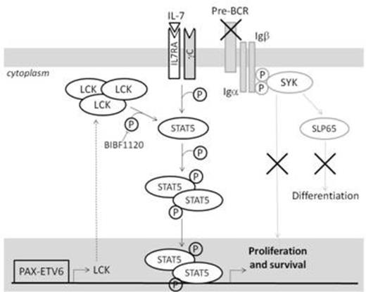
Strikingly, in addition to LCK overexpression, PAX5/ETV6 induction in pre-BI cells dephosphorylates the inhibitory domain of LCK (LCK Y505) (mean difference in LCKY505/total LCK ratio: 63%, ranging from 38% to 81% in 3 different pre-BI lines), thus hyper-activating the LCK signaling. By phosphoflow analysis we also demonstrated LCK-dependent STAT5 hyper-phosphorylation after IL7 stimulation at all time points in time course experiments (mean fold change: 2.0, range 1.77-2.18). Downstream in the pathway, as a confirmation, we found by RQ-PCR the up-regulation of cMYC, a STAT5 target (FC: 2.65, p<0.01, at 30 minutes and 3.85, p<0.01, at 24h after IL-7 stimulation). Also CCND2, a cMYC target, was upregulated (FC: 2.27, p<0.001, at 30 minutes and 1.83, p<0.001, at 24h after IL-7 stimulation). Moreover, PAX5/ETV6 induced pre-BI cells show an increase in the replicative S phase cells at all time points after o.n. IL7 withdrawal (+58%, +39% and 61%, p<0.001, for PAX5/ETV6 after 0, 24 and 48 hours, respectively) and a faster proliferation rate, likely due to a different STAT5 activation profile. This mechanism is specific and reversible, as the LCK inhibitor BIBF1120 reduces STAT5 phosphorylation in PAX5/ETV6 cells (-11% and -46% after 24 and 48 hours, respectively), thus decreasing the fraction of S phase cells (-20%, p<0.01 and -31%, p<0.001, after 24 and 48 hours, respectively) and leading to a proliferation rate comparable to the control cells.
These results further sustain the role of PAX5 fusion proteins as aberrant transcriptional factors, leading to the over-expression and hyper-activation of LCK, with a role in the activation of signaling pathways alternative to BCR (i.e. STAT5 hyper-phosphorylation) to sustain survival of leukemic cells. The further comprehension of the signaling mechanisms activated by PAX5 fusion genes will be fundamental for the development of new strategies to arrest tumor proliferation.
Hypothesis on the role of PAX5 fusion proteins to sustain survival of leukemic cells through a signaling pathway alternative to BCR, by over-expression and hyper-activation of LCK and STAT5 hyper-phosphorylation.
Hypothesis on the role of PAX5 fusion proteins to sustain survival of leukemic cells through a signaling pathway alternative to BCR, by over-expression and hyper-activation of LCK and STAT5 hyper-phosphorylation.
No relevant conflicts of interest to declare.
Author notes
Asterisk with author names denotes non-ASH members.


This feature is available to Subscribers Only
Sign In or Create an Account Close Modal