Key Points
KT64/86 artificial antigen–presenting cells culture stimulation provides marked expansion of Tregs.
In the context of sirolimus, mycophenolate mofetil immunosuppression, adoptive transfer of Tregs resulted in low risk of acute GVHD.
Abstract
We studied the safety and clinical outcomes of patients treated with umbilical cord blood (UCB)-derived regulatory T cells (Tregs) that expanded in cultures stimulated with K562 cells modified to express the high-affinity Fc receptor (CD64) and CD86, the natural ligand of CD28 (KT64/86). Eleven patients were treated with Treg doses from 3-100 × 106 Treg/kg. The median proportion of CD4+FoxP3+CD127– in the infused product was 87% (range, 78%-95%), and we observed no dose-limiting infusional adverse events. Clinical outcomes were compared with contemporary controls (n = 22) who received the same conditioning regimen with sirolimus and mycophenolate mofetil immune suppression. The incidence of grade II-IV acute graft-versus-host disease (GVHD) at 100 days was 9% (95% confidence interval [CI], 0-25) vs 45% (95% CI, 24-67) in controls (P = .05). Chronic GVHD at 1 year was zero in Tregs and 14% in controls. Hematopoietic recovery and chimerism, cumulative density of infections, nonrelapse mortality, relapse, and disease-free survival were similar in the Treg recipients and controls. KT64/86-expanded UCB Tregs were safe and resulted in low risk of acute GVHD.
Introduction
Regulatory T cells (Tregs) are key modulators of the immune response and play an important role in self-tolerance.1 Thymic-derived Tregs express CD4+ CD25++, FoxP3 transcription factor, and low levels of CD127, the interleukin-7 receptor α-chain,2,3 and they depend on interleukin-2 (IL-2) for survival and proliferation.1
In a previous first-in-human clinical trial, we demonstrated the safety and preliminary effectiveness of ex vivo expanded umbilical cord blood (UCB)-derived Tregs in doses up to 10 × 106/kg recipient weight with a lower risk of acute graft-versus-host disease (GVHD) compared with identically treated historical controls (43% vs 61%, P = .05).4,5 With a minimum follow up of 2 years, no adverse effects on nonrelapse mortality (NRM) or relapse was detected.5 In an attempt to maximize the infused Treg dose, selected CD25+ UCB cells were expanded in the presence of K562 cells modified to express the high-affinity Fc receptor (CD64) and CD86, the natural ligand of CD28 (KT64/86).6 During preclinical development, we demonstrated that a single restimulation resulted in marked expansion of Tregs while maintaining Foxp3 expression and potent suppressor function. We here report on the safety profile, kinetics, and clinical outcomes in patients treated with higher doses of KT64/86-expanded UCB-derived Tregs.
Patients and methods
Patient inclusion criteria
Patient eligibility and UCB graft selection criteria were as reported in our initial Treg trial.4 Briefly, patients who had advanced or high-risk lympho-hematopoietic malignancy, ages 12 to 70 years received a double-UCB graft with each unit 4-6/6 locus HLA-matched to the patient and 4-6/6 HLA-matched and ABO compatible to the other unit. The grafts contained a combined nucleated cell dose of ≥3.0 × 10⁷ cells/kg, with each unit required to have a minimum 1.5 × 10⁷ cells/kg at cryopreservation.7 Patients were treated between November 2012 and October 2014.
Treatment and supportive care
The nonmyeloablative conditioning regimen consisted of cyclophosphamide (CY) 50 mg/kg on day –6, fludarabine (FLU) 40 mg/m2 daily on days –6 to –2, and a single fraction of total body irradiation 200 cGy on day –1.4,7 Criteria for nonmyeloablative conditioning included age ≥55 years, previous autologous transplant, ≥12 months of alkylating agent therapy or extensive radiation therapy preventing 1320 cGy total body irradiation, multiple comorbidities, or Karnofsky Performance Score >60% but <80%. All patients received sirolimus from day –3 to day +100, with tapering over 8 to 12 weeks, with a loading dose of 12 mg followed by 4 mg daily and a target trough level between 3 and 12 µg/mL in combination with mycophenolate mofetil (MMF) at 1.5 g IV or orally twice daily from day –3 to +30. Details on supportive care and flow cytometry are provided in supplemental Methods, available on the Blood Web site.
Study design
This study was a “fast-track” dose escalation trial8 designed to assess the safety profile and maximal tolerated dose of KT64/86-stimulated ex vivo expanded UCB-derived Treg. Planned dose levels were 3, 10, 30, 100, and 300 × 106 Treg/kg actual body weight. Dose escalation occurred with each successive patient unless a dose-limiting toxicity (DLT; defined later in “Endpoints and definitions”) was observed or Treg expansion was insufficient to meet the planned dose. Before Treg infusion on day +1, all patients received IV hydration and were premedicated with acetaminophen and diphenhydramine. No steroids were allowed starting 24 hours before the Treg infusion. No steroids or antithymocyte globulin were allowed starting within 24 hours of Treg infusion. No steroids or antithymocyte globulin were allowed starting within 24 hours of Treg infusion. Tregs were infused on day +1 to avoid overlap with any infusional toxicities associated with the double-UCB graft.
The protocol was approved by the Institutional Review Board of the University of Minnesota and registered at http://www.clinicaltrials.gov under NCT#00602693. All patients provided written informed consent following the principles of the Declaration of Helsinki before enrollment.
Treg manufacture
Tregs were isolated from a third UCB unit 4-6/6 HLA matched to the patient. No interunit matching was required between the Treg donor unit and the 2 HSC graft units. Enrichment of CD25+ cells was accomplished by positive selection with directly conjugated anti-CD25 magnetic microbeads (Miltenyi Biotec, Bergish Gladbach, Germany) and the CliniMACS device.9 Our validation run used an open selection system using a conjugated anti-CD25 magnetic microbeads concentration of 1:350. At the start of the clinical trial, we changed to a closed selection system and initially used the same concentration. This resulted in poor post-column CD25+ cell count recovery. During the process improvement investigation, we identified the conjugated anti-CD25 magnetic microbeads concentration as the root cause for poor CD25+ cell count recovery post-column, and changed to a concentration of 1:60. Isolated cells were stimulated with anti-CD3 mAb (MACS GMP CD3 Pure, Miltenyi Biotec) loaded KT64/86 artificial antigen-presenting cells (aAPCs)10,11 (produced and tested according to a Biologics Master File submitted to the Food and Drug Administration) at a 1:1 aAPC to Treg ratio for 18 ± 1 days. On day 3, cultures were supplemented with 300 IU/mL IL-2 (Proleukin, Chiron Corporation, Emeryville, CA). Cells were maintained at a density of 5.0 × 10⁵ viable nucleated cells/mL by splitting every 48 to 72 hours. On day 12 ± 1, cultures were washed and restimulated with frozen/thawed, anti-CD3 mAb-loaded KT64/86 aAPCs at a 1:1 aAPC to cell ratio. All products met lot release criteria that included: 7AAD viability ≥70%, CD4+CD25+ purity ≥60%, <10% CD4–/CD8+ cells, negative gram stain, and low endotoxin (≤5 EU/kg).
Statistical analysis
Descriptive statistics and plots were used to summarize patient and transplant characteristics, safety parameters, and details of the Treg products. To assess potential alterations in risks of outcomes after Treg infusion, we compared outcomes with 22 contemporary, nonrandomized controls that, except for the adoptive transfer of UCB-derived Tregs, were treated in an identical fashion using the same nonmyeloablative conditioning along with sirolimus and MMF immunoprophylaxis. Comparison of patient and transplant characteristics between groups was completed by the generalized Wilcoxon test for continuous variables and the χ2 or Fisher’s exact test for categorical variables. Spearman’s correlation coefficient was used to study the correlation between infused Treg cell dose and the level of detection using a trapezoidal rule to calculate the area under the curve over time to measure the level of detection. To evaluate immune reconstitution, a generalized linear mixed model was used on log-transformation of immune reconstitution parameters over time because of non-normally distributed data using an autoregressive correlation structure. The autoregressive correlation structure was used in the mixed models procedure after assessing correlation over time.12 Overall survival and disease-free survival were estimated by Kaplan-Meier curves. Comparisons were completed with the simple log-rank test.13 Nonrelapse mortality was estimated using cumulative incidence, treating relapse as a competing risk. The probability of relapse, neutrophil, and platelet recovery and GVHD were estimated by cumulative incidence, treating nonevent deaths as competing events.14 To analyze risks of multiple infections per patient, the rates of infections were estimated by infection density per 1000 patient days. Infections were evaluated through 100 days post-transplant. Comparisons were completed with the Mantel-Haenszel test for person-years data. All reported P values were two-sided. SAS 9.3 (SAS Institute, Cary, NC) and R 3.0.2 were used to perform all statistical analyses. Data analysis was conducted in January 2015.
Results
Patient and UCB graft characteristics
Eleven patients were treated: 2 were treated at 3 × 10⁶ Treg/kg, 4 at 10 × 106 Treg/kg, 1 at 30 × 106 Treg/kg, and 4 at the highest achievable dose of 100 × 106 Treg/kg. The demographics and UCB graft characteristics of the Treg recipients and 22 identically treated contemporary controls are summarized in Table 1. The 2 groups were similar in age, diagnosis, comorbidity index score, and pretransplant cytomegalovirus (CMV) serostatus. Treg recipients, however, were more likely to be female. The median follow-up of survivors was 20 months (range, 9-24) and 28 months (range, 8-32) for Treg and control recipients, respectively. The degree of HLA matching among patients, the Treg unit, and each of the 2 donor units is summarized in supplemental Table 1.
Patient and graft characteristics
| Variable . | Treg recipients . | Siro/MMF controls . |
|---|---|---|
| Number of patients | 11 | 22 |
| Median age, y (range) | 61 (45-68) | 60 (34-69) |
| Males | 6 (55%) | 20 (91%) |
| HCT Comorbidity Index | ||
| 0 | 3 (27%) | 8 (36%) |
| 1-2 | 4 (36%) | 9 (41%) |
| 3+ | 4 (36%) | 5 (23%) |
| Recipient CMV+ | 6 (55%) | 7 (32%) |
| Diagnosis | ||
| ALL/AML | 6 (54%) | 13 (59%) |
| CLL | 2 (18%) | 2 (9%) |
| NHL/HL | 2 (18%) | 5 (23%) |
| Other* | 1 (9%) | 2 (10%) |
| High-risk disease | 5 (45%) | 10 (45%) |
| HLA disparity† | ||
| 4/6 + 4/6 | 3 (27%) | 7 (32%) |
| 4/6 + 5/6 | 1 (9%) | 4 (18%) |
| 4/6 + 6/6 | 1 (9%) | 0 |
| 5/6 + 5/6 | 4 (36%) | 6 (27%) |
| 5/6 + 6/6 | 1 (9%) | 2 (9%) |
| 6/6 +6/6 | 1 (9%) | 3 (14%) |
| Median combined TNC × 107 cells/kg (range) (IQR) | 4.2 (3.0-9.2) (3.3-4.7) | 3.9 (2.8-7.7) (3.3-4.7) |
| Median combined CD34+ × 105 cells/kg (range) (IQR) | 5.2 (2.7-11.9) (4.3-8.1) | 4.9 (2.0-18.3) (3.3-8.3) |
| Median combined CD3+ × 107 cells/kg (range) (IQR) | 1.6 (1.0-4.0) (1.3-1.8) | 1.6 (1.0-7.6) (1.3-2.0) |
ALL, acute lymphoblastic leukemia; AML, acute myeloid leukemia; CD cluster of differentiation; CLL, chronic lymphocytic leukemia; CMV, cytomegalovirus; HCT, hematopoietic stem cell transplantation–specific comorbidity index; HL, non-Hodgkin lymphoma; HLA, human leukocyte antigen; IQR, interquartile range; NHL, non-Hodgkin lymphoma; Siro/MMF, sirolimus/mycohenolate mofetil; TNC, total nucleated cell dose.
Includes 1 patient with myeloma in each group and 1 patient with myeloproliferative disease.
HLA disparity considers the worst matched of the 2 donor units.
Treg product characteristics
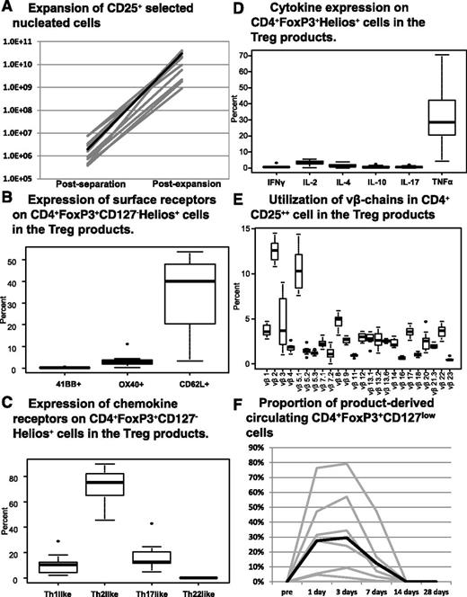
Thirteen Treg products were manufactured and 11 were infused. The cell doses for individual products at the various steps of processing are shown in Table 2. Because of a change in processing during the study, the anti-CD25 mAb conjugate concentration that was used in 9 products (products 1-7, 12-13) was suboptimal, resulting in poor recovery of CD25+ from the UCB unit. Despite this technical problem, adequate ex vivo expansion allowed us to reach cell doses up to 30 × 106/kg. However, 5 products did not reach the target dose level, with 4 of 5 failing to achieve the target cell dose of 100 × 106/kg (products 5-7 and 13; Table 2). Two products (products 12 and 13; Table 2) failed to reach the minimum required dose of 3 × 10⁶/kg for infusion; poor postcolumn yield and individual unit quality variation may have contributed to these 2 products’ poor expansion. Notably, 11 products performed as expected in the expansion culture. Once we corrected the antibody concentration for the initial CD25+ selection to 1:60 and were able to recover the usual yield of CD25+ (0.4% of post-thaw total nucleated cells [TNCs]) for the initiation of culture, we reproducibly achieved the target cell dose of 100 × 106/kg. In summary, in 11 of 13 products that were infused, the median number of TNCs of the product at the end of expansion culture was 40 billion (range, 1.88-42.6) corresponding to a median expansion of 13 000-fold (range, 1352 to 27 183-fold) (Figure 1A), of which the median proportion of CD4+CD25+ cells was 97% (range, 88-99) and with 87% (range, 78-95) expressing CD4+FoxP3+CD127–. The median proportion of CD4+FoxP3+CD127– that were Helios+ was 96% (range, 87-97), of which a median of 40% (range, 3-54) were CD62L+, with minimal expression of 4-1BB+ (0.06%; range, <0.01-0.65) and OX40 (2.8%; range, 0.3-11.1) (Figure 1B). The chemokine receptor expression of CD4+FoxP3+CD127–Helios+ cells showed Th2 phenotype differentiation with a median CXCR3–CCR6– expression of 75% (range, 46-90), whereas there was lower expression of Th1 (11%; range, 2-29), Th17 (13%; range, 5-43), and Th22-like (0.1%; range, <0.01-0.3) chemokine receptors (Figure 1C). After Phorbol myristate acetate (PMA) and calcium ionophore stimulation the median proportion of CD4+Foxp3+Helios+ cells that expressed tumor necrosis factor-alpha (TNFα) was 28% (range, 4-71), and IL-2 was 3.6% (range, 0.5-5.5); there was minimal expression of interferon-gamma (IFNγ) (0.6%; range, 0.3-3.2), IL-4 (1.1%; range, 0.4-21), IL-10 (0.3%; range, 0.1-2.5), and IL-17 (0.6%; range, 0.03-2.1) (Figure 1D). We also determined clonality on CD4+CD25++ cells in our product by studying the frequency of v beta-chain utilization and demonstrated that they were polyclonal, as shown in Figure 1E. In Table 3, we summarize the individual product phenotype and cytokine expression profile of non-CD25+Foxp3+CD127low cells. The median proportion of contaminant cells that were CD3+CD8hi was 0.14% (range, 0.03-1.36), CD4+CD8+ was 0.54% (range, 0.07-5.00), CD14 was 0.2% (range, 0-0.08), and CD19 was 0.03% (range, 0-0.44). There was similar expression of the natural Treg marker Helios in CD4+Foxp3+ (median, 89.9%) and CD4+Foxp3– cells (median, 68%), suggesting that they are still natural Tregs. Similar to CD25+Foxp3+CD127low cells, non-CD25+Foxp3+CD127low cells, there was low expression of effector cytokines (Table 3). Last, CD4+Foxp3+ cells and CD4+Foxp3– had similar low expression of the proliferation marker Ki-67 (6% [range, 1.8-16.7] vs 3.8% [range, 0-11.2]), demonstrating that they were are not rapidly dividing and suggesting that they were not likely to be activated T cells. In 6 products, the median in vitro suppressor function of CD3+ bead-stimulated T cells at a 1:4 T effector-to-Treg ratio was 53% (range, 45-71). The remaining 5 products samples were not adequately cryopreserved, resulting in suppression assays that were unevaluable.
Cell doses and proportions at the various steps of processing
| Product . | Absolute TNC donor UCB unit . | Postimmunomagnetic separation . | Post-expansion . | Target Treg cell dose (× 106 cells/kg) . | Actual Treg dose (× 106 cells/kg) . | |||||||
|---|---|---|---|---|---|---|---|---|---|---|---|---|
| Absolute TNC . | Percent TNC recovery . | Absolute CD4+ CD25+ . | Proportion of CD4+ CD25+ . | Fold expansion . | Absolute TNC . | Proportion of CD4+ CD25+ . | Absolute CD4+ CD25+ . | Proportion of CD4+ Foxp3+ CD127low . | ||||
| Infused products | ||||||||||||
| 1 | 1.15E+09 | 1.50E+06 | 0.13% | 4.35E+05 | 29% | 27183 | 4.08E+10 | 94% | 3.84E+10 | 83% | 3 | 3 |
| 2 | 1.06E+09 | 1.76E+06 | 0.17% | 1.55E+06 | 88% | 24213 | 4.26E+10 | 97% | 4.13E+10 | 83% | 3 | 3 |
| 3* | 1.10E+09 | 2.25E+06 | 0.20% | 2.05E+06 | 91% | 16365 | 3.68E+10 | 98% | 3.61E+10 | 81% | 10 | 10 |
| 4† | 1.02E+09 | 1.02E+06 | 0.10% | 9.15E+05 | 90% | 6951 | 6.10E+09 | 96% | 5.86E+09 | 86% | 30 | 30 |
| 5‡ | 9.10E+08 | 7.05E+05 | 0.08% | 4.63E+05 | 67% | 1352 | 9.46E+08 | 97% | 9.18E+08 | 82% | 100 | 10 |
| 6†‡ | 1.18E+09 | 4.81E+05 | 0.04% | 3.75E+05 | 78% | 4601 | 1.88E+09 | 97% | 1.82E+09 | 91% | 100 | 10 |
| 7‡ | 1.83E+09 | 8.00E+05 | 0.04% | 7.23E+05 | 90% | 5271 | 2.45E+09 | 91% | 2.23E+09 | 89% | 100 | 10 |
| 8 | 2.47E+09 | 7.92E+06 | 0.32% | 7.61E+06 | 96% | 16897 | 1.11E+10 | 88% | 9.77E+09 | 95% | 100 | 100 |
| 9 | 2.15E+09 | 8.91E+06 | 0.41% | 7.44E+06 | 84% | 13581 | 2.41E+10 | 95% | 2.29E+10 | 90% | 100 | 100 |
| 10 | 2.07E+09 | 1.08E+07 | 0.52% | 2.82E+06 | 26% | 13550 | 2.05E+10 | 98% | 2.01E+10 | 91% | 100 | 100 |
| 11 | 1.55E+09 | 3.96E+06 | 0.26% | 3.40E+06 | 86% | 17127 | 2.25E+10 | 99% | 2.23E+10 | 91% | 100 | 100 |
| Non-infused products | ||||||||||||
| 12‡ | 1.19E+09 | 5.60E+05 | 0.05% | 2.44E+05 | 44% | 174 | 9.77E+07 | NA | NA | NA | 3 | NA |
| 13‡ | 1.18E+09 | 1.16E+06 | 0.10% | 1.05E+06 | 90% | 246 | 2.30E+08 | 95% | 2.19E+08 | NA | 100 | NA |
TNC, total nucleated cells; UCB, umbilical cord blood.
This was the only product that was cryopreserved and posteriorly thawed and infused.
These two were the patients who developed acute GVHD. Patient 4 had the skin rash and patient 6 presented with late acute GVHD of the gastrointestinal tract.
These products failed to achieve the target cell dose level.
(A) Logarithmic scale of the absolute number of nucleated cells after CD25+ immunomagnetic separation and at the end of the KT64/86-stimulated culture, of which the median proportion of CD4+CD25+ cells was 97% (range, 88-99) and with 87% (range, 78-95) expressing CD4+FoxP3+CD127–. (B) The median proportion, interquartile range, and range of CD4+FoxP3+CD127lowHelios+ cells that expressed the surface markers CD62L, OX40, and 41BB. (C) The median proportion, interquartile range, and range of chemokine receptors expression on CD4+FoxP3+CD127lowHelios+ cells; CXCR3+ (Th1-like), CXCR3–CCR6– (Th2-like), CXCR3–CCR4+CCR6+CCR10– (Th17-like), and CXCR3–CCR4+CCR6+CCR10+ (Th22-like). (D) The median proportion, interquartile range, and range of CD4+FoxP3+Helios+ cells that after ionophore and PMA stimulation expressed IFNγ, TNFα, IL-2, IL-4, IL-10, and IL-17. (E) The median proportion, interquartile range, and range of utilization of 24 v beta-chains in CD4+CD25++ cells from the Treg product. (F) The proportion of CD4+FoxP3+CD127low cells derived from the expansion product detected in the PB of the patients with an informative HLA marker (n = 7). The black lines on all panels represent the median values.
(A) Logarithmic scale of the absolute number of nucleated cells after CD25+ immunomagnetic separation and at the end of the KT64/86-stimulated culture, of which the median proportion of CD4+CD25+ cells was 97% (range, 88-99) and with 87% (range, 78-95) expressing CD4+FoxP3+CD127–. (B) The median proportion, interquartile range, and range of CD4+FoxP3+CD127lowHelios+ cells that expressed the surface markers CD62L, OX40, and 41BB. (C) The median proportion, interquartile range, and range of chemokine receptors expression on CD4+FoxP3+CD127lowHelios+ cells; CXCR3+ (Th1-like), CXCR3–CCR6– (Th2-like), CXCR3–CCR4+CCR6+CCR10– (Th17-like), and CXCR3–CCR4+CCR6+CCR10+ (Th22-like). (D) The median proportion, interquartile range, and range of CD4+FoxP3+Helios+ cells that after ionophore and PMA stimulation expressed IFNγ, TNFα, IL-2, IL-4, IL-10, and IL-17. (E) The median proportion, interquartile range, and range of utilization of 24 v beta-chains in CD4+CD25++ cells from the Treg product. (F) The proportion of CD4+FoxP3+CD127low cells derived from the expansion product detected in the PB of the patients with an informative HLA marker (n = 7). The black lines on all panels represent the median values.
Individual Treg product phenotype of non-CD4+FoxP3+CD127low cells and cytokine profile CD4+FoxP3+CD127low and non-CD4+FoxP3+CD127low cells
| . | Infused product . | Noninfused products . | Median all products . | |||||||||||
|---|---|---|---|---|---|---|---|---|---|---|---|---|---|---|
| Product No. . | 1 . | 2 . | 3 . | 4 . | 5 . | 6 . | 7 . | 8 . | 9 . | 10 . | 11 . | 12 . | 13 . | |
| Phenotype | ||||||||||||||
| CD4–CD8++ | 0.14 | 0.06 | 0.80 | 0.04 | 0.04 | 0.77 | 1.36 | 0.23 | 0.06 | 0.88 | 0.03 | NA | 0.13 | 0.14 |
| CD4+CD8+ | 0.18 | 0.48 | 0.55 | 0.07 | 0.07 | 1.19 | 0.18 | 1.55 | 0.53 | 0.84 | 5.00 | NA | 2.40 | 0.54 |
| CD14+ | NA | NA | NA | NA | NA | 0.02 | 0.00 | 0.00 | 0.01 | 0.02 | 0.03 | NA | 0.08 | 0.02 |
| CD19+ | 0.06 | 0.44 | 0.08 | 0.11 | 0.01 | 0.03 | 0.00 | 0.00 | 0.00 | 0.00 | 0.02 | NA | 0.07 | 0.03 |
| CD4+FoxP3+ | ||||||||||||||
| HELIOS+ | 93.9 | 88.3 | 85.6 | 92.7 | 95.7 | NA | NA | NA | NA | 66.8 | 98.5 | NA | 97.6 | 89.9 |
| KI-67+ | 5.5 | 7.4 | 4.0 | 4.6 | 6.7 | 3.7 | 1.8 | 3.0 | 4.3 | 9.7 | 4.4 | NA | 16.7 | 6.0 |
| CD4+Foxp3– | ||||||||||||||
| HELIOS+ | 72.9 | 55.4 | 65.0 | 34.5 | 59.0 | NA | NA | NA | NA | 72.0 | 95.4 | NA | 90.2 | 68.1 |
| KI-67– | 3.4 | 2.3 | 1.4 | 1.4 | 2.4 | 0.6 | 0.0 | 3.4 | 1.3 | 11.2 | 2.4 | NA | 15.4 | 3.8 |
| Cytokine expression | ||||||||||||||
| CD4+Foxp3+Helios+ | ||||||||||||||
| IL2+ | 3.70 | 3.83 | 4.64 | 4.12 | 3.57 | 2.37 | 5.05 | 5.48 | 2.18 | 2.71 | 3.42 | 0.11 | NA | 3.6 |
| IFNγ+ | 0.55 | 0.26 | 0.39 | 0.55 | 0.88 | 0.59 | 0.67 | 3.18 | 0.47 | 0.92 | 0.39 | 0.25 | NA | 0.6 |
| TNFα+ | 27.50 | 28.90 | 21.20 | 15.80 | 43.40 | 41.20 | 34.90 | 67.90 | 70.50 | 20.10 | 27.90 | 4.30 | NA | 28.4 |
| IL4+ | 2.33 | 1.48 | 1.00 | 1.05 | 1.18 | 0.81 | 3.84 | 2.41 | 0.39 | 1.84 | 1.04 | 0.02 | NA | 1.1 |
| IL10+ | 0.19 | 0.14 | 0.11 | 1.79 | 0.28 | 0.38 | 1.18 | 2.52 | 0.86 | 0.28 | 0.42 | 0.26 | NA | 0.3 |
| IL17+ | 1.02 | 0.19 | 0.23 | 0.77 | 1.40 | 0.78 | 0.84 | 2.08 | 0.39 | 0.26 | 0.25 | 0.03 | NA | 0.6 |
| CD4+Foxp3–Helios– | ||||||||||||||
| IL2+ | 0.40 | 0.15 | 0.09 | 0.61 | 0.08 | 0.04 | 0.14 | 0.15 | 0.04 | 0.15 | 0.11 | 0.27 | NA | 0.1 |
| IFN+ | 0.19 | 0.15 | 0.04 | 0.99 | 0.18 | 0.09 | 0.14 | 1.06 | 0.22 | 0.07 | 0.30 | 0.05 | NA | 0.2 |
| TNFα+ | 4.57 | 2.62 | 4.76 | 3.15 | 2.77 | 5.41 | 8.09 | 12.90 | 15.20 | 3.98 | 3.02 | 0.40 | NA | 4.3 |
| IL4+ | 0.12 | 0.29 | 0.07 | 0.19 | 0.14 | 0.15 | 0.11 | 0.89 | 0.11 | 0.06 | 0.19 | 0.01 | NA | 0.1 |
| IL10+ | 0.09 | 0.02 | 0.00 | 0.12 | 0.05 | 0.06 | 0.21 | 0.35 | 0.29 | 0.08 | 0.04 | 0.08 | NA | 0.1 |
| IL17+ | 0.24 | 0.19 | 0.02 | 0.20 | 0.24 | 0.15 | 0.20 | 1.33 | 0.18 | 0.02 | 0.15 | 0.01 | NA | 0.2 |
CD, cluster of differentiation; FoxP3, forkhead box P3; IFNγ, interferon-γ; IL, interleukin; NA, not available; TNAα, tumor necrosis factor-α; Treg, regulatory T cells.
Treg infusional toxicity
As summarized in Table 4, infusional toxicities were monitored before the Treg product infusion, and then at 24 and 48 hours post-infusion. Toxicities were mild and no dose-limiting toxicity was observed in doses up to 100 × 106/kg.
Infusional toxicity after infusion of 11 Treg products
| Adverse event . | Pre-infusion . | 24 hours . | 48 hours . |
|---|---|---|---|
| Hypertension grade 1 | 1 | 1 | 0 |
| Hypertension grade 2 | 2 | 2 | 1 |
| Hypertension grade 3 | 1 | 0 | 0 |
| Pain grade 1 | 1 | 2 | 0 |
| Tachycardia grade 1 | 1 | 0 | 1 |
| Bradycardia grade 1 | 1 | 0 | 0 |
| Renal grade 1 | 1 | 1 | 0 |
| Anxiety grade 1 | 1 | 0 | 0 |
Treg, regulatory T cells.
Detection of product-derived Tregs in the peripheral blood
An informative HLA marker (unique to the Treg donor unit) was available in 7 of 11 patients. The presence of the Treg product in the peripheral blood (PB) was tracked by flow cytometry, with progressively greater detection correlating with higher infused Treg cell doses (Figure 1F; P = .02). Despite a higher peak, greater infused cell doses did not yield longer persistence of PB Tregs; none persisted after 14 days.
Acute graft-vs-host disease
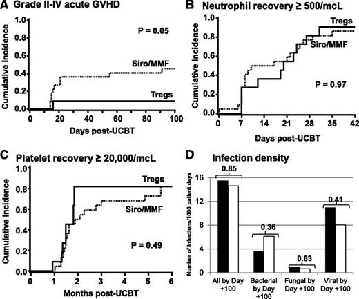
In our prior study, recipients of 3 × 10⁶ Treg/kg (n = 24), the incidence of grade II-IV acute GVHD was significantly (P = .05) reduced from 43% compared with 61% in the controls, but was not eliminated,4,5 suggesting potential efficacy. Preclinical studies demonstrated a marked reduction in murine acute GVHD at T effector:Treg ratios of 1:1 and 1:3,15 necessitating infusion of Treg doses of >15 × 106/kg to achieve a 1:1 ratio for the median infused CD3 cell dose in double-UCB grafts. Among 11 recipients of 3-100 × 106 Treg/kg, the cumulative incidence of grade II-IV acute GVHD at 100 days was 9% (95% confidence interval [CI], 0-25) (Figure 2A). One patient who was scored as having acute GVHD in our analysis developed a limited skin rash (50% of body surface) on day +16 with a skin biopsy not diagnostic for acute GVHD. The patient was treated with steroids, which were tapered rapidly over 4 weeks without recurrence of rash or development of other possible GVHD symptoms. However, one other patient developed biopsy-proven grade III gastrointestinal acute GVHD on day +119 and responded to systemic steroids. In contrast, 10 of 22 controls developed grade II-IV acute GVHD, with 4 cases of grade II and 6 cases of grade III-IV acute GVHD, for a cumulative incidence of grade II-IV GVHD of 45% (95% CI, 24-67) (P = .05) and grade III-IV of 27% (95% CI, 9-46) (P = .06). None of the Treg recipients and 3 (14%) patients in the control group developed chronic GVHD to date.
(A-C) Cumulative incidences of grade II-IV acute GVHD at day +100 (A), neutrophil recovery ≥500/μL by day +42 (B), and platelet recovery ≥20 000/μL at day +180 (C) for Treg recipients (solid line) and Siro/MMF controls (dotted line). (D) The density of infections at day +100 in Treg recipients (▪) and Siro/MMF controls (□).
(A-C) Cumulative incidences of grade II-IV acute GVHD at day +100 (A), neutrophil recovery ≥500/μL by day +42 (B), and platelet recovery ≥20 000/μL at day +180 (C) for Treg recipients (solid line) and Siro/MMF controls (dotted line). (D) The density of infections at day +100 in Treg recipients (▪) and Siro/MMF controls (□).
Hematopoietic recovery
The cumulative incidence of neutrophil recovery by day +42 was 91% (95% CI, 67-99) in Treg recipients compared with 86% (95% CI, 69-97) in controls (Figure 2B). There was one primary graft failure in the 11 Treg recipients (9%) vs 3 in 22 (14%) controls. The cumulative incidence of platelet recovery >20 000/μL by day +180 was 82% (95% CI, 55-100) for Treg recipients and 82% (95% CI, 59-100) for controls (Figure 2C). The median combined unit bone marrow (BM) chimerism at day +21 was 90% (range, 66-100) for Treg recipients and 90% (95% CI, 19-100) (P = .99) for controls, and at day +100 was 100% (range, 100-100) for Treg recipients and 100% (range, 97-100) for controls (P = .77). The prevalence of dual-donor unit chimerism in the BM at day +21 in Treg recipients was 38% (95% CI, 4-72) and in PB CD3+ cells was 33% (95% CI, 0-71) compared with 28% (95% CI, 7-49) and 20% (95% CI, 0-40) in controls (P = .71 for BM and P = .59 for PB). At day +100, the prevalence of dual chimerism in the BM was 29% (95% CI, 0-63) and in PB CD3+ cells was 29% (95% CI, 0-63) in Treg recipients, whereas in controls it was 18% (95% CI, 0-36) in the BM and 19% (95% CI, 0-38) in PB CD3+ cells (P = .65 for BM and P = .64 for PB).
Infections and immune reconstitution
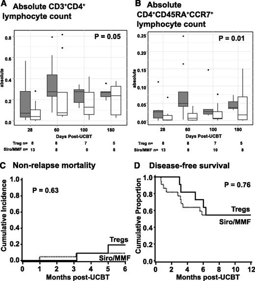
In the Treg group, there were a total of 17 infection events before day +100 in 9 patients vs 31 events in 14 Siro/MMF controls. The density of bacterial, viral, and fungal infections (infections per 1000 patient days) was similar in the 2 groups (Figure 2D). There were 2 CMV and 9 HHV-6 reactivations in Treg recipients compared with 1 CMV and 8 HHV-6 reactivations in controls. By day +180, there was significantly faster recovery of the absolute number of CD4+ (Figure 3A; P = .05) and a subset of naïve CD4+CD45RA+CCR7+ (Figure 3B; P = .01) lymphocytes in Treg recipients compared with controls. In contrast, recovery was similar for other CD4+ subsets and CD8+ and its subsets, CD19+ and CD56+ cells between the 2 groups (data not shown).
(A-D) The absolute number of CD3+CD4+ (A) and CD4+CD45+CCR7+ (B) lymphocytes subsets at days +28, +60, +100, and +180 after transplantation for Treg recipients (gray square) and Siro/MMF controls (□). The cumulative incidence of NRM at 6 months (C) and the probability of disease-free survival at 1 year (D) for Treg recipients (solid line) and Siro/MMF controls (dotted line).
(A-D) The absolute number of CD3+CD4+ (A) and CD4+CD45+CCR7+ (B) lymphocytes subsets at days +28, +60, +100, and +180 after transplantation for Treg recipients (gray square) and Siro/MMF controls (□). The cumulative incidence of NRM at 6 months (C) and the probability of disease-free survival at 1 year (D) for Treg recipients (solid line) and Siro/MMF controls (dotted line).
Relapse, nonrelapse mortality, survival, and disease-free survival
Like infection, it is possible the Treg cells might increase the risk of disease relapse. However, thus far, relapse has occurred in only 3 of 9 (33%) surviving patients treated with Treg compared with 8 of 20 (40%) in controls. At 6 months, 2 Treg patients and 2 controls died of nonrelapse causes, resulting in similar cumulative incidence of NRM (Figure 3C). Disease-free survival at 1 year is 55% (95% CI, 23-78) among Treg recipients and 55% (95% CI, 32-72) in the control group (P = .76) (Figure 3D). The estimated overall survival at 1 year was 81% (95% CI, 42-95) among Treg recipients and 61% (95% CI, 37-79) in the control group (P = .30). The causes of death in the Treg group were graft rejection and acute respiratory distress syndrome (1 each), whereas in the control group causes of death were graft rejection (n = 1), acute GVHD (n = 2), and relapse (n = 5).
Discussion
This report extends our initial observations on the adoptive transfer of ex vivo expanded UCB-derived Tregs. The principal observations are: (1) feasibility of KT64/86 aAPCs to produce large numbers of cGMP-grade Tregs up to 100 × 106/kg; (2) tolerable infusional safety profile of Treg infusions at 100 × 106/kg; (3) low risk of acute GVHD, NRM, and chronic GVHD; (4) similar infection densities; and (5) faster CD4 and naïve CD4 T-cell recovery compared with identically treated controls without Treg. Longer follow-up will allow us to confirm these preliminary findings showing no adverse effect of Tregs on infection, relapse, chronic GVHD, and survival.
Compared with our initial anti-CD3/anti-CD28 mAb-bead–stimulated UCB Treg expansion methodology,4 the current methodology introduced stimulation with KT64/86 aAPCs and restimulation of the culture with the aAPCs on day 12. This resulted in considerably more (a >60-fold mean expansion benefit) cGMP-grade Tregs available for adoptive transfer, confirming in large scale the promise of our reported preclinical studies.6 The high efficiency of this expansion culture resulted in very large culture volumes at the Treg dose of 100 × 106/kg, which allowed a Treg ratio to CD3+ cells of up to 7:1 in double-UCB grafts. With these high ratios and very low incidence of acute GVHD, we did not pursue further dose escalation. At the highest Treg dose level tested, no notable toxicities were observed and no maximum tolerated dose has yet been defined. Thus, we demonstrate that the KT64/86-stimulated Treg expansion method is robust and reproducible, the postcolumn initial CD25+ cell recovery number appears critical to achieve sufficient expansion, and infusion of these products was well tolerated.
Current GMP manufacture is always more complex, and having a proportion of products fail to meet the target cell dose was not unexpected. During our product development, we typically used larger units that we had stored in our cell therapy laboratory. Based on that work, the units for the Treg product were selected based on HLA matching to the patient and to allow postinfusion detection by flow cytometry, rather than cell dose, resulting in the selection of smaller units. Because all donor units were obtained from a clinical cord blood bank, there was expected interunit variability in clinical units. As we conducted the clinical trial, we observed products fail to achieve the target cell dose in a frequency >10%, which would have been expected based on our experience expanding Tregs using anti-CD3/CD28 mAb-loaded beads.4 Interunit variability may have contributed to the poor expansions observed in the 2 products that did not even meet the minimal dose for infusion. Thus, we halted enrollment to the trial and reviewed that data on all cGMP Tregs products. We identified a drop in the postimmune magnetic separation CD25+ cell recovery when we introduced a closed immune magnetic separation system that required higher concentration of the anti-CD25 antibody conjugate. We speculate that using a smaller cord blood unit precipitated the technical limitation we observed. Process improvement led us to optimize the anti-CD25 antibody conjugate and improved CD25+ cell recovery, and subsequently there were no failures to achieve target cell doses.
In our extensive characterization of the Treg products, we demonstrated that CD4+FoxP3+CD127– cells were Helios+ characterizing thymic-derived Tregs.16 There was high expression of CD62L (l-selectin), which is consistent of naïve T cells.15 We also found that our product was predominantly CXCR3–CCR6–, consistent with the Th2 suppressor phenotype.17 Our product cells expressed high levels of TNFα after potent stimulation of cytokine production by PMA and ionomycin. Under these conditions Tregs expressed low levels of IL-2 and other cytokines. The finding of PMA and ionomycin-driven TNFα expression in these Th2 suppressor phenotypic Treg products is of uncertain significance in our patients who also received sirolimus and MMF to blunt T-effector cell responses. Our Treg product was polyclonal on the basis of v beta-chain repertoire. Overall, our product characterization is was consistent with thymus-derived Tregs.
With the very low incidence of grade II-IV acute GVHD (9%), KT64/86-expanded UCB thymic–derived Tregs appear to potent suppressors with clinical activity. This incidence of acute GVHD was also lower than that observed in our initial study, in which we observed 43% in recipients bead-stimulated UCB Treg patients and 61% seen in historical controls, most of whom (17/23 patients) received a different immune prophylaxis that consisted of cyclosporine A/MMF.4 Notably, the 1 Treg patient developed biopsy-proven acute GVHD very late, suggesting that it may have been delayed by the Tregs. These data suggest that the T effector–to–Treg ratios of 1:1 or greater are needed, similar to that predicted by the xenogeneic GVHD model.15
In this study, the risk of infections was similar in Treg recipients and Siro/MMF controls. In particular, of HHV-6 was also similar to previously reported rates in UCB transplant recipients.18 Nonetheless, recognizing the potential immune suppressive effects and growing interest in adoptive transfer of Treg clinical trials in HCT, solid organ transplant, and autoimmune diseases, we suggest that careful monitoring for viral reactivation is appropriate.
In summary, with the adoptive transfer of KT64/86 ex vivo–expanded UCB-derived Tregs, we observed a low risk of acute GVHD, and similar NRM and survival compared with contemporary nonrandomized Siro/MMF controls. This expansion methodology permits clinical achievement of a targeted Treg-to-CD3+ cell ratio (≥1:1) that suppresses acute GVHD and its associated morbidity and mortality, and potentially improves immune reconstitution.
The online version of this article contains a data supplement.
The publication costs of this article were defrayed in part by page charge payment. Therefore, and solely to indicate this fact, this article is hereby marked “advertisement” in accordance with 18 USC section 1734.
Acknowledgments
The authors acknowledge Stefanie Hage and Jill L. Aughey from the University of Minnesota Clinical Trials Office for research nurse and regulatory support during the execution of this clinical trial; Diane Kadidlo and Fran Rabe from the University of Minnesota Medical Center Cell Therapy Laboratory and Molecular & Cellular Therapeutics for their assistance with cell therapy development and production; The Saint Louis Cord Blood Bank for kindly providing the UCB units for Treg manufacture; and the faculty, advanced practice providers, unit and clinic nurses, pharmacists, and social workers of the Blood and Marrow Transplant Program who cared for the patients at the University of Minnesota Medical Center.
This work was supported in part by National Institutes of Health (NIH) National Cancer Institute grants P30 CA77598, P01 CA65493 (C.G.B., J.S.M., D.H.M., P.B.M., B.R.B., J.E.W.), and R01 CA105216 (C.H.J.), Leukemia and Lymphoma Society Scholar in Clinical Research Award grant CDP-2417-11 (C.G.B.), the Children’s Cancer Research Fund (J.E.W., T.E.D.), NIH National Heart, Lung, and Blood Institute grant R01 HL11879 (B.R.B.), and contract HHSN268201000008C (J.S.M., D.H.M., K.L.H., J.C., J.E.W.), and Leukemia and Lymphoma Translational Research grant R6029-07 (B.R.B.).
Authorship
Contribution: C.G.B. conceived and designed the study, analyzed data, and drafted the manuscript; J.S.M. conceived the study, analyzed data, and wrote the manuscript; D.H.M. performed the study and wrote the manuscript; K.L.H. performed the study and wrote the manuscript; T.E.D. designed the study, analyzed data, and wrote the manuscript; D.S., J.C., M.R.V., M.L.M., B.L.L., J.L.R., C.H.J., and D.J.W. performed the study and wrote the manuscript; C.L. designed the study, analyzed data, and wrote the manuscript; P.B.M. conceived the study and wrote the manuscript writing; B.R.B. conceived and designed the study, performed and analyzed data, and wrote the manuscript; J.E.W. conceived and designed the study, performed and analyzed data and wrote the manuscript; and all authors gave final approval of the manuscript.
Conflict-of-interest disclosure: The authors declare no competing financial interests.
Correspondence: Claudio G. Brunstein, Department of Medicine, Mayo Mail Code 480, 420 Delaware St, S.E., Minneapolis, MN 55455; e-mail: bruns072@umn.edu.


This feature is available to Subscribers Only
Sign In or Create an Account Close Modal