Abstract
Introduction: Maintenance therapy with lenalidomide (LEN) is considered standard of care in the US, supported by meta analysis showing that LEN maintenance is associated with significantly longer progression free survival (PFS) and overall survival (OS). In a previously reported study of MM patients (pts) treated with extended courses of LEN, we have described a polyclonal Ig and FLC elevation exceeding the upper limit of normal (up to 3-4 X the ULN) and associated with striking polyclonal bone marrow (BM) plasmacytosis (up to 20% of plasma cells (PCs)), occurring with a relatively high frequency (20% of pts) and associated with better outcome (Zamarin, Leukemia, 2013). Importantly, we cautioned about the risk of misinterpretation of this polyclonal Ig response and plasmacytosis. Since Immuno-histochemical staining (IHS) may often be an unreliable tool to distinguish between a reactive polyclonal plasmacytosis from monoclonal disease, especially when the percentage of PCs in the BM is relatively low (5 -15%), we aimed at confirming the polyclonal nature of this often unrecognized and paradoxical response to prolonged LEN exposure, and at validating the important role of BM assessment by flow cytometry (FACS) in this setting.
Methods: Pts analyzed in this study were enrolled at MSKCC on three randomized, first line therapy, clinical trials (NCT01109004, NCT01208662, and NCT00807599). All pts were kept on LEN maintenance until POD, toxicity, or major event. BM aspirate and biopsy with IHS were performed as per standard of care. Eight-fluorochrome FACS method (minimum of 1 x 10⁶ cells) was used for 2014 samples employing a surface marker cocktail (CD117 PC5.5, CD19 BV421, CD138 APC, CD56 PC7, CD45 APC-H7, CD38 BV510) and cytoplasmic Kappa FITC/Lambda PE staining. Ten-fluorochrome (minimum of 5 x 10⁶ WBCs) method was used in 2015 and performed employing a surface marker cocktail (CD117 PC5.5, CD19 PC7, CD138 APC, CD56 APC-R700, CD45 APC-H7, CD81 Pacific Blue, CD38 BV510, CD27 BV605) and cytoplasmic Kappa FITC/Lambda PE staining.
Results: Among 147 pts enrolled in the three trials, 55 achieved serologic CR after first line therapy. Of those, 46 had a BM biopsy with FACS analysis available. Among these 46 patients, bone marrow plasmacytosis as assessed by light microscopy of BM aspirate and biopsy ranged from 2% to 15% (32 pts had PCs ≤ 5%, 14 PCs > 5%). Among the 14 pts with > 5% PCs, 2 had PCs = 15%, 9 had PCs = 10%, and 3 between 6% and 9%. By FACS, 9 of 14 pts showed no clonal or phenotypically aberrant PCs. The percentage of aberrant PCs among all PCs in the other 5 pts remained low (≤7.3%). Among the 2 pts with PC = 15%, the percentage of aberrant PCs among all PCs was 0% and 7.3%. In pts with PC = 10%, the percentage of aberrant PCs among all PCs was 0% (6 pts), or ranged from 0.17% to 5.1% (3 pts).
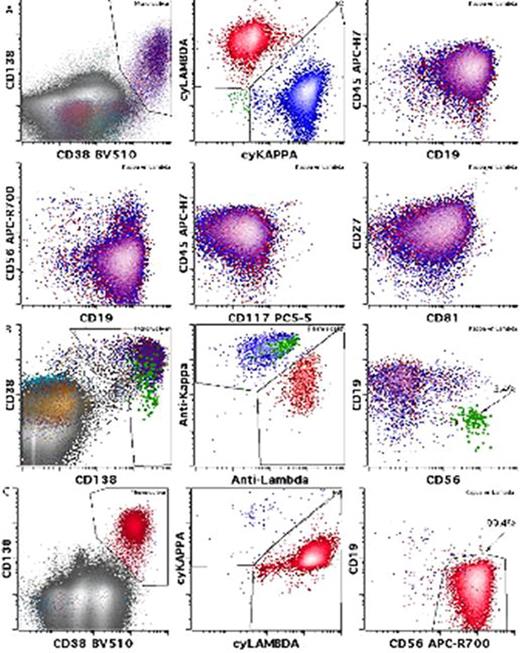
Figure 1 depicts representative pt cases with BM plasmacytosis estimated at 15% by light microscopy. PCs were gated based on CD38 and CD138 expression as well as forward and side scatter (not shown). Blue dots represent kappa LC expression; Red lambda LC. Proportion of aberrant PCs among total PCs in the sample is quantitated and shown using high sensitivity MRD detection with 10-color panel with 6-million event acquisitions. Panel A shows a pt with 15% PC in the BM with no residual disease confirming the polyclonal nature of the plasmacytosis. Panel B shows a pt with 15% PC exhibiting a low proportion of aberrant PCs (green) among a predominant background of normal polytypic PCs. In contrast, Panel C shows a pt with early relapsed disease with 15% PCs exhibiting a high proportion of aberrant PCs (99% of all PCs).
Conclusions: These results confirm the common and striking polyclonal plasmacytosis (up to 15% PCs in this cohort) associated with prolonged LEN exposure, and the importance of FACS in the assessment of BM plasmacytosis in these MM pts. In the setting of current MM response criteria established by the IMWG and NCCN guidelines, mandating a BM plasmacytosis <5% in addition to negative serum and urine immunofixation for CR determination, as well as an increase in plasmacytosis from baseline by 25% with an absolute value ≥10% for determination of disease progression, FACS analysis with assessment of clonality and percentage of aberrant PCs among total PCs, becomes an essential tool to distinguish reactive polyclonal patterns from monoclonal disease persistence or recurrence.
Hassoun:Novartis: Consultancy; Binding Site: Research Funding; Celgene: Research Funding; Takeda: Consultancy, Research Funding. Roshal:BD Biosciences: Consultancy. Landau:Prothena: Honoraria, Membership on an entity's Board of Directors or advisory committees; Takeda: Membership on an entity's Board of Directors or advisory committees, Research Funding; Amgen: Research Funding; Janssen: Consultancy; Spectrum Pharmaceuticals: Membership on an entity's Board of Directors or advisory committees, Research Funding. Korde:Medscape: Honoraria. Landgren:Merck: Honoraria; BMS: Honoraria; Takeda: Honoraria; Amgen: Honoraria, Research Funding; Celgene: Honoraria, Research Funding; Medscape Myeloma Program: Honoraria.
Author notes
Asterisk with author names denotes non-ASH members.


This feature is available to Subscribers Only
Sign In or Create an Account Close Modal