Key Points
Human CD4+CD161+ T cells display unique properties including MDR1-mediated drug efflux capacity and quiescence.
CD4+CD161+ T cells are enriched within the long-lived viral-specific Th1 memory repertoire in humans.
Abstract
The establishment of long-lived pathogen-specific T cells is a fundamental property of the adaptive immune response. However, the mechanisms underlying long-term persistence of antigen-specific CD4+ T cells are not well-defined. Here we identify a subset of memory CD4+ T cells capable of effluxing cellular toxins, including rhodamine (Rho), through the multidrug efflux protein MDR1 (also known as P-glycoprotein and ABCB1). Drug-effluxing CD4+ T cells were characterized as CD161+CD95+CD45RA−CD127hiCD28+CD25int cells with a distinct chemokine profile and a Th1-polarized pro-inflammatory phenotype. CD4+CD161+Rho-effluxing T cells proliferated vigorously in response to stimulation with anti-CD3/CD28 beads and gave rise to CD161− progeny in vitro. These cells were also capable of self-renewal and maintained their phenotypic and functional characteristics when cultured with homeostatic cytokines. Multidrug-effluxing CD4+CD161+ T cells were enriched within the viral-specific Th1 repertoire of healthy donors and patients with acute myeloid leukemia (AML) and survived exposure to daunorubicin chemotherapy in vitro. Multidrug-effluxing CD4+CD161+ T cells also resisted chemotherapy-induced cytotoxicity in vivo and underwent significant expansion in AML patients rendered lymphopenic after chemotherapy, contributing to the repopulation of anti-CMV immunity. Finally, after influenza vaccination, the proportion of influenza-specific CD4+ T cells coexpressing CD161 was significantly higher after 2 years compared with 4 weeks after immunization, suggesting CD161 is a marker for long-lived antigen-specific memory T cells. These findings suggest that CD4+CD161+ T cells with rapid efflux capacity contribute to the maintenance of viral-specific memory T cells. These data provide novel insights into mechanisms that preserve antiviral immunity in patients undergoing chemotherapy and have implications for the development of novel immunotherapeutic approaches.
Introduction
The adaptive immune response is distinguished by a broad range of long-lived pathogen-specific T cells that are ready to act on their second encounter with specific pathogens. After contact with antigen, naive T cells proliferate vigorously in an antigen-specific manner and acquire effector functions. A subset of antigen-specific memory T cells with slow proliferative potential under normal homeostatic conditions is thought to reside within the KLRG1−CD127+ memory precursor compartment and to persist for life.1-3 Studies in mice have shown that virus-specific T cells rely on interleukin 7 (IL-7) and IL-15, rather than antigenic stimulation and/or major histocompatibility complex (MHC) interaction, for their survival.3 Otherwise, very little is known about the mechanisms responsible for the long-term persistence of virus-specific cytotoxic T cells under normal or perturbed physiological conditions in humans, such as those seen after chemotherapy. Patients with acute myeloid leukemia (AML) undergoing repetitive cycles of cytotoxic chemotherapy experience severe, although short-lived, lymphocytopenia, yet rarely suffer serious viral reactivations such as cytomegalovirus (CMV) disease.4 This observation suggests the existence of chemoresistant populations of virus-specific memory CD8+ and CD4+ T cells with the ability to survive, expand, and repopulate the memory pool, preserving immunity against infectious agents. Furthermore, CMV-specific T cells of recipient origin were reported to contribute greatly to the mixed chimerism status and to protection from CMV-related events after reduced-intensity conditioning for allogeneic stem cell transplantation.5 These findings provide strong evidence that after chemotherapy, some CMV-specific T cells can escape deletion and provide protective immunity.
Cell-mediated immunity arises from the priming of naive T cells recognizing foreign peptides in the context of host MHC molecules. Murine studies have reported the existence of approximately 20 to 200 naive CD4+ T cells specific for any given antigenic epitope.6 Starting from a single activated T cell, the immune system uses different dynamic mechanisms to produce a variety of cellular descendants, generating diversity among the progeny.7 Accordingly, a novel T-cell subset named “stem cell–like” memory T cells and representing the earliest developmental stage of memory T cells was first described in murine CD8+ T cells.8 Despite expressing naive T-cell markers, memory T cells have high self-renewal capacity and the ability to give rise to other subsets.8-10 Another study proposed a subset of memory CD8+ T cells (CD45RA−CD95+) with the ability to rapidly efflux cytotoxic drugs through the ATP-binding cassette (ABC) superfamily multidrug-effluxing protein ABCB1, and defined phenotypically by high expression of CD161 to have stem-like properties.11,12 A subsequent study, however, suggested that ABCB1+CD161hiCD8+ T cells may in fact represent a subset of mucosal associated invariant T cells.13
Whereas much of our understanding of T-cell memory has been attained through studies of CD8+ T cells, recent reports have identified the existence of CD4+ T cells with stem-like properties within Th17 cells, suggesting cell fate diversification results in the generation of T cells with stem-like phenotype, even within more differentiated T-cell subsets.14,15
Here we describe the existence of a specialized subset of effector memory CD4+ T cells with rapid-efflux capacity. This unique CD4+ T-cell subset can proliferate, differentiate, and self-renew and is enriched within the long-lived viral-specific Th1 memory T-cell repertoire. Our findings shed light on some of the mechanisms used by T cells to preserve long-term immunity.
Materials and methods
Peripheral blood samples
Peripheral blood (PB) samples were collected after informed consent from healthy donors and newly diagnosed CMV-seropositive patients with AML undergoing treatment with daunorubicin (50-60 mg/m2 × 3 doses over the course of 5 days) and cytarabine (100 mg/m2 × 20 doses over the course of 10 days) at our institution from May 2009 to December 2011. Patient characteristics are detailed in supplemental Table 1, available on the Blood Web site.
Peptides and cytokine production
Intracellular cytokine detection was performed as described previously.16 In brief, PBMCs were stimulated with or without a pool of overlapping MHC class II CMVpp65 peptides (1 μg/mL; JPT Peptides Technologies, Berlin, Germany) for 6 hours. Phorbol 12-myristate 13-acetate (50 ng/mL) and ionomycin (2 μg/mL; Sigma Aldrich, St Louis, MO) were used as positive control. A more detailed account of the intracellular cytokine assay is included in the corresponding section in the supplemental Data.
Surface receptors and intracellular staining
Multiparameter flow cytometry was used to assess the phenotype and function of CD4+ and CD8+ T cells. Details of monoclonal antibodies are included in the corresponding section in the supplemental Data.
Rhodamine123 and daunorubicin efflux assays
Freshly isolated PBMCs from healthy controls were suspended in efflux buffer containing RPMI 1640 (GIBCO, United Kingdom) and 1% bovine serum albumin (Sigma-Aldrich, St. Louis, MO) and loaded with either 10 μg/mL Rhodamine 123 (Rho) (Sigma-Aldrich) for 30 minutes on ice or 2.5 mM daunorubicin (Sigma-Aldrich) for 20 minutes at 37°C/5% CO2. For additional information, see the corresponding section in the supplemental Methods.
Isolation of effluxing and noneffluxing CD4+ T-cell subsets
PBMCs were isolated using the density gradient separation method (Lymphoprep), and untouched CD4+ T cells were enriched using magnetic bead selection with magnetic-activated cell sorting kits (Miltenyi Biotec Ltd, Auburn, CA). Purified CD4+ T cells were labeled with Rho followed by Live Dead Aqua, CD3 V450, CD4 APC-H7, CD161 APC, CD62L PerCP-Cy5.5, and CD45RA PE-Cy7. Labeled CD4+ T cells were sort-purified on a fluorescence-activated cell sorter Aria III instrument. For additional information, see the corresponding section in the supplemental Methods.
In vitro expansion of CMV-specific CD4+ T cells
Sort-purified CD4+CD161+ and CD4+CD161− T cells were cocultured with CMV pp65 peptide-pulsed autologous B cells as antigen-presenting cells in the presence of IL-4 (60 ng/mL) and IL-7 (10 ng/mL) or IL-2 (50 IU/mL). Media were changed every 2 to 4 days for 10 to 12 days. Expansion of CMV-specific CD4+ T cells was assessed on the basis of the production of interferon (IFN)-γ and tumor necrosis factor (TNF)-α in response to CMV peptide rechallenge, followed by intracellular staining.
Proliferation, differentiation, and self-renewal assays
Carboxyfluorescein diacetate succinimidyl ester-dilution assays and Ki-67 staining were used to assess T-cell proliferation, as described previously.17 Sort-purified CD161+Rholo, CD161+Rhohi, and CD161− central memory (Tcm) and effector memory (Tem) cells were labeled with 2.5 µM CFSE (eBioscience, San Diego, CA) and stimulated with plate-bound CD3 and CD28 antibodies (1 µg/mL each; both from Biolegend, San Diego, CA) in the presence or absence of IL-7 (10 ng/mL) and IL-15 (10ng/mL; R&D, Minneapolis, MN) for 10 days, or with IL-7 (10 ng/mL) and IL-15 (10 ng/mL) only.
CMV Rho-effluxing assay and stimulation of sort-purified subsets with peptide-pulsed antigen-presenting cells
PBMCs were isolated from CMV-seropositive donors and stimulated with a pool of overlapping MHC class II CMVpp65 peptides (1 μg/mL; JPT Peptides Technologies) in a 24-well plate (1 × 10⁷ cells/well) for 4 hours. Cells were harvested, washed with PBS, and then labeled with Rho. T cells were enriched using the IFN-γ secretion assay-cell enrichment and detection kit (Miltenyi Biotec), using the manufacturer’s instructions. For additional information, see the corresponding section in the supplemental Methods.
Intracellular staining for transcription factors
CD4+ T cells were sort-purified into the 3 subsets of effluxing, noneffluxing, and CD161− CD4+ T cells, as described earlier. To assess Bcl-2 and Bcl-X expression, sort-purified subsets were stained with CD62L PerCP-Cy5.5 and CD45RA PE-CY7 (Biolegend) and then fixed and permeabilized using transcription buffer (BD Biosciences, San Jose, CA), as per the manufacturer’s instructions. Expression of ABCB1 and Ki-67 was measured in PBMCs after fixation and permeabilization. For additional information, see the corresponding section in the supplemental Methods.
Quantitative reverse transcription polymerase chain reaction analysis
Quantitative reverse transcription polymerase chain reaction to detect T-bet and RORC2 expression was performed in purified CMV-specific CD4+ T-cell subsets with sequence-specific TaqMan probes. Total RNA was purified with the RNeasy Mini Kit (Qiagen, Germantown, MD), following the manufacturer’s instructions. For additional information, see the corresponding section in the supplemental Methods.
Confocal immunofluorescence microscopy
Sort-purified CD4+ T cells were centrifuged onto slides using a cell concentrator (Cytofuge 2; IRIS International, Chatsworth, CA), fixed with 3% methanol-free formaldehyde, and then permeabilized with Triton 0.3% (Sigma Aldrich). For additional information, see the corresponding section in the supplemental Methods.
In vitro induction of apoptosis with daunorubicin
PBMCs from healthy CMV-seropositive donors were cultured in RPMI 1640 medium (GIBCO, United Kingdom) supplemented with 10% fetal calf serum, gentamicin, and L-glutamine in the presence or absence of 0.1 mM daunorubicin for 44 to 48 hours at 37°C/5% CO2, and apoptotic cells were identified by Annexin V staining per the manufacturer’s instructions (BD Bioscience, United Kingdom). Data analysis was performed with FlowJo software (Treestar, Ashland, OR).
Statistical analysis
All values are reported as medians and ranges or as means ± standard error of the mean. Statistical significance was performed with Prism (GraphPad, La Jolla, CA) by unpaired or paired 2-tailed t-test analysis and by nonparametric 2-way ANOVA, as appropriate. A probability of P ≤ .05 was considered statistically significant.
Results
A subset of CD4+CD161+ T cells rapidly effluxes daunorubicin through ATP-dependent transporters and resists apoptosis
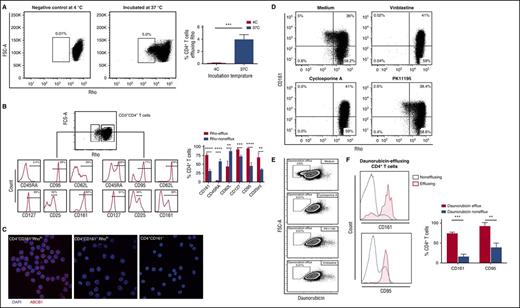
Expression of ABC-superfamily multidrug efflux proteins is one of the most important mechanisms whereby hematopoietic stem cells resist chemotherapy-induced cytotoxicity.18 Peripheral blood natural killer cells, B cells, and T cells also express functional ABCB1,19-21 although the exact role of the ABCB1 pump in lymphocyte survival has not been clearly defined. Recent studies show a role for the ABCB1 pump in the persistence of antigen-specific CD8+ memory T cells after chemotherapy,12 and in the resistance of proinflammatory Th17 cells to glucocorticoid treatment.22 Thus, we investigated whether CD4+ T cells also have similar specialized functions by assessing ABCB1-mediated efflux of the fluorescent ABCB1 substrate, Rho. This analysis identified a subset of CD4+ T cells with rapid Rho-effluxing capacity (median, 3.7%; range, 2.5%-5%) (Figure 1A). The rapidly effluxing CD4+ T cells (Rholo) were phenotypically similar to the recently described rapidly effluxing CD8+ T cells; in addition to the capacity for rapid efflux of Rho,12 they showed high expression of CD95, CD127, CD45RO, and CD161 (Figure 1B; supplemental Figure 1). Confocal microscopy confirmed high expression of ABCB1 on the cell membrane of the CD4+CD161+Rholo subset compared with CD4+CD161+Rhohi and CD4+CD161− T cells (Figure 1C).
A subset of CD4+T cells efflux Rhodamine123 and daunorubicin. PBMCs were incubated with Rho for 30 to 60 minutes at 37°C, after which multicolor FACS analysis was performed on gated CD4+Rholo (effluxing subset) and CD4+Rhohi cells (non-effluxing subset). (A) Representative FACS plot showing Rho efflux by CD4+ T cells after incubation at 37°C for 30-60 min (right) compared with negative control (left); cumulative data from 5 independent experiments are shown; ***P < .001. (B) Histograms comparing the phenotypes of effluxing (left) and non-effluxing (right) subsets within the total CD4+ T cell compartments. Note enrichment of Rho effluxing CD4+ T cell subsets within the CD161+ population; cumulative data from 4 independent experiments are shown; **P < .01, ***P < .001 and ****P < .0001. (C) Sort-purified CD4+CD161+Rholo, CD4+CD161+Rhohi and CD4+CD161- T cell subsets were stained for ABCB1 and examined by laser scanning confocal microscopy. Representative images from CD4+CD161+Rholo (left), CD4+CD161+Rhohi (middle) and CD4+CD161- (Right) T cell subsets are shown. ABCB1 (red) is only expressed on CD4+CD161+Rholo. DAPI (blue) was used as counterstaining. (D) FACS plots showing the abrogation of Rho efflux by CD4+CD161+ T cells after coincubation with inhibitors of ABCB1 and ABCC1 (cyclosporine A, PK11195 and vinblastine). (E) PBMCs were coincubated with fluorescent anthracycline (daunorubicin) for 30 to 60 min at 37°C. FACS plots show daunorubicin efflux by CD4+ T cells and complete abrogation of efflux upon addition of ABCB1 inhibitors (cyclosporin A and PK11195) and ABCC1 (vinblastine). (F) Histograms show that the gated daunorubicin-effluxing CD4+ T cells (red histogram) are enriched within the CD95+ CD161+ memory T-cell compartment compared with their non-effluxing counterpart (blue histogram); cumulative data from 3 independent experiments are shown; **P < .01 and ***P < .001.
A subset of CD4+T cells efflux Rhodamine123 and daunorubicin. PBMCs were incubated with Rho for 30 to 60 minutes at 37°C, after which multicolor FACS analysis was performed on gated CD4+Rholo (effluxing subset) and CD4+Rhohi cells (non-effluxing subset). (A) Representative FACS plot showing Rho efflux by CD4+ T cells after incubation at 37°C for 30-60 min (right) compared with negative control (left); cumulative data from 5 independent experiments are shown; ***P < .001. (B) Histograms comparing the phenotypes of effluxing (left) and non-effluxing (right) subsets within the total CD4+ T cell compartments. Note enrichment of Rho effluxing CD4+ T cell subsets within the CD161+ population; cumulative data from 4 independent experiments are shown; **P < .01, ***P < .001 and ****P < .0001. (C) Sort-purified CD4+CD161+Rholo, CD4+CD161+Rhohi and CD4+CD161- T cell subsets were stained for ABCB1 and examined by laser scanning confocal microscopy. Representative images from CD4+CD161+Rholo (left), CD4+CD161+Rhohi (middle) and CD4+CD161- (Right) T cell subsets are shown. ABCB1 (red) is only expressed on CD4+CD161+Rholo. DAPI (blue) was used as counterstaining. (D) FACS plots showing the abrogation of Rho efflux by CD4+CD161+ T cells after coincubation with inhibitors of ABCB1 and ABCC1 (cyclosporine A, PK11195 and vinblastine). (E) PBMCs were coincubated with fluorescent anthracycline (daunorubicin) for 30 to 60 min at 37°C. FACS plots show daunorubicin efflux by CD4+ T cells and complete abrogation of efflux upon addition of ABCB1 inhibitors (cyclosporin A and PK11195) and ABCC1 (vinblastine). (F) Histograms show that the gated daunorubicin-effluxing CD4+ T cells (red histogram) are enriched within the CD95+ CD161+ memory T-cell compartment compared with their non-effluxing counterpart (blue histogram); cumulative data from 3 independent experiments are shown; **P < .01 and ***P < .001.
To substantiate our findings that efflux occurs through ABCB1, we used specific pump-blocking agents. The addition of vinblastine (a competitive inhibitor of ABCB1 and ABCC1) and other inhibitors with more specificity for ABCB1, such as cyclosporine A and PK11195,12 completely abrogated or substantially reduced the efflux of Rho by CD4+CD161+ T cells (Figure 1D). We next asked whether such T cells are also capable of effluxing fluorescent anthracycline “daunorubicin,” another substrate of ABCB1, used mainly for the treatment of AML.23 We found that memory CD4+CD161+CD95+ T cells was also capable of rapidly effluxing daunorubicin, a property that was completely blocked by the addition of ABCB1 and ABCC1 inhibitors (Figure 1E-F).
CD4+CD161+Rholo T cells are a differentiated subset with distinct phenotype and pro-inflammatory polarization
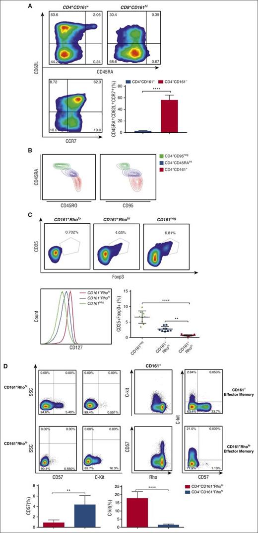
We next used multiparameter flow cytometry to delineate in more detail the surface phenotype of CD4+CD161+Rholo T cells compared with CD4+CD161+Rhohi and CD4+CD161− T cells. CD4+CD161int and CD8+CD161hi T cells showed remarkable phenotypic similarities, including high expression of CD127, CD95, and CD28 (supplemental Figure 2A). CD4+CD161+ T cells were almost equally divided into effector and central memory subsets (CD45RA−CD62L− and CD45RA−CD62L+, respectively), with no representation within the naive T-cell compartment (Figure 2A). A third, naive-like CD45RAdimCD62L+CCR7+ subset was clearly identified within the CD4+CD161+ population (Figure 2A). Additional analysis revealed striking phenotypic similarities between this subset and stem-like memory T cells, defined as CD45RAintCD45ROintCCR7+CD62L+CD95int-hi (Figure 2B). Interestingly, stem-cell-like CD4+ memory T cells did not exhibit rapid effluxing activity, although they had high expression of CD161 on their surface (supplemental Figure 2B). These data suggest that expression of CD161 on naive CD4+CD161− T cells may only occur after antigenic encounter, with acquisition of ABCB1 function occurring in later stages of differentiation.
Comprehensive phenotypic characterization of CD4+CD161+RholoT cells by multiparameter flow cytometry. PBMCs were stained with a panel of surface markers including monoclonal antibodies against CD16, TCR Vα24, CD8β, and γδ TCR to exclude natural killer, natural killer T, CD8dim, and γδ T cells, respectively. (A) Multiparameter analysis of gated CD4+CD161+ T cells revealed a mixture of effector (CD45RA−CD62L−CCR7−), central memory (CD45RA−CD62L+CCR7+), and naive-like (CD45RAintCD62L+ CCR7+) subsets; cumulative data from 5 independent experiments are shown. ***P < .001. (B) Contour plots showing expression of memory markers, CD45RO (left) and CD95 (right), on CD4+CD95neg, CD4+CD161+CD45RAint, and CD4+CD161+ T cells. (C) Regulatory T cells lack ABCB1 activity. Representative fluorescence-activated cell sorter (FACS) plots showing the frequency of CD25+Foxp3+ T cells (upper) in CD4+CD161+Rholo (left), CD4+CD161+Rhohi (middle), and CD4+CD161− (right) T cells; cumulative data from 9 independent experiments are shown in right lower panel. **P < .01; ****P < .0001. Histogram in lower left panel compares the mean fluorescence intensity (MFI) of CD127 among CD4+CD161+Rholo (red), CD4+CD161+Rhohi (blue), and CD4+CD161− (green) T-cell subsets. (D) Expression of c-kit and CD57 on CD4+CD161+Rhohi and CD4+CD161+Rholo T cells is mutually exclusive. Representative FACS plots show that c-kit expression is restricted to the rapidly effluxing CD4+CD161+ subset, whereas CD57 expression is restricted to the CD4+CD161+Rhohi T-cell subset with no detectable expression on CD4+CD161+Rholo T cells; cumulative data from 4 independent experiments are shown. **P < .01; ****P < .0001.
Comprehensive phenotypic characterization of CD4+CD161+RholoT cells by multiparameter flow cytometry. PBMCs were stained with a panel of surface markers including monoclonal antibodies against CD16, TCR Vα24, CD8β, and γδ TCR to exclude natural killer, natural killer T, CD8dim, and γδ T cells, respectively. (A) Multiparameter analysis of gated CD4+CD161+ T cells revealed a mixture of effector (CD45RA−CD62L−CCR7−), central memory (CD45RA−CD62L+CCR7+), and naive-like (CD45RAintCD62L+ CCR7+) subsets; cumulative data from 5 independent experiments are shown. ***P < .001. (B) Contour plots showing expression of memory markers, CD45RO (left) and CD95 (right), on CD4+CD95neg, CD4+CD161+CD45RAint, and CD4+CD161+ T cells. (C) Regulatory T cells lack ABCB1 activity. Representative fluorescence-activated cell sorter (FACS) plots showing the frequency of CD25+Foxp3+ T cells (upper) in CD4+CD161+Rholo (left), CD4+CD161+Rhohi (middle), and CD4+CD161− (right) T cells; cumulative data from 9 independent experiments are shown in right lower panel. **P < .01; ****P < .0001. Histogram in lower left panel compares the mean fluorescence intensity (MFI) of CD127 among CD4+CD161+Rholo (red), CD4+CD161+Rhohi (blue), and CD4+CD161− (green) T-cell subsets. (D) Expression of c-kit and CD57 on CD4+CD161+Rhohi and CD4+CD161+Rholo T cells is mutually exclusive. Representative FACS plots show that c-kit expression is restricted to the rapidly effluxing CD4+CD161+ subset, whereas CD57 expression is restricted to the CD4+CD161+Rhohi T-cell subset with no detectable expression on CD4+CD161+Rholo T cells; cumulative data from 4 independent experiments are shown. **P < .01; ****P < .0001.
CD4+CD161+Rholo memory T cells expressed higher levels of CD127 than did the CD4+CD161+Rhohi T-cell population (Figure 2C). These cells also expressed CD25 (IL-2Rα), with the vast majority enriched within the CD25int population. Interestingly, CD4+CD25+CD127loFoxp3+ regulatory T cells were devoid of ABCB1 pump activity (Figure 2C; supplemental Figure 2C). C-kit expression was restricted to the rapidly effluxing CD4+CD161+ subset, whereas CD57, a marker of terminal differentiation, was expressed exclusively on CD4+CD161+Rhohi T cells with no detectable expression on CD4+CD161+Rholo T cells (Figure 2D).
CD4+CD161+Rholo T cells have a distinct chemokine receptor profile, with preferential enrichment of Th17 cells with Th1-like features
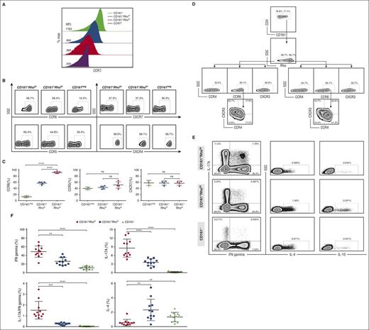
To gain insight into the homing properties and lineage commitment of CD4+CD161+Rholo T cells, we examined their chemokine receptor profile. The majority of the rapidly effluxing CD4+ T cells did not express CCR7, indicating that they have a predilection to home to the periphery, rather than lymph nodes (Figure 3A). Strikingly, CCR6 was expressed on most (>90%) rapidly effluxing cells, in contrast to CD161+ noneffluxing and CD161− subsets (Figure 3B-C). CD4+CD161+Rholo T cells were enriched among CCR6+CXCR3+CCR4− cells, previously reported to define Th17 cells with a pathogenic signature,22 whereas CD4+CD161+Rhohi T cells were characterized by a largely CCR6+CXCR3−CCR4+ phenotype, corresponding to the Th17 subset with a nonpathogenic gene signature22 (Figure 3D). In keeping with the phenotype, sort-purified CD4+CD161+Rholo T cells produced higher amounts of IL-17A and IFN-γ than did the CD4+CD161+Rhohi or CD4+CD161− T-cell subsets after ex vivo stimulation with anti-CD3/CD28 beads, with no significant production of IL-10 or IL-4 compared with CD4+CD161+Rhohi or CD4+CD161− T cells (Figure 3E-F).
Distinct chemokine receptor profile of CD4+CD161+RholoT cells. (A) Histogram comparing the MFI of CCR7 among CD4+CD161+Rholo (red), CD4+CD161+Rhohi (blue), and CD4+CD161− (green) T cells. (B) Representative FACS plots depicting the chemokine receptor profile, including CCR6, CCR5 and CXCR7 expression, on CD4+CD161+Rholo (left), CD4+CD161+Rhohi (middle), and CD4+CD161− (right) T cells; cumulative results from 5 independent experiments are shown in C. Each dot represents 1 individual. (D) Representative FACS plot depicting expression of CCR6, CCR4, and CXCR3 on CD4+CD161+Rholo (lower left) and CD4+CD161+Rhohi (lower right) T cells. (E) FACS-purified CD161+Rholo (top), CD161+Rhohi (middle), and CD161− (bottom) CD4+ T cells were stimulated with phorbol 12-myristate 13-acetate/ionomycin in the presence of brefeldin A for 6 hours. Indicated cytokines were analyzed by intracellular staining. (F) Dot plots comparing cytokine production among T-cell subsets with a low-, high-, or no-efflux capacity. **P < .01; ***P < .001; ****P < .0001.
Distinct chemokine receptor profile of CD4+CD161+RholoT cells. (A) Histogram comparing the MFI of CCR7 among CD4+CD161+Rholo (red), CD4+CD161+Rhohi (blue), and CD4+CD161− (green) T cells. (B) Representative FACS plots depicting the chemokine receptor profile, including CCR6, CCR5 and CXCR7 expression, on CD4+CD161+Rholo (left), CD4+CD161+Rhohi (middle), and CD4+CD161− (right) T cells; cumulative results from 5 independent experiments are shown in C. Each dot represents 1 individual. (D) Representative FACS plot depicting expression of CCR6, CCR4, and CXCR3 on CD4+CD161+Rholo (lower left) and CD4+CD161+Rhohi (lower right) T cells. (E) FACS-purified CD161+Rholo (top), CD161+Rhohi (middle), and CD161− (bottom) CD4+ T cells were stimulated with phorbol 12-myristate 13-acetate/ionomycin in the presence of brefeldin A for 6 hours. Indicated cytokines were analyzed by intracellular staining. (F) Dot plots comparing cytokine production among T-cell subsets with a low-, high-, or no-efflux capacity. **P < .01; ***P < .001; ****P < .0001.
Taken together, our data indicate that CD4+CD161+Rholo T cells have a distinct, polarized pro-inflammatory phenotype with a tendency to traffic to inflammatory sites.24-26 Their surface phenotype and cytokine profile suggest this subset largely comprises Th17 cells enriched with a Th1 signature.
Rapid Rho-effluxing CD161-expressing CD4+ T cells are not terminally differentiated and are able to fully proliferate and differentiate in vitro
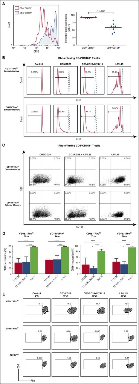
We next examined the proliferative and differentiation potentials of the rapidly effluxing CD4+CD161+ T cells in response to in vitro stimulation. Compared with their CD161− counterpart, CD4+CD161+ T cells proliferated more slowly in response to stimulation with anti-CD3/CD28 beads, suggesting these cells are relatively quiescent (Figure 4A). Addition of the homeostatic cytokines, IL-7 and IL-15, however, led to vigorous proliferation of CD4+CD161+Rholo, CD4+CD161+Rhohi Tcm and Tem, and CD161− T-cell subsets (Figure 4B; supplemental Figure 3A-B). Approximately 50% of each stimulated subset differentiated into CD161− T cells (Figure 4C-D; supplemental Figure 3C-D).
CD4+CD161+T cells with rapid efflux activity have the capacity to proliferate and differentiate. (A) Representative histograms showing the proliferative response of CD161+ (blue) and CD161− (red) CD4+ T cells to stimulation with anti-CD3/CD28 beads; cumulative data from 8 independent experiments are shown. ***P < .001. (B) Representative histograms showing the proliferative response of FACS-purified CD4+CD161+Rholo T cells that were further selected based on Tcm (CD45RA−CD62L+) (upper) or Tem (CD45RA−CD62L−) phenotype (bottom). Each FACS-sorted subset was stimulated under 3 different conditions: anti-CD3/anti-CD28 alone, anti-CD3/anti-CD28+IL-7/IL-15 and IL-7/IL-15 alone for 10 days. Proliferation was assessed by the CFSE dilution assay on day 10 of culture. (C) Expression levels of CD161 on sort-purified CD161+Rholo CD4+ Tcm and Tem cells after stimulation with anti-CD3/anti-CD28 alone, anti-CD3/anti-CD28+IL-7/IL-15, and IL-7/IL-15 alone on day 10. (D) Quantitative analysis compares CD161 expression on CD161+Rholo (left) and CD161+Rhohi (right) by different proliferative stimulus for Tcm and Tem subsets. (E) Homeostatic cytokines, IL-7 and IL-15, instruct self-renewal and maintain retention of rapid effluxing function. Sort-purified CD161+Rholo, CD161+Rhohi, and CD161−CD4+ T cells were stimulated with anti-CD3/anti-CD28, anti-CD3/anti-CD28+IL-7/IL-15, and IL-7/IL-15 alone for 10 days. Representative FACS plots showing Rho efflux by CD161+Rholo, CD161+Rhohi, and CD161−CD4+ T cells after incubation at 37°C for 30 to 60 minutes compared with negative control on day 10.
CD4+CD161+T cells with rapid efflux activity have the capacity to proliferate and differentiate. (A) Representative histograms showing the proliferative response of CD161+ (blue) and CD161− (red) CD4+ T cells to stimulation with anti-CD3/CD28 beads; cumulative data from 8 independent experiments are shown. ***P < .001. (B) Representative histograms showing the proliferative response of FACS-purified CD4+CD161+Rholo T cells that were further selected based on Tcm (CD45RA−CD62L+) (upper) or Tem (CD45RA−CD62L−) phenotype (bottom). Each FACS-sorted subset was stimulated under 3 different conditions: anti-CD3/anti-CD28 alone, anti-CD3/anti-CD28+IL-7/IL-15 and IL-7/IL-15 alone for 10 days. Proliferation was assessed by the CFSE dilution assay on day 10 of culture. (C) Expression levels of CD161 on sort-purified CD161+Rholo CD4+ Tcm and Tem cells after stimulation with anti-CD3/anti-CD28 alone, anti-CD3/anti-CD28+IL-7/IL-15, and IL-7/IL-15 alone on day 10. (D) Quantitative analysis compares CD161 expression on CD161+Rholo (left) and CD161+Rhohi (right) by different proliferative stimulus for Tcm and Tem subsets. (E) Homeostatic cytokines, IL-7 and IL-15, instruct self-renewal and maintain retention of rapid effluxing function. Sort-purified CD161+Rholo, CD161+Rhohi, and CD161−CD4+ T cells were stimulated with anti-CD3/anti-CD28, anti-CD3/anti-CD28+IL-7/IL-15, and IL-7/IL-15 alone for 10 days. Representative FACS plots showing Rho efflux by CD161+Rholo, CD161+Rhohi, and CD161−CD4+ T cells after incubation at 37°C for 30 to 60 minutes compared with negative control on day 10.
We subsequently examined whether the ex vivo expanded CD4+CD161+Rholo retained their effluxing capacity. Stimulation of CD4+CD161+Rholo cells with anti-CD3/CD28 beads abrogated their ability to efflux and drove their differentiation into CD161− noneffluxing T cells, whereas culture with IL-7/IL-15 alone favored retention of effluxing properties (Figure 4E). Notably, however, culture with IL-7/IL-15 did not induce the acquisition of effluxing properties in sort-purified noneffluxing CD4+CD161+Rhohi and CD161− T cells (Figure 4E), despite inducing upregulation of CD161 in vitro (supplemental Figure 3C-D). This suggests that homeostatic cytokines may play role in the maintenance, but not the induction, of rapid effluxing function.
We next addressed whether conditions of lymphopenia-driven homeostasis, as seen after chemotherapy, can drive the proliferation of CD4+CD161+ T cells. We found a significant increase in the absolute number of CD4+CD161+ T cells in PB samples collected from patients with AML recovering from daunorubicin-based induction chemotherapy compared with samples collected before chemotherapy (174.5 ± 28.3 vs 89.5 ± 25.8 cells/μL; P = .026). These data suggest that CD4+CD161+ T cells with rapid efflux capacity can survive chemotherapy in vivo and repopulate the memory compartment.
CMV-specific CD4+ T cells are enriched within the CD4+CD161+Rholo T-cell subset and survive induction chemotherapy in patients with AML
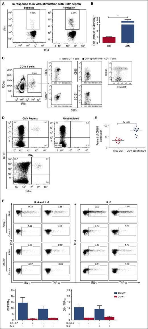
Given the body’s long-term need for virus-specific CD4+ T cells, we hypothesized that rapidly effluxing CD4+CD161+ T cells would be favored to survive toxic insults in vivo. We therefore determined CMV-specific CD4+ T-cell frequencies in the PB of patients with AML after treatment with intensive chemotherapy. although the frequencies of CMV-specific CD4+ T cells did not differ significantly in patients with AML at diagnosis (range, 0.03%-0.87%), compared with healthy controls, there was a nearly 5-fold increase in the frequencies of CMV-specific CD4+ T cells (range, 0.06%-2.61%) after recovery from the first cycle of remission induction chemotherapy, including daunorubicin (median, 4.98-fold increase; range, 1.6-fold to 11.7-fold; P = .036 (Figure 5A-B). This result suggests that a subset of CMV-specific memory CD4+ T cells can in fact preferentially resist, and therefore survive, the lymphocytotoxic effects of daunorubicin, and perhaps other lymphocytotoxic chemotherapeutic agents.
Characterization of CMV-specific CD4+T cells in healthy donors vs AML patients. PBMCs were stimulated with an overlapping pool of MHC class II CMVpp65 peptides. Cells were gated on IFN-γ-producing CD4+ T cells and examined for the coexpression of a panel of surface markers as outlined in “Materials and methods” (n = 11). (A) A representative FACS plot depicting IFN-γ-producing CMV-specific CD4+ T cells in a patient with AML before (baseline) and after recovery from chemotherapy (remission). (B) Significant fold increase in CMV-specific CD4+ T cells in patients with AML after daunorubicin treatment (*P = .036). (C) A representative FACS plot showing the phenotype of IFN-γ-producing CMV-specific CD4+ T cells (black) overlaid on total CD4+ T cells (gray). CMV-specific T cells are predominantly CD95+, CD45RA−, CD62L−, and CD127+. (D) A representative FACS plot showing that the majority of IFN-γ+ and TNF-α+ CMV-specific CD4+ T cells also express CD161. (E) CD161 expression on CMV-specific CD4+ and total CD4+ T cells. The majority of CMV specific CD4+ T cells coexpress CD161. (F) Representative FACS plots show the frequency of CMV-specific CD4+ T cells derived from CD161+ (top) and CD161− (bottom) T cells after stimulation with CMVpp65-loaded autologous B cells in the presence of IL-4 and IL-7 (left) or IL-2 (right) for 10 days. Bar graphs depict cumulative results for absolute frequency of CMV-specific T cells from 4 independent experiments. Data are shown as mean ± SE.
Characterization of CMV-specific CD4+T cells in healthy donors vs AML patients. PBMCs were stimulated with an overlapping pool of MHC class II CMVpp65 peptides. Cells were gated on IFN-γ-producing CD4+ T cells and examined for the coexpression of a panel of surface markers as outlined in “Materials and methods” (n = 11). (A) A representative FACS plot depicting IFN-γ-producing CMV-specific CD4+ T cells in a patient with AML before (baseline) and after recovery from chemotherapy (remission). (B) Significant fold increase in CMV-specific CD4+ T cells in patients with AML after daunorubicin treatment (*P = .036). (C) A representative FACS plot showing the phenotype of IFN-γ-producing CMV-specific CD4+ T cells (black) overlaid on total CD4+ T cells (gray). CMV-specific T cells are predominantly CD95+, CD45RA−, CD62L−, and CD127+. (D) A representative FACS plot showing that the majority of IFN-γ+ and TNF-α+ CMV-specific CD4+ T cells also express CD161. (E) CD161 expression on CMV-specific CD4+ and total CD4+ T cells. The majority of CMV specific CD4+ T cells coexpress CD161. (F) Representative FACS plots show the frequency of CMV-specific CD4+ T cells derived from CD161+ (top) and CD161− (bottom) T cells after stimulation with CMVpp65-loaded autologous B cells in the presence of IL-4 and IL-7 (left) or IL-2 (right) for 10 days. Bar graphs depict cumulative results for absolute frequency of CMV-specific T cells from 4 independent experiments. Data are shown as mean ± SE.
CMV-specific CD4+ T cells consisted mainly of effector-memory T cells, with high expression of CD95 and low expression of CD45RA and the adhesion molecule l-selectin (CD62L) (Figure 5C). A significantly higher proportion of CMV-specific CD4+ memory T cells from patients with AML (n = 11) expressed CD161 (mean 84.98% ± 2.7%) vs only 33.83% ± 3.2% of the total CD4+ T-cell population (P < .001) (Figure 5D-E).
Finally, to confirm that CMV-specific CD4+ T cells are enriched within the CD161+ subpopulation, we sort-purified CD4+ T cells into CD161+ and CD161− subsets and stimulated them in vitro with CMV pulsed autologous B cells and cytokines. As shown in Figure 5F, IFN-γ and TNF-α producing CMV-specific CD4+ T cells were predominately expanded from the CD161+ subset, further supporting the notion that CMV-specific memory T cells are enriched within the CD4+CD161+ T-cell repertoire.
CMV-specific CD4+CD161+ T cells are Th1-skewed, have rapid-effluxing ability, and are resistant to daunorubicin-induced lymphocytotoxicity
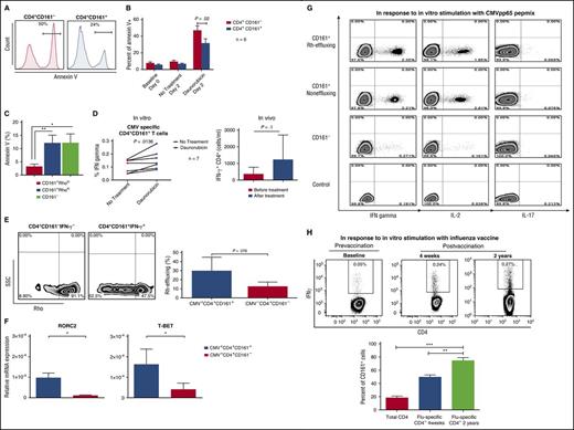
We next conducted studies on blood samples from 7 CMV-seropositive healthy donors to test the prediction that CMV-specific CD4+CD161+ T cells rely on a multidrug efflux mechanism to resist chemotherapy. After in vitro incubation with daunorubicin for 48 hours, significantly fewer CD161+CD4+ T cells underwent apoptosis compared with their CD161− counterpart (mean, 31.3% ± 5.3 vs 46.7% ± 5.9% annexin V+; P = .02) (Figure 6A-B). To confirm that daunorubicin resistance is limited to the rapidly effluxing CD161+ population, we sort-purified CD4+ T cells into CD161−, CD161+Rhohi, and CD161+Rholo subsets and treated them with daunorubicin for 48 hours. CD4+CD161+ T cells with effluxing capacity (Rholo) were significantly more resistant to daunorubicin-induced apoptosis, as measured by Annexin V staining, compared with Rhohi and CD161− populations (Figure 6C). In addition, we enumerated CMV-specific T cells in the culture. In vitro treatment with daunorubicin resulted in a relative increase in CMV-specific CD4+CD161+ T cells, supporting the notion that CMV-specific CD4+CD161+ T cells display resistance to daunorubicin-induced cytotoxicity in vitro (Figure 6D). We also found an increase in the absolute number of CMV-specific CD4+ T cells as measured by IFN-γ response in patients with AML recovering from daunorubicin-based induction chemotherapy compared with samples collected before chemotherapy (Figure 6D). Altogether, these data support the existence of a drug-resistant subset with antigenic specificity within the CD4+CD161+Rholo T-cell compartment.
CMV-specific CD4+CD161+T cells are resistant to daunorubicin-induced lymphotoxicity in vitro, have drug-effluxing capability, and reside within the Th1 rather than Th17 subset. PBMCs were exposed to daunorubicin in vitro and apoptosis was assessed by annexin V staining. (A) Histograms show the percentage of annexin V+ cells in CD4+CD161− and CD4+CD161+ subsets. (B) CD4+CD161+ T cells were significantly more resistant to apoptosis after 2 days of in vitro treatment with daunorubicin (P = .02), as assessed by annexin V staining compared with their CD161− counterpart (n = 6). (C) CD161+RhloCD4+ T cells are resistant to daunorubicin-induced cytotoxicity in vitro. Sort-purified CD161+Rholo, CD161+Rhohi, and CD161− CD4+ T cell subsets were cultured for 48 hours in the presence and absence of daunorubicin. Bar plots show cumulative data comparing annexin V+ cell frequency within the CD4+CD161+Rholo, CD4+CD161+Rhohi, and CD4+CD161− populations. Data represent the mean ± SE from 3 separate experiments. (D) PBMCs from 7 CMV-seropositive healthy donors were incubated in vitro with daunorubicin for 48 hours and then stimulated with CMVpp65 peptide at a concentration of 1 µg/mL for 4 to 6 hours; cumulative data depict a relative increase in the frequencies of IFNγ-producing CD4+CD161+ CMV-specific T cells after in vitro treatment with daunorubicin; data from 7 different experiments are shown. *P = .0136. The absolute number of CMV-specific CD4+ T cells as measured by IFN-γ response in patients with AML recovering from daunorubicin-based induction chemotherapy compared with samples collected before chemotherapy. (E) PBMCs from CMV-seropositive healthy donors were stimulated with CMVpp65 peptide at a concentration of 1µg/mL for 4 to 6 hours, followed by incubation with Rho. IFN-γ secreting cells were enriched using the IFN-γ cell secretion and enrichment kit (Miltenyi). Rho pumping was assessed in CD4+CD161+IFN-γ secreting T cells compared with CD4+CD161−IFN-γ− cells. Bar graph shows proportion of CD4+CD161+ IFN-γ+ T cells vs proportion of CD4+CD161−IFN-γ− T cells capable of rapidly effluxing Rho (n = 5). (F) CMV-specific CD4+ T cells were enriched after in vitro stimulation with CMVpp65 pepmix, using an IFN-γ cell secretion and enrichment kit. IFN-γ-expressing cells were then sort-purified based on CD161 expression into CD4+CD161+IFN-γ+ and CD4+CD161−IFN-γ+T cells, and T-bet and RORC2 mRNA expression was analyzed by quantitative reverse transcription polymerase chain reaction. Bar graphs show cumulative results from 3 healthy individuals. *P < .05. (G) Sort-purified CD161+Rholo, CD161+Rhohi, and CD161− T-cell subsets from CMV-seropositive healthy donors (n = 3) were stimulated with CMVpp65 peptide-loaded monocytes at a 2:1 ratio for 6 hours. Cytokine production was analyzed by intracellular staining, assessing production of IFN-γ, IL-2, and IL-17A. (H) PBMCs were collected from healthy donors approximately 4 weeks and 2 years after influenza (Flu) vaccination, and Flu-specific CD4+ T cells were enumerated after in vitro stimulation with influenza antigen by intracellular IFN-γ assay. A representative experiment shows the frequencies of IFN-γ-producing Flu-specific CD4+ T cells before vaccination (left), 4 weeks (middle), and 2 years (right) after vaccination. Bar graph compares CD161 expression between Flu-specific-IFN-γ producing CD4+ T cells at 4 weeks and 2 years. **P < .01; ***P < .001).
CMV-specific CD4+CD161+T cells are resistant to daunorubicin-induced lymphotoxicity in vitro, have drug-effluxing capability, and reside within the Th1 rather than Th17 subset. PBMCs were exposed to daunorubicin in vitro and apoptosis was assessed by annexin V staining. (A) Histograms show the percentage of annexin V+ cells in CD4+CD161− and CD4+CD161+ subsets. (B) CD4+CD161+ T cells were significantly more resistant to apoptosis after 2 days of in vitro treatment with daunorubicin (P = .02), as assessed by annexin V staining compared with their CD161− counterpart (n = 6). (C) CD161+RhloCD4+ T cells are resistant to daunorubicin-induced cytotoxicity in vitro. Sort-purified CD161+Rholo, CD161+Rhohi, and CD161− CD4+ T cell subsets were cultured for 48 hours in the presence and absence of daunorubicin. Bar plots show cumulative data comparing annexin V+ cell frequency within the CD4+CD161+Rholo, CD4+CD161+Rhohi, and CD4+CD161− populations. Data represent the mean ± SE from 3 separate experiments. (D) PBMCs from 7 CMV-seropositive healthy donors were incubated in vitro with daunorubicin for 48 hours and then stimulated with CMVpp65 peptide at a concentration of 1 µg/mL for 4 to 6 hours; cumulative data depict a relative increase in the frequencies of IFNγ-producing CD4+CD161+ CMV-specific T cells after in vitro treatment with daunorubicin; data from 7 different experiments are shown. *P = .0136. The absolute number of CMV-specific CD4+ T cells as measured by IFN-γ response in patients with AML recovering from daunorubicin-based induction chemotherapy compared with samples collected before chemotherapy. (E) PBMCs from CMV-seropositive healthy donors were stimulated with CMVpp65 peptide at a concentration of 1µg/mL for 4 to 6 hours, followed by incubation with Rho. IFN-γ secreting cells were enriched using the IFN-γ cell secretion and enrichment kit (Miltenyi). Rho pumping was assessed in CD4+CD161+IFN-γ secreting T cells compared with CD4+CD161−IFN-γ− cells. Bar graph shows proportion of CD4+CD161+ IFN-γ+ T cells vs proportion of CD4+CD161−IFN-γ− T cells capable of rapidly effluxing Rho (n = 5). (F) CMV-specific CD4+ T cells were enriched after in vitro stimulation with CMVpp65 pepmix, using an IFN-γ cell secretion and enrichment kit. IFN-γ-expressing cells were then sort-purified based on CD161 expression into CD4+CD161+IFN-γ+ and CD4+CD161−IFN-γ+T cells, and T-bet and RORC2 mRNA expression was analyzed by quantitative reverse transcription polymerase chain reaction. Bar graphs show cumulative results from 3 healthy individuals. *P < .05. (G) Sort-purified CD161+Rholo, CD161+Rhohi, and CD161− T-cell subsets from CMV-seropositive healthy donors (n = 3) were stimulated with CMVpp65 peptide-loaded monocytes at a 2:1 ratio for 6 hours. Cytokine production was analyzed by intracellular staining, assessing production of IFN-γ, IL-2, and IL-17A. (H) PBMCs were collected from healthy donors approximately 4 weeks and 2 years after influenza (Flu) vaccination, and Flu-specific CD4+ T cells were enumerated after in vitro stimulation with influenza antigen by intracellular IFN-γ assay. A representative experiment shows the frequencies of IFN-γ-producing Flu-specific CD4+ T cells before vaccination (left), 4 weeks (middle), and 2 years (right) after vaccination. Bar graph compares CD161 expression between Flu-specific-IFN-γ producing CD4+ T cells at 4 weeks and 2 years. **P < .01; ***P < .001).
Whether CMV-specific CD4+CD161+ T cells can directly efflux Rho was also addressed. In general, quantification of CMV-specific CD4+ and CD8+ T cells relies on detection of cytokines as a readout. Although intracellular cytokine staining after stimulation of PBMCs with viral peptides provides resolution at the single-cell level, this is at the expense of loss of viability, hampering the ability to examine the functional properties of cells using Rho efflux assay. Moreover, the generation of reliable MHC class II tetramer for sort-purification of CMV-specific CD4+ T cells has been more challenging than generation of MHC class I tetramers.27 As a consequence, we exploited the IFN-γ capture assay to “catch” and enrich live CMV-specific CD4+ T cells for the Rho efflux assay. As shown in Figure 6E, CMV-specific CD4+CD161+ T cells were capable of significant and rapid Rho efflux, supporting the hypothesis that a subset of CMV-specific CD4+ T cells uses the ABCB1 pump to survive chemotherapy.
The T-box transcription factor T-bet is a central regulator of the Th1 immune response and represses Th17 differentiation by antagonizing RORγt activation.28 To gain insight into the lineage commitment of CMV-specific CD4+ T cells and to delineate their transcriptional profile, we measured RORC2 and T-bet expression by real-time PCR in cytokine-captured and sort-purified CMV-specific CD4+CD161+ and CD4+CD161− T cells. CMV-specific CD4+CD161+ T cells expressed significantly higher levels of T-bet than RORC2 compared with their CD161− counterpart (Figure 6F). Consistent with this result, stimulation of sort-purified CD161+ effluxing, CD161+ noneffluxing, and CD161−CD4+ T cells with CMV pepmix resulted in IFN-γ and IL-2 production with no discernible IL-17A response (Figure 6G). These findings indicate that CMV-specific CD4+CD161+Rholo T cells are committed long-lived Th1 cells with rapid effluxing properties.
CD161 is a marker of long-lived antigen-specific memory CD4+ T cells
To verify that CD161 is a marker of long-lived memory CD4+ T cells, we analyzed the phenotype of influenza-specific CD4+ T cells in paired samples collected at approximately 4 weeks and 2 years after influenza vaccination from 4 healthy donors. Vaccination resulted in induction of influenza-specific CD4+ T cells in all healthy controls, as assessed by IFN-γ production after overnight in vitro stimulation, with or without seasonal influenza vaccine (A/Brisbane/59/2007[H1N1]). Although the frequencies of influenza-specific CD4+ T cells remained stable and were not significantly different at 4 weeks and 2 years after vaccination (0.15% ± 0.05% vs 0.21% ± 0.04%; P = .32; n = 4; Figure 6H), the proportion of influenza-specific CD4+ T cells that coexpressed CD161 was significantly higher at 2 years compared with 4 weeks postvaccination (72.8% ± 3.1 vs 49.3% ± 3.5%; P = .005), confirming that CD161 is a marker of long-lived virus-specific memory CD4+ T cells.
CD4+CD161+Rholo T cells display relative quiescence and maintain c-kit expression in response to stimulation
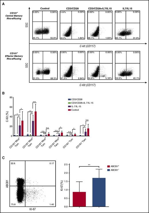
The mechanism by which CD4+CD161+Rholo T cells resist daunorubicin-induced apoptosis is undoubtedly complex, involving more than an active drug efflux pump. We therefore sought additional factors that could promote the survival of this rapidly effluxing T-cell subset. C-kit+ T cells are remarkably enriched within the CD4+CD161+Rholo population (Figure 2D). Signaling through c-kit promotes cellular survival and induces expression of the anti-apoptotic molecule, Bcl-2 and Bcl-x, by downstream activation of phosphatidylinositol 3′-kinase and Akt pathways.29,30 We found that the expression of Bcl-2 and Bcl-x was significantly higher in the CD4+CD161+Rholo Tcm and Tem fractions compared with the CD4+CD161+Rhohi or CD4+CD161− fractions (supplemental Figure 4A-C). Moreover, the rapidly effluxing CD4+CD161+ T cells maintained c-kit expression when stimulated with IL-7/IL-15, whereas TCR ligation induced rapid downregulation of c-kit expression (Figure 7A-B; supplemental Figure 4D-E). In addition, Rho-effluxing (ABCB1+) CD161+CD4+ T cells expressed less Ki-67 compared with the noneffluxing (ABCB1−) counterpart (Figure 7C), supporting the relative quiescence of the RholoCD4+CD161+ T-cell population. Taken together, these data suggest that multiple mechanisms of resistance, including relative quiescence, high levels of Bcl-2 and Bcl-x proteins, and conserved c-kit expression, in addition to ABCB1 activity, may contribute to the resistance of CD4+CD161+Rholo T cells to lymphocytotoxins, thus affording superior survival.
CD4+CD161+drug-effluxing T cells maintain c-kit expression after stimulation. CD4+CD161+ T cells were sort-purified according to rhodamine effluxing capacity (Rholo and Rhohi) and were stained for c-kit expression after stimulation with anti-CD3/CD28 beads. (A) Representative FACS plots depict c-kit expression on CD4+CD161+Rholo T cells in response to stimulation. (B) Cumulative data from 5 independent experiments show the c-kit expression on CD4+CD161+Rholo, CD4+CD161+Rhohi, and CD4+CD161−. Statistics were calculated using Student t test. *P < .05; **P < .01; ***P < .001; ****P < .0001; ns = not significant. (C) CD4+CD161+ T cells were stained for ABCB1, followed by intracellular staining for Ki67. Representative dot plots show the expression of Ki67 on ABCB1+ vs ABCB1− CD161+CD4+ T cells. Cumulative data demonstrate the higher expression of Ki67 on ABCB1− noneffluxing CD161+CD4+ T cells. Mean ± SE of 3 separate experiments were shown.
CD4+CD161+drug-effluxing T cells maintain c-kit expression after stimulation. CD4+CD161+ T cells were sort-purified according to rhodamine effluxing capacity (Rholo and Rhohi) and were stained for c-kit expression after stimulation with anti-CD3/CD28 beads. (A) Representative FACS plots depict c-kit expression on CD4+CD161+Rholo T cells in response to stimulation. (B) Cumulative data from 5 independent experiments show the c-kit expression on CD4+CD161+Rholo, CD4+CD161+Rhohi, and CD4+CD161−. Statistics were calculated using Student t test. *P < .05; **P < .01; ***P < .001; ****P < .0001; ns = not significant. (C) CD4+CD161+ T cells were stained for ABCB1, followed by intracellular staining for Ki67. Representative dot plots show the expression of Ki67 on ABCB1+ vs ABCB1− CD161+CD4+ T cells. Cumulative data demonstrate the higher expression of Ki67 on ABCB1− noneffluxing CD161+CD4+ T cells. Mean ± SE of 3 separate experiments were shown.
Discussion
Memory T cells confer immediate protection as well as the capacity to mount more rapid and effective secondary immune responses to antigenic challenge. Under circumstances in which the immune system is compromised, for instance, after treatment with lymphocytotoxic chemotherapy, patients with AML still appear to be protected from opportunistic viral infections such as CMV. This strongly suggests that a number of still unidentified mechanisms have evolved to maintain long-term T-cell memory. Here we show that a subpopulation of CD4+CD161+ memory T cells exploit their rapid effluxing properties to preserve long-term immunity. The ABCB1-mediated drug-efflux capacity of CD4+CD161+ memory T cells facilitated their resistance to chemotherapy, an effect that could be abrogated by the addition of competitive inhibitors of ABCB1 and ABCC1. Moreover, with respect to expression of cytokines and chemokine receptors, we found that rapidly effluxing CD4+CD161+ T cells are enriched within Th17 cells with Th1-like properties, as defined by a CCR6+CXCR3+CCR4lo phenotype.22
Although rapidly effluxing CD4+ T cells had the phenotypic characteristics of more differentiated T cells with relatively lower expression of CD62L compared with their noneffluxing counterpart, they preferentially expressed c-kit, a stem cell marker31 ; lacked expression of CD57, a marker of terminal differentiation; and displayed relative quiescence with low Ki-67 expression. These findings suggest that CD4+CD161+Rholo T-cell differentiation is governed by distinct mechanisms that lead to the generation of a differentiated T-cell subset with unique functions and a surface phenotype favorable for proliferation and self-renewal. Indeed, we showed that human CD4+CD161+Rholo Tcm and Tem, sort-purified ex vivo, are capable of rapid proliferation and differentiation in vitro. TCR ligation and CD28 costimulation in the presence of IL-7/IL-15 triggered vigorous proliferation and differentiation, with the emergence of CD161− c-kit− noneffluxing progeny.32 In contrast, when stimulated with homeostatic cytokines such as IL-7 and IL-15 alone, and without TCR ligation, CD4+CD161+Rholo T cells maintained their surface phenotype and rapid effluxing function.
IL-7 and IL-15 levels are increased in the sera of patients after lymphodepleting chemotherapy and drive T-cell proliferation.33 The relative resistance of CMV-specific CD4+ T cells to daunorubicin-induced apoptosis, together with our findings of persistence and a relative increase in CMV-specific CD4+ T cells in the PB of patients with AML after recovery from chemotherapy, support our prediction that such T cells have the ability to survive lymphocytotoxins, respond to homeostatic cytokines, and proliferate. Using the IFN-γ capture assay to directly select CMV-specific T cells from the PB of healthy CMV-seropositive donors, we confirmed that CMV-specific T cells display remarkable and rapid Rho-effluxing capability. These findings provide a direct mechanism whereby CMV-specific CD4+ T cells could survive lymphocytotoxic insult.
CMV-specific CD4+ T cells display considerable diversity with respect to their surface phenotype, function, and transcriptional profile based on CD161 and rhodamine-efflux capability (ie, CD161−, CD161+Rhohi, and CD161+Rholo). Transcriptional regulation dictates the cytokine profile and surface phenotype of T cells.34 T-bet is essential for commitment to the Th1 lineage and the production of IFN-γ.35,36 Transcriptional analysis of sort-purified CMV-specific CD4+CD161+Rholo or Rhohi and CD4+CD161− fractions revealed preferential T-bet expression and production of IFN-γ and IL-2 by CD4+CD161+Rholo T cells in response to stimulation with CMVpp65 with no detectable IL-17A. These data indicate that drug-effluxing CMV-specific CD4+CD161+ T cells reside within the Th1, rather than the Th17, lineage.
Finally, the long-term survival capacity of CD161-expressing, drug-effluxing T cells make them especially attractive candidates for adoptive therapy against viruses as well as cancer. Thus, the development of strategies to generate antigen-specific CD161-expressing memory T cells with superior survival and functional attributes might offer a highly effective approach to immunotherapy. One plausible approach would be to develop a system to generate large numbers of CD161+Rholo CD4+ T cells that self-renew and maintain their phenotype and drug-effluxing capacity for trials of adoptive T-cell therapy.
The online version of this article contains a data supplement.
The publication costs of this article were defrayed in part by page charge payment. Therefore, and solely to indicate this fact, this article is hereby marked “advertisement” in accordance with 18 USC section 1734.
Acknowledgments
This work was funded in part by National Institutes of Health, National Cancer Institute grants P01 CA148600-02 and RO1 CA061508-18. The flow studies were performed in the Flow Cytometry & Cellular Imaging Facility, which is supported in part by the National Institutes of Health through M. D. Anderson’s Cancer Center Support Grant CA016672.
Authorship
Contribution: A.A. and M.M. performed experiments, participated in the design and interpretation of the analysis, and wrote the manuscript. A.K., Y.-O.A., and R.B. performed experiments, participated in interpretation of the analysis, and commented on the manuscript. M.R.V., P.M., A.J.B., E.L., L.L., D.A.-J., K.S., H.S., K.K., N.I., B.A., D.M., R.E.C., and E.J.S. provided advice on experiments and analysis and commented on the manuscript. K.R. designed and directed the study and wrote the manuscript.
Conflict-of-interest disclosure: The authors declare no competing financial interests
Correspondence: Katayoun Rezvani, Department of Stem Cell Transplantation and Cellular Therapy, MD Anderson Cancer Center, 1515 Holcombe Blvd, Box 448, Houston, TX 77030-4009; e-mail: krezvani@mdanderson.org.
References
Author notes
A.A. and M.M. contributed equally to this study.

This feature is available to Subscribers Only
Sign In or Create an Account Close Modal