Abstract
The molecular approach for the analysis of leukemia associated chromosomal translocations has led to the identification of prognostic relevant subgroups. In pediatric acute lymphoblastic leukemia (ALL), the most common translocations, t(9; 22) and t(4; 11), have been associated with a poorer clinical outcome. Recently the TEL gene at chromosome 12p13 and the AML1 gene at chromosome 21q22 were found to be involved in the translocation t(12; 21)(p13; q22). By conventional cytogenetics, however, this chromosomal abnormality is barely detectable and occurs in less than 0.05% of childhood ALL. To investigate the frequency of the molecular equivalent of the t(12; 21), the TEL/AML1 gene fusion, we have undertaken a prospective screening in the running German Berlin-Frankfurt-Münster (BFM) and Italian Associazione Italiana Ematologia Oncologia Pediatrica (AIEOP) multicenter ALL therapy trials. We have analyzed 334 unselected cases of pediatric ALL patients consecutively referred over a period of 5 and 9 months, respectively. The overall incidence of the t(12; 21) in pediatric ALL is 18.9%. The 63 cases positive for the TEL/AML1 chimeric products ranged in age between 1 and 12 years, and all but one showed CD10 and pre-B immunophenotype. Interestingly, one case displayed a pre-pre–B immunophenotype. Among the B-lineage subgroup, the t(12; 21) occurs in 22.0% of the cases. Fifteen of 61 (24.6%) cases coexpressed at least two myeloid antigens (CD13, CD33, or CDw65) in more than 20% of the gated blast cells. DNA index was available for 59 of the 63 TEL/AML1 positive cases; a hyperdiploid DNA content (≥1.16) was detected in only four patients, being nonhyperdiploid in the remaining 55. Based on this prospective analysis, we retrospectively evaluated the impact of TEL/AML1 in prognosis by identifying the subset of B-lineage ALL children enrolled in the closed German ALL-BFM-90 and Italian ALL-AIEOP-91 protocols who had sufficient material for analysis. A total of 342 children were investigated for the presence of TEL/AML1 fusion gene and 99 cases (28.9%) were positive. The patients expressing the TEL/AML1 fusion mRNA appeared to have a better event-free survival (EFS) than the patients who lacked this chimeric product. Whereas three of the TEL/AML1 positive cases (3.0%) have relapsed to date, 27 patients without TEL/AML1 rearrangement (11.1%) suffered from relapse. To date, the only subset of B-lineage ALL with a favorable prognosis has been the hyperdiploid group (DNA index ≥1.16 <1.6). Our findings reinforce the need to include the molecular screening of the t(12; 21) translocation within ongoing prospective ALL trials to prove definitively its prognostic impact.
TWENTY-FIVE YEARS AGO, the prognosis of children suffering from acute lymphoblastic leukemia (ALL) was very poor, and most of them survived only 2 or 3 months after diagnosis. With current chemotherapy protocols, more than 90% of children with ALL achieve complete remission (CR) and by far, the majority of them will remain free of disease (continuous complete remission, CCR) during follow-up.1,2 To further increase the cure rate and to reduce unnecessary toxicity, efforts have been made to identify clinical and biological features of prognostic significance at diagnosis.3 4
Cytogenetic abnormalities of leukemic cells have helped identify patients with defined clinical and therapeutic responses.5 Hyperdiploidy (>50 chromosome) identifies a subset of B-lineage ALL with a favorable prognosis.6 By contrast, the presence of the t(9; 22) and t(4; 11) translocations confers a poor prognosis.5
Until recently, the translocation t(12; 21) was considered to be of limited prognostic value due to its apparent rarity, being barely detectable in less than 0.05% of the patients.5 Initial attempts to identify the t(12; 21) translocation by fluorescence in-situ hybridization technique (FISH) have indicated that its prevalence is largely underestimated.7,8 The TEL gene on chromosome 12p139 and the AML1 gene on chromosome 21q22 reviewed in Nucifora and Rowley10 have been recently shown to be involved in the t(12; 21) translocation,11 and a polymerase chain reaction (PCR) technique has become available for the molecular detection of the translocation.12-14
Because the prevalence of the t(12; 21) in pediatric ALL has been exclusively reported on a retrospective or limited series of patients, we planned to investigate the presence of the TEL/AML1 fusion transcripts in a series of ALL patients consecutively enrolled in two contemporary ongoing studies of the German (Berlin-Frankfurt-Münster [BFM]) and Italian (Associazione Italiana Ematologia Oncologia Pediatrica [AIEOP]) multicenter therapy trials over a period of 5 and 9 months, respectively. Based on the clinical and biological features obtained in the prospective series, we analyzed the presence of the t(12; 21) translocation in 342 ALL patients enrolled and treated with a traditional BFM schedule in the contemporary closed studies ALL-BFM 90 and ALL-AIEOP 91, for which an adequate clinical follow-up was available. The aim of the retrospective study was to evaluate whether TEL/AML1 fusion genes retained some prognostic value in ALL children who are considered at relatively good prognosis according to current standard criteria. The results of this intergroup study indicate that the presence of TEL/AML1 fusion product identifies a subgroup of B-cell precursor ALL with a good outcome, and that in addition with well established clinical features (age and white blood cell count), it may help to enroll ALL patients in less intensive chemotherapy regimens.
MATERIALS AND METHODS
Patients
ALL-AIEOP 95 and ALL-BFM 95 studies.From May 1995 to January 1996, 226 untreated patients with ALL younger than 15 years were enrolled for the ALL 95 protocols of the AIEOP study group. From May to September 1996, 212 untreated patients with ALL younger than 18 years were enrolled for the ALL 95 protocols of the BFM study group. Bone marrow (BM) and/or blood samples were successfully analyzed by reverse transcriptase-polymerase chain reaction (RT-PCR) for the molecular detection of the t(4; 11),15 t(9; 22),16 and t(12; 21)11 translocations in 347 cases (79.2%). In the remaining cases, either the inadequacy of BM samples and, more frequently, the quality of mRNA preparations hampered the molecular analyses. Data will be further referred to 334 cases negative for the BCR/ABL and MLL/AF4 fusion mRNAs.
Diagnosis of ALL was established according to standard morphologic, cytochemical, and immunological criteria.17 A portion of each BM sample was centralized to confirm the diagnosis, for immunophenotypic molecular analyses and for DNA content evaluation. Informed consent was obtained from patients for both biological and treatment procedures.
ALL-AIEOP 91 and ALL-BFM 90 studies.For the retrospective study, 342 cryopreserved BM samples from children with ALL enrolled in the ALL-AIEOP 91 and ALL-BFM 90,18,19 studies were analyzed. The selection of cases was based on the availability of BM cells and the inclusion in a predefined “no high-risk” ALL population accounting for about 80% of the entire ALL population. Although cases analyzed were selected based on the availability of cryopreserved material, no significant differences in clinical features or outcome were observed between analyzed and unanalyzed patients treated during the same period (data not shown). Children were eligible to this retrospective study if they had been enrolled in the Standard Risk (SR) and Intermediate Risk (IR) ALL-AIEOP 91 and ALL-BFM 90 protocols (84% and 86% of ALL patients, respectively). SR included patients with low tumor burden at diagnosis, as defined by the BFM Risk Index (RI)18 less than 0.8, good response to corticosteroid and none of the following: T phenotype, no complete remission (CR) at the end of protocol Ia, age <1 year (only for AIEOP), t(9; 22) translocation, t(4; 11) translocation (only for AIEOP), and signs of central nervous system (CNS) leukemia (only for AIEOP). High-risk (HR) included children with one of the following: poor response to corticosteroid, no CR at the end of protocol Ia, t(9; 22) translocation, RI>1.7 (only for AIEOP), t(9; 22) translocation, t(4; 11) translocation (only for AIEOP), and signs of CNS leukemia (only for AIEOP). All of the remaining patients were eligible for the IR. Our target population also excluded children older that 14 years (treated only in BFM protocols).
Immunophenotype and DNA Content Analyses
The immunophenotype was determined using a panel of monoclonal antibodies (MoAbs) including CD10, CD19, CD3, CD7, HLA-Dr, CD13, CD33, and CDW65.
Reactivity with MoAbs was assessed by indirect immunofluorescence. According to surface antigen expression, the B-cell precursor ALL were classified as pre-pre-B ALL (CD19+, CD10−, CD20−, cytoplasmic immunoglobulin-cyIgM-), common (CD19+, CD10+, cyIgM−), pre-B (CD19+, CD10+/−, cyIg+).17
DNA content of blast cells was evaluated and DNA index was calculated according to the guidelines provided by the Committee on Nomenclature of the Society for Analytical Cytology.20
RT-PCR Assay
RNA was extracted and RT-PCR performed as previously described.21,22 Samples were analyzed for the presence of the MLL-AF4,15 BCR-ABL,16 and the TEL/AML1 fusion gene products. The sequence of amplification primers for the TEL/AML1 product are the previously described set,11 B12 (5′-CGTGGATTTTTCAATACATGTCTCA-3′) for TEL and AM3 (5′-AACTGCCTTCGTCTCTATCTTTGTCCTTGG-3′) for AML1, with minor modifications according to the published TEL sequence.9 Fifty-six cases negative for the der21 TEL/AML1 product were reamplified in the same PCR conditions, using a more 5′ TEL olygonucleotide, TEL 318 (5′-CAATAGATGGATCTTTTCGTCTATTCGTATCTTC-3′) and the 3′ AML1 olygonucleotide (AM3).11 After amplification, 10 μL of PCR products were run on a 2.5% agarose gel stained with ethidium bromide and visualized under an ultraviolet (UV) lamp. Representative PCR products were cloned into the plasmid vector pMOS (Amersham, Buckinghamshire, England) and sequenced by the dideoxynucleotide chain termination method modified for use with double-stranded DNA templates.
Treatment Protocols
Treatment schedules for the SR and IR patients in the two studies (ALL-BFM 90 and ALL-AIEOP 91) have been previously reported.18 19 Briefly, all patients received 7 days of steroid therapy. Induction therapy included vincristine, prednisone, daunorubicin, L-asparaginase (L-ASP), and intrathecal (IT) chemotherapy (protocol Ia), followed by cyclophosphamide, cytarabine, 6-mercaptopurine (6-MP), and IT chemotherapy (protocol-Ib), only for IR patients. Consolidation therapy consisted of intermediate (2 g/m2) or high-dose MTX (HD-MTX) (5 g/m2) with leucovorin rescue plus oral 6-MP and IT chemotherapy. BFM patients were randomly selected to receive L-ASP (25,000 IU/m2) or not after each course of HD-MTX (total four doses). Reinduction therapy consisted of protocol II; it was similar to induction therapy except that it was shorter and daunorubicin, prednisone, and 6-MP were substituted with doxorubicin, dexamethasone, and 6-thioguanine, respectively. AIEOP patients were randomly selected to receive L-ASP at standard dose (10,000 IU/m2) twice a week or, at high dose (25,000 IU/m2) weekly for 20 weeks. Cranial radiotherapy (CRT) was given to BFM IR patients only. Continuation therapy consisted of 6-MP and MTX; IT chemotherapy was given to AIEOP patients only. The duration of treatment was 24 months.
Statistical Analysis
Event-free survival (EFS) was estimated according to Kaplan-Meier. The starting point was the date of diagnosis and the end point was death during induction, relapse, or death in continuous complete remission (CCR). Time was censored at last follow-up, if no failure was observed. Follow-up was updated in December 1995 and November 1996 for the AIEOP and BFM group, respectively. The univariate comparison of the EFS of different groups of patients was performed by means of the log-rank test. A multivariate approach was adopted to evaluate differences in the clinical outcome of positive and negative TEL/AML1 cases, adjusting for group (AIEOP and BFM) and known prognostic features. The Cox model23 was applied with a variable indicating the TEL/AML1 status (positive; negative) and other covariates indicating the source group (AIEOP; BFM), WBC count at diagnosis (WBC <20,000/μL; 20 to 100,000/mm3; ≥ 100,000/μL), age at diagnosis (in years: < 6; ≥ 6), sex, immunophenotype (common; pre-B or pre-pre-B). No major departures from the proportional hazards assumption were detected by graphical checking. We tested for interaction of the variable related to TEL/AML1 with the indicator of the source group, by introducing a first order term in the analysis. The Pearson′s χ2 test was used to assess the association between different characteristics. The SAS package (SAS Institute, Cary, NC) was used for the analysis.
RESULTS
Screening of TEL/AML1 Transcripts by RT-PCR
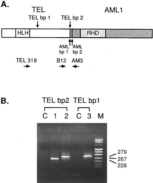
PCR using B12 and AM3 primers was expected to generate a 267-bp TEL/AML1 when the TEL gene was fused to exon 2 of the AML1 gene (Fig 1). In all but eight cases, identical RT-PCR products were amplified including a main product of 267-bp and a shorter and less abundant one of 228-bp (as shown in Fig 1, lane 2). DNA sequencing showed that the differently sized PCR fragment (228-bp) was caused by alternative splicing of 39 bp from AML1 exon 2 (data not shown). In eight patients, only the later product was detected. Due to the presence of alternative TEL breakpoints occurring at a more 5′ site,13 56 cases were analyzed for the presence of TEL/AML1 fusion transcripts by using TEL 318 and AM3 primers. In none of 56 cases was a fragment amplified.
Representative analysis of RT-PCR amplification of the TEL/AML1 chimeric transcripts in t(12; 21) ALL samples. (A) The relative position of the primers used to amplify the TEL/AML1 fusion cDNAs resulting from the most common breakpoint within the TEL gene (TEL bp-2) and the one occuring at a more 5′ site (TEL bp-1) is shown. The amplified fragment in the lane corresponding to patient n.1 is slighty shorter than usual (as in case n.2), lacking the 39 bp-AML1 exon 2. The sequence analysis of the cDNA product obtained from patient n.3, showed the junction of the exon 4 of the TEL gene to exon 2 of the AML1 gene.
Representative analysis of RT-PCR amplification of the TEL/AML1 chimeric transcripts in t(12; 21) ALL samples. (A) The relative position of the primers used to amplify the TEL/AML1 fusion cDNAs resulting from the most common breakpoint within the TEL gene (TEL bp-2) and the one occuring at a more 5′ site (TEL bp-1) is shown. The amplified fragment in the lane corresponding to patient n.1 is slighty shorter than usual (as in case n.2), lacking the 39 bp-AML1 exon 2. The sequence analysis of the cDNA product obtained from patient n.3, showed the junction of the exon 4 of the TEL gene to exon 2 of the AML1 gene.
Clinical Characteristics of TEL/AML1-Positive Cases
A total of 334 children consecutively referred to the AIEOP and BFM centers over a period of 9 and 5 months, respectively, were successfully investigated for the presence of TEL/AML1 fusion transcripts. Table 1 summarizes the main clinical and biological features at diagnosis. Sixty-three of 334 (18.9%) harbored a t(12; 21) translocation. All TEL/AML1-positive cases ranged in age between 1 and 12 years, with a peak of incidence between 1 and 5 years (76.2%). All cases showed a B-cell precursor phenotype, being present in all but one either common or pre-B, and one case displaying a pre-pre-B immunophenotype. Among the B-lineage subgroup, the t(12; 21) occurs in 22.0% of the cases. By contrast, no T-ALL or B-cell ALL with a mature phenotype, was positive. Interestingly, when the coexpression of CD10 and at least two myeloid antigens of CD13, CD33, and CDw65 was evaluated, a statistically (P < .001) significant association was present between this characteristic and the TEL/AML1 gene expression (as shown in Table 2). DNA content was available in 59 of the 63 TEL/AML1-positive cases; nonhyperdiploid DNA index was detected in 55 (93.2%), being ≥ 1.16 and < 1.6 in two cases and >1.6 in another two. According to the presenting features, 94% of the TEL/AML1-positive cases were enrolled in the IR group and 6% in the SR (data not shown). Moreover, all TEL/AML1-positive cases lacked evidence for BCR/ABL and MLL/AF4 fusion mRNAs.
Comparison of the Clinical and Laboratory Features of Patients With and Without TEL/AML1 Gene Expression (ALL-AIEOP95 and ALL-BFM95)
| . | TEL/AML1 Positive . | TEL/AML1 Negative . | ||
|---|---|---|---|---|
| . | (n = 63, 18.9%) . | (n = 271, 81.1%) . | ||
| . | . | . | . | . |
| Sex | ||||
| Male | 29 | 46% | 163 | 60% |
| Female | 34 | 54% | 108 | 40% |
| Age (yr) | ||||
| <1 | 0 | 13 | 5% | |
| 1-5 | 48 | 76% | 152 | 56% |
| 6-9 | 13 | 21% | 57 | 21% |
| 10-14 | 2 | 3% | 42 | 15% |
| 15-18 | 0 | 7 | 3% | |
| WBC | ||||
| <20,000 | 40 | 64% | 139 | 51% |
| 20-100,000 | 14 | 22% | 65 | 24% |
| ≥100,000 | 5 | 8% | 38 | 14% |
| Not known | 4 | 6% | 29 | 11% |
| Immunophenotype | ||||
| Common | 54 | 86% | 154 | 57% |
| Pre-B | 8 | 13% | 55 | 20% |
| Pre-pre-B | 1 | 1% | 8 | 3% |
| B mature | 0 | 6 | 2% | |
| T-ALL | 0 | 38 | 14% | |
| Not known | 0 | 10 | 4% | |
| DNA index | ||||
| <1 | 3 | 5% | 7 | 3% |
| 1 | 52 | 83% | 142 | 52% |
| >1 <1.16 | 0 | 38 | 14% | |
| ≥1.16 <1.6 | 2 | 3% | 46 | 17% |
| ≥1.6 | 2 | 3% | 2 | 1% |
| Not known | 4 | 6% | 36 | 13% |
Comparison of Myeloid Antigen Expression in B-Lineage ALL Patients With and Without TEL/AML1 Gene Expression (ALL-AIEOP95 and ALL-BFM95)
| . | TEL/AML1 Positive . | TEL/AML1 Negative . | |||
|---|---|---|---|---|---|
| . | n = 61* . | n = 199* . | |||
| . | . | . | . | . | |
| My-positive† | 15 | 24.6% | 6 | 3.0% | P < .01 |
| My-negative | 46 | 75.4% | 193 | 97.0% | |
| . | TEL/AML1 Positive . | TEL/AML1 Negative . | |||
|---|---|---|---|---|---|
| . | n = 61* . | n = 199* . | |||
| . | . | . | . | . | |
| My-positive† | 15 | 24.6% | 6 | 3.0% | P < .01 |
| My-negative | 46 | 75.4% | 193 | 97.0% | |
Immunological data on the coexpression of myeloid antigens was available in 61 of 63 (96.8%) TEL/AML1-positive cases and in 199 of 217 (91.7%) TEL/AML1-negative ALL cases.
As defined by the expression of at least two of the following antigens: CD13, CD33, and CDw65, in more than 20% of the gated blast cell population.
Clinical Outcome of TEL/AML1-Positive Cases
Based on the results of this analysis on a large prospective series, we retrospectively evaluated the prognostic impact of the t(12; 21) translocation in the closed German ALL-BFM 90 and Italian ALL-AIEOP 91 protocols that have sufficient follow-up for the analysis. Among the 342 children whose frozen BM materials were analyzed, 99 cases (28.9%) were positive and displayed almost comparable features as in the prospective series (Table 3). The major difference was the distribution of TEL/AML1-positive patients with respect to treatment protocols: whereas in ALL-BFM 90 and ALL-AIEOP 91 protocols, age was not considered as stratification criteria within SR and IR-risk subgroups, in ALL-BFM 95 and ALL-AIEOP 95 protocols, all children older than 6 years were enrolled in IR-risk treatment protocol. This retrospective series includes all patients without high-risk features (see Material and Methods) for whom a marrow sample was available for molecular analysis.
Clinical and Laboratory Features of Patients Enrolled in the ALL-AIEOP91 and ALL-BFM90 Protocols Analyzed for the Prognostic Impact of the t(12; 21) Translocation
| . | TEL/AML1 Positive . | TEL/AML1 Negative . | ||
|---|---|---|---|---|
| . | n = 99 (28.9%) . | n = 243 (71.1%) . | ||
| . | . | . | . | . |
| Sex | ||||
| Male | 53 | 54% | 129 | 53% |
| Female | 46 | 46% | 114 | 47% |
| Age (yr) | ||||
| <1 | 0 | 7 | 3% | |
| 1-5 | 74 | 75% | 159 | 65% |
| 6-9 | 19 | 19% | 34 | 14% |
| 10-14 | 6 | 6% | 43 | 18% |
| WBC | ||||
| <20,000 | 65 | 66% | 159 | 66% |
| 20-100,000 | 20 | 20% | 56 | 23% |
| ≥100,000 | 14 | 14% | 28 | 12% |
| Immunophenotype | ||||
| Common | 84 | 85% | 183 | 75% |
| Pre-B | 15 | 15% | 49 | 20% |
| Pre-pre-B | 0 | 6 | 2% | |
| Not known | 0 | 5 | 3% | |
| Protocol | ||||
| Standard risk | 27 | 27% | 61 | 25% |
| Intermediate risk | 72 | 73% | 182 | 75% |
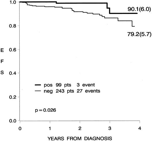
The estimated EFS was 90.1% (standard error [SE], 6.0) and 79.2% (SE, 5.7) at 4 years from diagnosis for the TEL/AML1 positive and negative cases, respectively, with a significant difference according to the univariate comparison (log-rank test P = .026) (Fig 2). The multivariate analysis was performed to adjust the evaluation of the prognostic impact on EFS of TEL/AML1 by other known prognostic factors and by the data source (AIEOP, BFM). The results of the Cox model are shown in Table 4, with all the variables included in the original model. The positivity of TEL/AML1 seems to maintain its beneficial impact on prognosis: positive TEL/AML1 patients had one third the risk of failure, as compared with negative TEL/AML1 patients. This result is of borderline significance (estimated hazard ratio = 0.29, P = .04). Other factors that significantly influenced prognosis were age, WBC, and immunophenotype. Specifically, children more than 6 years old experienced a worse prognosis (hazard ratio, 2.5; P = .02). Also, children with more than 100,000/μL WBC count at diagnosis had a fourfold increase of the failure rate (hazard ratio, 3.9; P < .01) with respect to children with low WBC (<20,000/μL). Pre-B or pre-pre-B phenotype ALL was unfavorably related to prognosis with respect to common ALL (hazard ratio, 2.8; P < .01). The EFS of patients treated in the two groups, AIEOP and BFM, did not differ significantly; the interaction between TEL/AML1 status and group was also not significant (and was thus excluded from the model), indicating that the prognostic impact of TEL/AML1 is in the same direction in the two groups.
Kaplan-Meier curve, showing the EFS of 342 patients enrolled in the ALL-AIEOP 91 and ALL-AIEOP 90 protocols, according to the presence of TEL/AML1 gene expression.
Kaplan-Meier curve, showing the EFS of 342 patients enrolled in the ALL-AIEOP 91 and ALL-AIEOP 90 protocols, according to the presence of TEL/AML1 gene expression.
Results of the Cox Model
| . | HR4-150 . | Estimated 95% CI . | P4-151 . |
|---|---|---|---|
| TEL/AML1 | |||
| Negative | 1 | ||
| Positive | 0.29 | 0.09-0.97 | 0.044 |
| Sex | |||
| Male | 1 | ||
| Female | 0.72 | 0.34-1.53 | 0.396 |
| Age (yr) | |||
| <6 | 1 | ||
| ≥6 | 2.49 | 1.18-5.24 | 0.016 |
| WBC | |||
| <20,000 | 1 | ||
| 20-100,000 | 0.65 | 0.22-1.95 | 0.443 |
| ≥100,000 | 3.94 | 1.61-9.67 | 0.003 |
| Immunophenotype | |||
| Common | 1 | ||
| Pre-B or pre-pre-B | 2.84 | 1.34-6.00 | 0.006 |
| Protocol | |||
| BFM | 1 | ||
| AIEOP | 1.35 | 0.58-3.13 | 0.487 |
Abbreviations: HR, hazard ratio; CI, confidence interval.
In the HR column, 1 identifies the reference category, an HR < 1 (>1) indicates that the risk of failure (relapse or death) in the corresponding group is less (more) than the risk of failure in the reference group.
P according to the Wald test.
DISCUSSION
In the present study, we used RT-PCR for the detection of TEL/AML1 fusion transcripts in 334 consecutive children with ALL enrolled in the German (BFM) and Italian (AIEOP) multicenter trials. We found an overall incidence of 18.9% unexceptionally limited to B-lineage ALL patients, accounting for up to 22.0% within the cases with common and pre-B phenotype. This incidence, exceeds dramatically the incidence of the translocation t(12; 21)(p13; q22), barely detectable by karyotyping alone.5 The presented intergroup series, which included unselected ALL cases, confirm and further extend previous observations indicating that the fusion of TEL and AML1 genes, as a result of a cryptic t(12; 21), represents the most common genetic lesion in pediatric ALL.12 13
Comparing the data obtained by Southern blotting and RT-PCR, patients with TEL gene rearrangements detected by DNA analysis, but without specific TEL/AML1 transcripts tested by RT-PCR, have been reported.13,24 The possibility that different TEL/AML1 breakpoints may occur outside the primer-spanning region causing false negative PCR results, has been extensively evaluated. None of the 56 cases analyzed, by using different sets of primers that encompass the site of fusion described for the t(5; 12)(q33; p13) translocation,9 showed the presence of TEL/AML1 fusion products. Alternatively, the TEL gene could have been fused to other genes than AML1, as shown in cases of translocation t(5; 12)(q33; p13), t(10; 12)(q24; p13), or t(12; 22)(p13; q11).9,25,26 In this study, we found only a limited heterogeneity of TEL/AML1 fusion products, with the presence of 39-bp AML1 exon sequence, alternatively present within the fusion transcript.12
As previously reported, our findings confirm that TEL/AML1-positive ALL is characterized by young age, with more than 76% of the cases aged between 1 and 5 years, lack of hyperleukocytosis (64% of the patients have less than 20 × 109/L WBC at diagnosis) and nonhyperdiploid DNA content (in 88% of the cases). Immunophenotypic analysis showed a remarkable association between the presence of TEL/AML1 fusion transcripts and the expression of myeloid antigens (at least two myeloid antigens, CD13, CD33, or CDw65, in more than 20% of the gated blast cells), occurring in 15 of 61 TEL/AML1-positive (24.6%) cases as compared with six of 199 (3.0%) negative cases. Although in several studies, mainly in adults, expression of myeloid antigens in ALL has been found to be associated with poor outcome,27,28 this feature was not prognostically important in the ALL-AIEOP 91 (G. Basso, unpublished observation, 1996) and ALL-BFM 90 studies.29
The presenting features associated with the expression of TEL/AML1 fusion transcripts (age, WBC, phenotype) known to be associated with a good prognosis raised the issue of the clinical significance of this molecular lesion as an independent prognostic factor. The outcome of ALL patients harboring the t(12; 21) translocation, retrospectively enrolled over 10 years time and treated on a variety of intensive chemotherapy, was recently evaluated.13 The TEL/AML1 positive group had a superior outcome, although the difference was not statistically significant because of the small number of patients studied.13 Moreover, although reported in limited series,30,31 a similar incidence of TEL/AML1 fusion transcripts was observed in ALL relapsing patients, as well as in those being in CCR. More recently the association between TEL/AML1 positivity and lack of relapse in ALL patients was reported in a series of 76 childhood ALL observed with a median follow-up of 8.3 years.32 Although the study showed a statistically significant difference, a high proportion of the patients selected within the TEL/AML1 negative displayed high-risk features, possibly amplifying the differences in clinical outcome observed with respect to TEL/AML1 gene expression.32
The need to evaluate a large group of patients receiving uniform treatment and to compare the outcome of TEL/AML1 with a cohort of B-cell precursor ALL known to have a good outcome, prompted us to investigate TEL/AML1 expression in ALL patients enrolled and treated with a traditional BFM schedule in two contemporary closed studies, ALL-BFM 90 and ALL-AIEOP 91.18,19 The two protocols were almost comparable, the main difference being the CNS directed therapy, which included CRT (ALL-BFM 90), whereas in the ALL-AIEOP 91, CRT was replaced by extended IT chemotherapy.18,19 Although all patients were consecutively enrolled in the two studies, we selected 342 cases based on the availability of frozen material and the absence of any known high-risk features. The differences in EFS between the TEL/AML1-positive ALL and other B-lineage were found to be statistically significant. However, it should be considered that the clinical and biological features of the three relapsing patients among the TEL/AML1-positive cases did not differ with respect to age, WBC, and phenotype, from the ones observed among other B-lineage ALL (data not shown). Therefore, it appears that the apparent improved prognosis of cases with TEL/AML1 should be evaluated further to identify whether TEL/AML1 expression alone or in association with other good prognostic features, turn out to be a strong predictor for an excellent outcome, as recently suggested.32 To date, the only subset of B-lineage ALL cases with a favorable prognosis have been the hyperdiploid group, and DNA index (> 1.16) is currently included as a criteria to identify a subgroup of ALL patients considered for less intense chemotherapy.6
In conclusion, our findings, promising in the possibility to identify a second subgroup of B-lineage cases with favorable outcome, reinforce the need to include the molecular screening of the t(12; 21) translocation as part of diagnostic ALL procedures, to identify a marker for minimal residual disease detection, and to fully elucidate its prognostic significance in childhood ALL.
ACKNOWLEDGMENT
We thank all of the clinicians of the Italian (AIEOP) and German (BFM) centers for providing BM samples and D.Silvestri for excellent data management.
A.B. and G.C. contributed equally to this work.
Supported in part by Fondazione Tettamanti (Monza, Italy) and by grants and fellowship of the Associazione Italiana per la Ricerca sul Cancro (Milano, Italy; to A.B. and M.G.V.) and Consiglio Nazionale delle Ricerche (Roma, Italy; PF ACRO, Grants No. 92.02140.PF.39 to A.B. and 96.006494 to M.G.V.), as well as the Deutsche Krebshilfe (Bonn, Germany) and the Forschungshilfe Station Peiper (Giessen, Germany).
Address reprint requests to Andrea Biondi, MD, Clinica Pediatrica Università di Milano, Ospedale S.Gerardo, v.Donizetti, 106, 20052 Monza (MI), Italy.


This feature is available to Subscribers Only
Sign In or Create an Account Close Modal