Key Points
Srsf2P95H/+ mutation within hemopoietic stem cells is required to initiate myeloid-biased hemopoiesis.
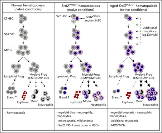
Mutation of Srsf2 is sufficient to initiate the development of MDS/MPN in vivo in the setting of native hemopoiesis.
Abstract
Mutations in SRSF2 occur in myelodysplastic syndromes (MDS) and MDS/myeloproliferative neoplasms (MPN). SRSF2 mutations cluster at proline 95, with the most frequent mutation being a histidine (P95H) substitution. They undergo positive selection, arise early in the course of disease, and have been identified in age-related clonal hemopoiesis. It is not clear how mutation of SRSF2 modifies hemopoiesis or contributes to the development of myeloid bias or MDS/MPN. Two prior mouse models of Srsf2P95H mutation have been reported; however, these models do not recapitulate many of the clinical features of SRSF2-mutant disease and relied on bone marrow (BM) transplantation stress to elicit the reported phenotypes. We describe a new conditional murine Srsf2P95H mutation model, where the P95H mutation is expressed physiologically and heterozygously from its endogenous locus after Cre activation. Using multiple Cre lines, we demonstrate that during native hemopoiesis (ie, no BM transplantation), the Srsf2P95H mutation needs to occur within the hemopoietic stem-cell–containing populations to promote myelomonocytic bias and expansion with corresponding transcriptional and RNA splicing changes. With age, nontransplanted Srsf2P95H animals developed a progressive, transplantable disease characterized by myeloid bias, morphological dysplasia, and monocytosis, hallmarks of MDS/MPN in humans. Analysis of cooccurring mutations within the BM demonstrated the acquisition of additional mutations that are recurrent in humans with SRSF2 mutations. The tractable Srsf2P95H/+ knock-in model we have generated is highly relevant to human disease and will serve to elucidate the effect of SRSF2 mutations on initiation and maintenance of MDS/MPN.
Introduction
Myelodysplastic syndrome (MDS) represents a spectrum of hematological cancers with a high rate of progression to acute leukemia, where recurrent somatic mutations in the RNA splicing machinery have been identified.1-3 In MDS, >80% of patients have a mutation in the spliceosome, and in phenotypically overlapping malignancies such as MDS/myeloproliferative neoplasms (MPN) and chronic myelomonocytic leukemia (CMML), spliceosome mutations are also common.4,5 Spliceosomal mutations arise early in disease, including as initiating events.6 Mutations generally occur in 1 splicing gene and are heterozygous, and different spliceosomal mutations associate with distinct clinical phenotypes. How mutations in the spliceosome alter normal hemopoiesis and contribute to disease pathogenesis remains unclear.
Mutations in SRSF2 occur in MDS (∼12% to 15%), CMML (28% to 47%), and mastocytosis,3,4,7 clustering at proline 95 (supplemental Figure 1A, available on the Blood Web site). Features of SRSF2 mutation–associated disease include myeloid bias and an increased risk of transformation to acute leukemia.8-10 SRSF2 mutations undergo positive selection and occur in age-related clonal hemopoiesis.11-13 P95 mutations alter messenger RNA splicing by changing the RNA-binding affinity of SRSF2, leading to misregulation of exon inclusion.14 This leads to modest overall changes in global messenger RNA splicing, with a subset of misspliced transcripts proposed to be relevant for MDS, including FYN, EZH2, and Csf3r.14-16
The first murine Srsf2P95H/+ conditional mutation model reported a mild dysplastic phenotype characterized by leukopenia and anemia, only observed after bone marrow (BM) transplantation. De novo excised animals did not develop dysplasia/disease.15,17 These phenotypes showed variable penetrance, given the lack of leukopenia/anemia in an ensuing study with the same allele.15,17 In a second Srsf2P95H model, animals also did not develop disease after 90 weeks of monitoring.16 Neither model reported myeloid-biased hemopoiesis, a feature of SRSF2 mutation in humans, or MDS in nontransplanted models. Divergent consequences on phenotypic hemopoietic stem cells (HSCs)/progenitors were concluded. As a result, how SRSF2 mutation alters normal hemopoiesis and promotes malignancy remains uncertain.
Here we describe a new Srsf2P95H model, where the P95H mutation is conditionally expressed physiologically and heterozygously from its endogenous locus. This model recapitulates the core characteristics of human disease associated with SRSF2 mutation and demonstrates that the primary effect of SRSF2(P95H) on normal hemopoiesis is to promote myeloid bias from HSCs.
Methods
Experiments were approved by the Animal Ethics Committee (#033/12 and 001/16; St. Vincent’s Hospital, Melbourne, Australia).
Animal models
Srsf2P95H/+ mice (C57BL/6NTac-Srsf2tm2874(P95H)Arte) were generated by Taconic Biosciences (Cologne, Germany). A full description of the targeting construct and genotyping procedures is provided in the supplemental Methods. hScl-CreERT 18-20, Rosa26-eYFP (Jackson Laboratory strain #00614818-20 ), LysM-Cre (strain #00478121-23 ), and Rosa26-CreERT2 (strain #00846324-26 ) mice have been described. B6.SJL-PtprcaPep3b/BoyJArc mice were purchased from the Animal Resources Centre (Perth, Australia). Heterozygous CD45.1/CD45.2 C57BL/6 mice were bred at St. Vincent’s Hospital. Food contained 400 mg/kg of tamoxifen citrate (Sigma) in standard mouse chow (Specialty Feeds, Perth, Australia).25,26 All mouse lines were on a C57BL/6 background.
Flow cytometric analysis
BM transplantation
All transplantations were performed in lethally irradiated (2 × 5 Gy; 3 hours apart) recipients (B6.SJL-PtprcaPep3b/BoyJArc [Animal Resources Centre]; 3-6 recipients per transplantation cohort) through IV injections. Recipients received antibiotics (Baytril) in the drinking water for 3 weeks after irradiation. Experiments were performed in duplicate or as indicated.
RNA sequencing
RNA was isolated from lin−cKit+eYFP+ BM cells from hScl-CreERTR26eYFPSrsf2+/+ and hScl-CreERTR26eYFPSrsf2P95H/+ animals treated 20 weeks prior with tamoxifen. RNA was ribosome depleted, subjected to library preparation (Kapa Stranded RNA-Seq Library Kit; Kapa Biosystems), and sequenced on the Illumina platform by Novogene (Hong Kong). Differential expression was assessed using voom with sample weighting.28 Quantitative Set Analysis for Gene Expression was used to determine gene set enrichment29 against the Molecular Signatures Database collection.20,30 Data sets were deposited in the Gene Expression Omnibus (GSE99852). RNA splicing was assessed using rMATS.14,16,31 Results are provided in supplemental Data Set 2.
Exome analysis
Exome sequencing was performed on genomic DNA isolated from whole BM and matched ear tissue from 3 independent animals (2 R26-CreERT2Srsf2P95H/+, 1 hScl-CreERTR26eYFP Srsf2P95H/+) from 50 to 60 weeks post–tamoxifen treatment (time of euthanasia when moribund). Exome capture was performed with the Agilent SureSelect Mouse Exon system and sequenced on the Illumina platform by BGI Ltd (Hong Kong). Raw and analyzed data are available in the Gene Expression Omnibus (GSE99850).
Statistics
The results were analyzed using the unpaired Student t test or analysis of variance (with multiple comparison correction) unless otherwise stated. P < .05 was considered significant. All data are presented as mean ± standard error of the mean.
Results
To model SRSF2P95H mutation, we generated a conditional Srsf2P95H knock-in allele (P95H). Murine Srsf2 has 2 closely adjacent genes at each end of the locus, so a whole-locus duplication approach was used. The proline 95 (CCG) to histidine (CAT) mutation was introduced into a duplicated Srsf2 exon 1, generating a conditional point mutant allele (Figure 1B). In the presence of Cre, the wild-type (WT) Srsf2 allele is deleted and the mutant Srsf2P95H allele is transposed into the endogenous locus. All animals were heterozygous for the knock-in allele. The Srsf2P95H/+ animals had a mildly elevated mean cell volume at baseline, before Cre activation (supplemental Figure 1D). There was low-level expression of the mutant Srsf2P95H allele in the absence of Cre, with a slight increase in B cells and CD4 T cells in the PB but no change in myeloid populations (supplemental Figure 1E-J). When the Srsf2P95H allele was activated in vivo, it was expressed heterozygously, genomically, and transcriptionally (Figure 1A; supplemental Figure 1C).
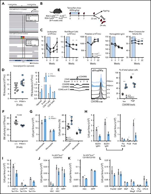
Widespread expression of Srsf2P95H/+promotes leukocytosis and myeloid bias. (A) Srsf2P95H transcript expression in whole BM from a control and P95H animal. (B) Experimental outline and timeframe. (C) PB indices over 22 weeks after Cre activation. (D) PB leukocyte counts and lineage distribution at 24 weeks after Cre activation (n ≥ 6 per genotype per time point). (E) Histograms and expression of CD45RB isoform on splenocytes (n = 5+/+; n = 3 P95H+). (F) BM cellularity per femur. (G) Number and percentage of granulocytes and monocytes per femur. (H) Number of B cells and B-cell progenitor populations per femur. (I) Erythroid populations per femur. Number of phenotypic long-term (LT) and short-term (ST) stem-cell and multipotent progenitor (MPP) populations per femur based on lin−cKit+Sca1+CD34/Flt3 (J) or lin−cKit+Sca1+CD105/CD150 (K) phenotype. (L) Numbers of myeloerythroid progenitors per femur. (E-I) n = 5+/+; n = 3 P95H/+. Presented as mean ± standard error of the mean. Student t test. *P < .05, **P < .01, ***P < .001. CFU-E, colony-forming unit erythroid; GMP, granulocyte macrophage progenitor; IgM, immunoglobulin M; MkP, megakaryocyte progenitor; preMegE, premegakaryocyte erythroid progenitor.
Widespread expression of Srsf2P95H/+promotes leukocytosis and myeloid bias. (A) Srsf2P95H transcript expression in whole BM from a control and P95H animal. (B) Experimental outline and timeframe. (C) PB indices over 22 weeks after Cre activation. (D) PB leukocyte counts and lineage distribution at 24 weeks after Cre activation (n ≥ 6 per genotype per time point). (E) Histograms and expression of CD45RB isoform on splenocytes (n = 5+/+; n = 3 P95H+). (F) BM cellularity per femur. (G) Number and percentage of granulocytes and monocytes per femur. (H) Number of B cells and B-cell progenitor populations per femur. (I) Erythroid populations per femur. Number of phenotypic long-term (LT) and short-term (ST) stem-cell and multipotent progenitor (MPP) populations per femur based on lin−cKit+Sca1+CD34/Flt3 (J) or lin−cKit+Sca1+CD105/CD150 (K) phenotype. (L) Numbers of myeloerythroid progenitors per femur. (E-I) n = 5+/+; n = 3 P95H/+. Presented as mean ± standard error of the mean. Student t test. *P < .05, **P < .01, ***P < .001. CFU-E, colony-forming unit erythroid; GMP, granulocyte macrophage progenitor; IgM, immunoglobulin M; MkP, megakaryocyte progenitor; preMegE, premegakaryocyte erythroid progenitor.
Srsf2P95H causes myeloid bias
We used R26-CreERT2Srsf2+/+ and R26-CreERT2Srsf2P95H/+ animals to elicit widespread expression of P95H. Unless stated, experiments evaluated native hemopoiesis without prior BM transplantation. Tamoxifen-containing food was administered to activate P95H expression (Figure 1B). In the PB, P95H animals developed macrocytosis and reduced red blood cells (RBCs; Figure 1C), with leukocyte counts stable for the first 12 weeks but increasing by ∼20 weeks after P95H expression (Figure 1C).
At 22 weeks after tamoxifen, there were increased absolute numbers of myeloid cells and CD4 T cells but no change in B lymphocytes in the PB, indicative of myeloid bias/myeloproliferation (Figure 1D). The expression of CD45RB, a characterized splicing target of Srsf2,32 was measured on total splenocytes. In Srsf2+/+, ∼80% of splenocytes expressed CD45RB. Srsf2P95H/+ splenocytes had reduced expression of CD45RB, demonstrating altered protein isoform expression in vivo (Figure 1E). In the BM, there was a trend to reduced overall cellularity (Figure 1F). The granulocyte and monocyte percentages significantly increased in Srsf2P95H/+ (Figure 1G), while B lymphopoiesis was compromised from the pro–B-cell stage onward (Figure 1H). BM erythropoiesis was reduced, with compensatory splenic erythropoiesis (Figure 1I; supplemental Figure 2F). There was either no significant difference or decreased numbers of phenotypic LT-HSCs in the P95H BM, using multiple phenotypic methods (Figure 1J-L). The ST-HSCs and MPPs were reduced, with the myeloid progenitor populations least affected (Figure 1J-L; supplemental Figure 2B). Myeloid and erythroid cells increased, and B/T cells were reduced in the spleen, but overall weight/cellularity were not different (supplemental Figure 2C-G). All subsets were reduced in the thymus (supplemental Figure 2H). Therefore, Srsf2P95H/+ causes myeloid-biased hemopoiesis.
Srsf2P95H mutation is required within the HSCs
To determine if the cell population where P95H was activated would alter the phenotype, hScl-CreERTR26-eYFPki/ki was used.18 Unlike R26-CreER, this is specific to the HSC-containing populations and not expressed within the BM stroma/nonhemopoietic tissues. The hScl-CreERT–enabled establishment of mosaic hemopoiesis without prior BM transplantation, with eYFP+ cells marking the P95H mutants and the eYFP− cells retaining WT Srsf2. Thus, it allowed an assessment of P95H cells compared with WT cells from the same BM microenvironment under native conditions.
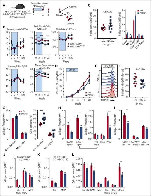
The PB demonstrated a phenotype comparable to R26-CreERT2, with the P95H animals developing leukocytosis, macrocytosis, and reduced RBC numbers (Figure 2A-B). At 20 weeks postactivation, a myelomonocytic expansion and increase in CD4 T cells without a reduction in B lymphocytes were present in the PB (Figure 2C). The PB eYFP+ percentage was equivalent between the genotypes (Figure 2D). Expression of CD45RB was reduced (Figure 2E). BM cellularity was similar, but the myeloid cell percentage was significantly increased in the P95H (Figure 2F-G; supplemental Figure 6A). Erythroid and B cells were reduced in the P95H BM. The LT-HSCs and ST-HSCs/MPPs showed either a reduction or trend to a reduction in the P95H BM (Figure 2J-K), with the myeloid progenitors relatively spared (Figure 2L; supplemental Figure 6A). Splenic and thymic hemopoiesis showed trends comparable to the R26-CreER, with a more subtle overall effect reflecting the contribution of WT cells (data not shown).
HSC-specific expression of Srsf2P95H/+causes myeloid bias. (A) Experimental outline and timeline. (B) PB indices over 20 weeks after Cre activation. (C) PB leukocyte counts and lineage distribution at 20 weeks after Cre activation (n = 7-23 per genotype per time point). (D) PB eYFP levels over time (n = 7-23 per genotype per time point). (E) Histograms of CD45RB isoform expression on splenocytes of the indicated genotypes (n = 4+/+; n = 5 P95H/+). (F) BM cellularity per femur. (G) Number and frequency of granulocytes and monocytes per femur. (H) Number of B cells and B-cell progenitor populations per femur. (I) Erythroid populations per femur. Number of phenotypic LT-HSC, ST-HSC, and MPP populations per femur using the (J) lin−cKit+Sca1+CD34/Flt3 or lin−cKit+Sca1+CD105/CD150 (K) phenotype. (L) Numbers of myeloerythroid progenitors per femur. (F-K) n = 7+/+; n = 8 P95H/+. Presented as mean ± standard error of the mean. Student t test. *P < .05, **P < .01, ***P < .001. CFU-E, colony-forming unit erythroid; GMP, granulocyte macrophage progenitor; IgM, immunoglobulin M; MkP, megakaryocyte progenitor; preMegE, premegakaryocyte erythroid progenitor.
HSC-specific expression of Srsf2P95H/+causes myeloid bias. (A) Experimental outline and timeline. (B) PB indices over 20 weeks after Cre activation. (C) PB leukocyte counts and lineage distribution at 20 weeks after Cre activation (n = 7-23 per genotype per time point). (D) PB eYFP levels over time (n = 7-23 per genotype per time point). (E) Histograms of CD45RB isoform expression on splenocytes of the indicated genotypes (n = 4+/+; n = 5 P95H/+). (F) BM cellularity per femur. (G) Number and frequency of granulocytes and monocytes per femur. (H) Number of B cells and B-cell progenitor populations per femur. (I) Erythroid populations per femur. Number of phenotypic LT-HSC, ST-HSC, and MPP populations per femur using the (J) lin−cKit+Sca1+CD34/Flt3 or lin−cKit+Sca1+CD105/CD150 (K) phenotype. (L) Numbers of myeloerythroid progenitors per femur. (F-K) n = 7+/+; n = 8 P95H/+. Presented as mean ± standard error of the mean. Student t test. *P < .05, **P < .01, ***P < .001. CFU-E, colony-forming unit erythroid; GMP, granulocyte macrophage progenitor; IgM, immunoglobulin M; MkP, megakaryocyte progenitor; preMegE, premegakaryocyte erythroid progenitor.
To determine if restricting P95H activation to the myeloid lineage would promote myeloid bias/myeloproliferation, LysM-Creki/+R26-eYFPki/ki cohorts were established (LysM-Cre is constitutively expressed). PB monitoring showed a mildly elevated MCV and leukocyte and platelet counts, but no evidence of morphological dysplasia (supplemental Figure 3A-D). Analysis of 20-week-old mice demonstrated that the PB and BM were largely normal (supplemental Figure 3E-K). There was a subtle increase in the percentage of granulocytes in the spleen, but no other phenotypes were apparent (supplemental Figure 3L-M). Therefore, expression of Srsf2P95H/+ within the HSC population is required to efficiently initiate myeloid-biased hemopoiesis.
Srsf2P95H HSC transplantation potential is determined by their competitor
To assess HSC activity, competitive transplantation of whole BM from Rosa26-CreERT2Srsf2P95H/+ and controls was performed (Figure 3A). In 2 independent experiments, the same outcome occurred: initial chimerism was low and heavily myeloid skewed; then, chimerism was rapidly reduced to low/negligible from the Srsf2P95H/+ cells (Figure 3B-E). The numbers of competitive repopulating units33 were reduced by 95% to 99% in the P95H BM (Figure 3B-C). The significantly impaired competitive repopulation of P95H HSCs is consistent with both prior models of Srsf2 mutations in mice,15,16 yet at odds with positive selection of SRSF2 mutations in humans.11-13
Srsf2P95H/+HSCs have a competitive advantage when transplanted with age- and microenvironment-matched support BM. (A) Experimental outline. (B-C) PB chimerism and repopulating units from 2 independent competitive transplantation experiments with BM from R26-CreER Srsf2+/+ (control) and R26-CreER Srsf2P95H/+ animals at 24 weeks after Cre activation (n = 3-5 recipients per transplantation cohort per experiment). (D-E) Fluorescence-activated cell sorting plots for PB CD11b/Gr-1 populations and lineage distribution at 4 weeks posttransplantation from R26-CreER model. (F) Experimental outline for hScl-CreER model transplantation. (G) PB leukocytes, mean cell volume (MCV), red blood cell counts, and hematocrit over 50 weeks posttransplantation. (H) PB chimerism as measured by CD45.2 staining. (I) eYFP levels within the CD45.2+ fraction for each genotype. (J) Percentage of eYFP+ granulocytes in the PB over time. Presented as mean ± standard error of the mean. Student t test or analysis of variance with repeated measures. *P < .05, **P < .01. BMT, BM transplantation; CRU, competitive repopulating unit.
Srsf2P95H/+HSCs have a competitive advantage when transplanted with age- and microenvironment-matched support BM. (A) Experimental outline. (B-C) PB chimerism and repopulating units from 2 independent competitive transplantation experiments with BM from R26-CreER Srsf2+/+ (control) and R26-CreER Srsf2P95H/+ animals at 24 weeks after Cre activation (n = 3-5 recipients per transplantation cohort per experiment). (D-E) Fluorescence-activated cell sorting plots for PB CD11b/Gr-1 populations and lineage distribution at 4 weeks posttransplantation from R26-CreER model. (F) Experimental outline for hScl-CreER model transplantation. (G) PB leukocytes, mean cell volume (MCV), red blood cell counts, and hematocrit over 50 weeks posttransplantation. (H) PB chimerism as measured by CD45.2 staining. (I) eYFP levels within the CD45.2+ fraction for each genotype. (J) Percentage of eYFP+ granulocytes in the PB over time. Presented as mean ± standard error of the mean. Student t test or analysis of variance with repeated measures. *P < .05, **P < .01. BMT, BM transplantation; CRU, competitive repopulating unit.
Transplantation of whole BM from the hScl-CreER model was also performed, although unlike R26-CreER, the eYFP− population within the same donor BM of each genotype was used as the competitor (Figure 3F). The eYFP levels, indicative of Srsf2P95H/+ expression, were comparable between genotypes. In this setting, LT chimerism from the eYFP+Srsf2P95H/+-expressing HSCs was achieved (Figure 3F-H). The P95H recipients had an early myeloid bias and progressive macrocytic anemia, consistent with the nontransplantation models (Figure 3G-J). The contribution to the donor-derived CD45.2 fraction from eYFP+Srsf2P95H/+ increased over time at the expense of the eYFP− cells (Figure 3I). Therefore, Srsf2P95H/+ altered HSC transplantation potential in a context-dependent manner. Importantly, it demonstrated that when transplanted with an age-/microenvironment-matched competitor, P95H cells can outcompete WT cells.
Myelomonocytic bias is intrinsic to Srsf2P95H/+ HSCs
To confirm the phenotype was intrinsic to P95H-expressing hemopoietic cells, whole BM was noncompetitively transplanted before activating expression of P95H from R26-CreER and hScl-CreER. Once hemopoiesis was reestablished, P95H expression was activated within the donor hemopoietic cells. R26-CreER recipients had high chimerism from both genotypes (Figure 4A-C). After tamoxifen, a relative leukopenia developed in the PB, with reduced B cells and increased myeloid cells in the Srsf2P95H/+ recipients (Figure 4D-E). Immature neutrophils and monocytes accumulated in the PB, as assessed using Ly6C/CD11b (Figure 4F).34 Within the BM and spleen, the P95H recipients recapitulated the myeloid bias observed in nontransplantation settings (Figure 4G-H). A similar phenotype arose in the hScl-CreER recipients (Figure 4I-O), where eYFP+ cells produced excess myeloid cells with a reduced contribution to the B lineage in the PB (Figure 4J-N; supplemental Figure 6B). The BM was highly similar to that observed in the nontransplanted animals, with a profound myeloid bias and suppression of other lineages (Figure 4O). In this setting, there was no increase in either proportion or number of phenotypic LT-HSCs but a preservation of ST-HSCs and myeloid progenitors (supplemental Figure 6B). Therefore, Srsf2P95H/+ mutation promotes hemopoietic cell–intrinsic myeloid bias.
Intrinsic myeloid bias and impaired repopulation of Srsf2P95H/+HSCs. (A) Experimental outline and timeframe for transplantation using R26-CreER model. (B) PB chimerism. (C) PB leukocyte counts in the recipients of R26-CreER Srsf2+/+ (control) and R26-CreER Srsf2P95H/+ BM over time. Number and percentage of donor-derived granulocytes (D) or B lymphocytes (E) in the PB over time. (F) Quantitation and representative fluorescence-activated cell sorting (FACS) plots of Ly6c/CD11b populations in the CD45.2 donor-derived PB populations: fraction I = Ly6Chi/Mac1−; fraction II = monocytes; fraction V = neutrophil precursors; fraction VII = macrophages. (G) Percentage of CD45.2+ granulocytes, macrophages, and B-cell populations in the BM, respectively. (H) Percentage of CD45.2+ granulocytes and macrophages in the spleen. (I) Experimental outline and timeframe for transplantation using hScl-CreER model. PB chimerism (J) and eYFP percentage (K) within the CD45.2 population over time. PB leukocyte counts (L) and BM cellularity (M) at 30 weeks posttransplantation. (N) Analysis of lineage distribution within the eYFP/CD45.2 population at 28 weeks after Cre activation. (O) FACS plots of the eYFP+ CD11b/Gr-1 populations in the BM of recipients of the indicated genotype. Presented as mean ± standard error of the mean; n = 5 recipients per genotype per transplantation model at initiation of transplantation. Student t test or analysis of variance with repeated measures. *P < .05, **P < .01, ***P < .001. BMT, BM transplantation.
Intrinsic myeloid bias and impaired repopulation of Srsf2P95H/+HSCs. (A) Experimental outline and timeframe for transplantation using R26-CreER model. (B) PB chimerism. (C) PB leukocyte counts in the recipients of R26-CreER Srsf2+/+ (control) and R26-CreER Srsf2P95H/+ BM over time. Number and percentage of donor-derived granulocytes (D) or B lymphocytes (E) in the PB over time. (F) Quantitation and representative fluorescence-activated cell sorting (FACS) plots of Ly6c/CD11b populations in the CD45.2 donor-derived PB populations: fraction I = Ly6Chi/Mac1−; fraction II = monocytes; fraction V = neutrophil precursors; fraction VII = macrophages. (G) Percentage of CD45.2+ granulocytes, macrophages, and B-cell populations in the BM, respectively. (H) Percentage of CD45.2+ granulocytes and macrophages in the spleen. (I) Experimental outline and timeframe for transplantation using hScl-CreER model. PB chimerism (J) and eYFP percentage (K) within the CD45.2 population over time. PB leukocyte counts (L) and BM cellularity (M) at 30 weeks posttransplantation. (N) Analysis of lineage distribution within the eYFP/CD45.2 population at 28 weeks after Cre activation. (O) FACS plots of the eYFP+ CD11b/Gr-1 populations in the BM of recipients of the indicated genotype. Presented as mean ± standard error of the mean; n = 5 recipients per genotype per transplantation model at initiation of transplantation. Student t test or analysis of variance with repeated measures. *P < .05, **P < .01, ***P < .001. BMT, BM transplantation.
Srsf2P95H initiates MDS/MPN in vivo
Nontransplanted animals from the hScl-CreER (Figure 5) and R26-CreER (supplemental Figure 4) models were aged after P95H expression. The time to illness was ∼8 to 12 months after P95H expression (Figure 5A). Both models demonstrated a similar phenotype. The animals became moribund and required euthanasia because of worsening macrocytosis/RBC counts, with a subset frankly anemic, and development of dermatitis/skin irritation on the snout and eyes (Figure 5C; supplemental Figure 4). In the PB, neutrophil and monocyte frequencies increased, with dysplastic neutrophils displaying hypersegmentation and/or abnormal segmentation (Figure 5B,D; supplemental Figure 4B-C). The increased PB monocytes and immature neutrophils were corroborated using Ly6C/CD11b staining (Figure 5E). Dysplastic cell frequency ranged from >10% to 50% in the PB and BM as assessed morphologically. Platelets were abundant, with occasional large/giant forms present. In the BM, a highly myeloid-biased differentiation was apparent, with a further suppression of B lymphopoiesis (Figure 5F-G). Morphological analysis of BM cytopspins confirmed the diagnosis of MDS/MPN,35 with reduced erythropoiesis including mild to moderate dyserythropoietic changes, accompanied by granulocytic hyperplasia, monocytosis, and dysplastic changes (supplemental Figure 4D). The LT-HSCs trended toward decreased in absolute number, and the granulocyte macrophage progenitor myeloid progenitors were preserved both in number and proportion (% P = .003; Figure 5H; supplemental Figures 4G and 6C). Granulocytes were increased in the spleen (Figure 5I), and CD45RB expression was further compromised (Figure 5I-J; supplemental Figure 4H). Noncompetitive secondary transplantations of whole BM from the moribund hScl-CreER (Figure 5K) and R26-CreER (supplemental Figure 4I) animals recapitulated the MDS/MPN, but neither group of recipients developed leukemia. Engraftment was only achieved in a subset of recipients, suggestive of decreased HSC frequency and/or transplantability in P95H, consistent with the phenotypic data. Srsf2P95H/+ expression in HSCs is sufficient to initiate MDS/MPN in vivo.
HSC-restricted activation of Srsf2P95H/+causes MDS/MPN. (A) Kaplan-Meier plot of disease-free survival for the hScl-CreER (blue) and R26-CreER (gray) models. (B) PB leukocyte counts and lineage distribution. (C) PB hemoglobin levels. (D) PB neutrophils from a control (age-/treatment-matched hScl-CreER R26eYFP Srsf2+/+ littermate) and 3 independent hScl-CreER R26eYFP Srsf2P95H/+ animals; May-Grunwald Giemsa stain. (E) Number of PB monocytes and neutrophil precursors over time after tamoxifen (n = 6-13 per genotype per time point; based on Ly6c/CD11b stain). (F) BM cellularity and number of granulocytes and monocytes per femur. (G) BM myeloid (Mac-1/Gr-1) and B lymphoid (B220/immunoglobulin M [IgM]) populations in BM. (H) Number of phenotypic LT-HSCs, ST-HSCs, and MPPs per femur and myeloerythroid progenitors per femur. (I) Number of granulocytes and monocytes per spleen. (J) CD45RB isoform expression on splenocytes. (K) PB CD45 chimerism from secondary transplantation of whole BM into lethally irradiated recipients (black circle = littermate control; blue squares = moribund Srsf2P95H/+); each line represents reconstitution of an individual recipient (n = 4); PB eYFP percentage in the CD45.2+ cells; PB leukocyte counts, representative PB FACS from recipients, and quantitation of lineage output from the CD45.2 population (4 weeks post–BM transplantation [BMT]). Presented as mean ± standard error of the mean. Student t test, with multiple comparisons correction (for panel E). *P < .05, **P < .01, ***P < .001.
HSC-restricted activation of Srsf2P95H/+causes MDS/MPN. (A) Kaplan-Meier plot of disease-free survival for the hScl-CreER (blue) and R26-CreER (gray) models. (B) PB leukocyte counts and lineage distribution. (C) PB hemoglobin levels. (D) PB neutrophils from a control (age-/treatment-matched hScl-CreER R26eYFP Srsf2+/+ littermate) and 3 independent hScl-CreER R26eYFP Srsf2P95H/+ animals; May-Grunwald Giemsa stain. (E) Number of PB monocytes and neutrophil precursors over time after tamoxifen (n = 6-13 per genotype per time point; based on Ly6c/CD11b stain). (F) BM cellularity and number of granulocytes and monocytes per femur. (G) BM myeloid (Mac-1/Gr-1) and B lymphoid (B220/immunoglobulin M [IgM]) populations in BM. (H) Number of phenotypic LT-HSCs, ST-HSCs, and MPPs per femur and myeloerythroid progenitors per femur. (I) Number of granulocytes and monocytes per spleen. (J) CD45RB isoform expression on splenocytes. (K) PB CD45 chimerism from secondary transplantation of whole BM into lethally irradiated recipients (black circle = littermate control; blue squares = moribund Srsf2P95H/+); each line represents reconstitution of an individual recipient (n = 4); PB eYFP percentage in the CD45.2+ cells; PB leukocyte counts, representative PB FACS from recipients, and quantitation of lineage output from the CD45.2 population (4 weeks post–BM transplantation [BMT]). Presented as mean ± standard error of the mean. Student t test, with multiple comparisons correction (for panel E). *P < .05, **P < .01, ***P < .001.
p53 loss does not promote leukemic conversion
A recurrent route of clonal evolution in SRSF2 mutation–driven human disease is isochromosome 17q, resulting in haploinsufficiency of TP53,36 and p53 deletion promoted leukemic progression in the NUP98-HOXD13 transgenic MDS mouse model.37,38 The R26-CreER and hScl-CreER lines were crossed to a p53 conditional allele (p53fl/fl). Both Cre models developed a similar phenotype to that described in Figures 1 and 2, except for a preservation of B cells, which is a p53-deficient phenotype in C57BL/6 mice (supplemental Figure 5A-D). Macrocytosis and reduced RBCs accompanied increased granulocytes, B lymphoid cells, and CD4 T cells in the PB (supplemental Figure 5D). Several animals in each cohort developed lymphoma, a p53-null phenotype, and were excluded. Aged animals developed anemia and dermatitis, as occurred in the p53+/+ animals, requiring euthanasia. These animals had an MDS/MPN phenotype comparable to p53+/+ animals. Upon secondary transplantation, the P95H p53-deficient cells reconstituted effectively and maintained stable LT reconstitution (supplemental Figure 5E-H). Secondary recipients recapitulated the MDS/MPN phenotype but with no evidence of leukemic transformation.
Myeloid-biased gene expression signatures and altered splicing
The most prominent effect of P95H was myeloid bias/myeloproliferation. To further understand this, we isolated lin−cKit+eYFP+ cells from the BM of the hScl-CreER model at 20 weeks after tamoxifen cessation for RNA sequencing. Myeloid bias/myeloproliferation was apparent at this time point, but it occurred before MDS/MPN development. There was reduced expression of lymphoid-related transcripts (Blk, Rag1, Lck, Ly6d) and transcripts that regulate myeloid function or lifespan (Socs3, Bcl2l11; Figure 6A).39,40 Transcripts previously associated with myeloid expansion (Hmga2)41 and/or HSC function (Grb10; Figure 6A)42 were increased in P95H cells. Quantitative polymerase chain reaction on lin−cKit+ cells from the R26-CreER model confirmed altered expression of these transcripts (supplemental data set 1). Pathway analysis corroborated the myeloid bias and identified the enrichment of gene signatures associated with human MDS/MPN43 and the activation of the p38-MAPK pathway (Figure 6B-C).44
Altered in vivo gene expression and splicing caused by Srsf2P95H/+. (A) Volcano plot of differential gene expression in BM lin−cKit+eYFP+ cells isolated 20 weeks after tamoxifen cessation (n = 3 per genotype). (B-C) Pathway enrichment using Quantitative Set Analysis for Gene Expression analysis of data set from panel A. (D) Analysis of abnormal splicing events by type (BM lin−cKit+eYFP+ cells; 20 weeks after tamoxifen cessation; n = 3 per genotype). (E) Plots of inclusion events of cassette exons and mutually exclusive exons in the WT and knock-in (KI) samples. Gray dots = <5% difference between genotypes; blue dots = P < .05 and >5% difference between genotypes; red dots = q < 0.05 and >5% difference between genotypes. (F) Ptprc/Cd45 isoform at the transcript (left) and protein level of the CD45RB isoform on splenocytes from R26-CreER Srsf2+/+ and R26-CreER Srsf2P95H/+ animals. Polymerase chain reaction (PCR) or quantitative PCR analysis of indicated alternative splicing of the indicated transcripts in WT and KI samples from either splenocytes from R26-CreER tamoxifen treated animals (G) or tamoxifen-treated Hoxb8-immortalized myeloid progenitors (H) (n = 2 per genotype per sample type). Data expressed as normalized fold inclusion rate of the cassette exon compared with the exclusion product (WT ratio normalized to 1). FC, fold change; FDR, false discovery rate.
Altered in vivo gene expression and splicing caused by Srsf2P95H/+. (A) Volcano plot of differential gene expression in BM lin−cKit+eYFP+ cells isolated 20 weeks after tamoxifen cessation (n = 3 per genotype). (B-C) Pathway enrichment using Quantitative Set Analysis for Gene Expression analysis of data set from panel A. (D) Analysis of abnormal splicing events by type (BM lin−cKit+eYFP+ cells; 20 weeks after tamoxifen cessation; n = 3 per genotype). (E) Plots of inclusion events of cassette exons and mutually exclusive exons in the WT and knock-in (KI) samples. Gray dots = <5% difference between genotypes; blue dots = P < .05 and >5% difference between genotypes; red dots = q < 0.05 and >5% difference between genotypes. (F) Ptprc/Cd45 isoform at the transcript (left) and protein level of the CD45RB isoform on splenocytes from R26-CreER Srsf2+/+ and R26-CreER Srsf2P95H/+ animals. Polymerase chain reaction (PCR) or quantitative PCR analysis of indicated alternative splicing of the indicated transcripts in WT and KI samples from either splenocytes from R26-CreER tamoxifen treated animals (G) or tamoxifen-treated Hoxb8-immortalized myeloid progenitors (H) (n = 2 per genotype per sample type). Data expressed as normalized fold inclusion rate of the cassette exon compared with the exclusion product (WT ratio normalized to 1). FC, fold change; FDR, false discovery rate.
Changes in RNA splicing were identified with rMATS,16,31 with on average <0.5% change in these event types (Figure 6D-E), consistent with reports from P95H-mutant primary mouse and human cells.14,16 Altered splicing of a range of transcripts, including those previously identified (Csf3r, Arhgap4, Tpm3, Hnrnpa2b1, and Fyn; supplemental data set 2),14-16,32 could be identified. Ezh2 was not misspliced. We validated the findings on independent cell types (splenocytes and Hox-immortalized cell line) and demonstrated reduced inclusion of exon 5 of the Cd45 transcript in Srsf2P95H/+ samples, with the concordant reduction of CD45RB (Figure 6F). Arhgap4 was misspliced in splenocytes; however, we did not observe changes in the splicing of Dot1l or Bcor (Figure 6G). Ezh2 was not misspliced in either additional cell type using multiple approaches (Figure 6G-H; supplemental data set 2).16
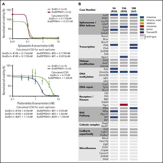
We examined the sensitivity of the myeloid progenitor cell lines to inhibitors of RNA splicing. Two different inhibitors were tested, but there were no substantive differences in sensitivity between WT and P95H cells (Figure 7A). This result is consistent with modest sensitization to splicing inhibition seen in cells from an Sf3b1K700E/+ mouse model.45 Therefore, P95H-mutant cells are not substantively sensitized to RNA splicing inhibitors.
Srsf2P95H/+ expression does not substantially sensitize to splicing inhibitors but does enable accumulation of human MDS/MPN-relevant mutations. (A) Response of Hox-immortalized cells of the indicated genotypes to the spliceosome inhibitors spliceostatin A (left) and pladienolide B (right) with calculated 50% inhibitory concentration (IC50). (B) Analysis of somatic mutations accumulating in the BM of moribund R26-CreER Srsf2P95H/+ (n = 2) and an hScl-CreER R26eYFP Srsf2P95H/+ animal (n = 1) by exome capture analysis. Mutations are defined against the list of most frequently occurring mutations in human MDS/MPN.
Srsf2P95H/+ expression does not substantially sensitize to splicing inhibitors but does enable accumulation of human MDS/MPN-relevant mutations. (A) Response of Hox-immortalized cells of the indicated genotypes to the spliceosome inhibitors spliceostatin A (left) and pladienolide B (right) with calculated 50% inhibitory concentration (IC50). (B) Analysis of somatic mutations accumulating in the BM of moribund R26-CreER Srsf2P95H/+ (n = 2) and an hScl-CreER R26eYFP Srsf2P95H/+ animal (n = 1) by exome capture analysis. Mutations are defined against the list of most frequently occurring mutations in human MDS/MPN.
Mutation acquisition in Srsf2P95H cells mirrors that of human MDS/MPN
As the aged Srsf2P95H/+ animals developed MDS/MPN, we assessed if the end-stage disease acquired additional mutations similar to those in human MDS/MPN. Exome sequencing was performed on 3 independent moribund animals where whole BM was compared with ear tissue from the same individual to discriminate somatic mutations. Mutations in Dnmt3a were present in all P95H BM samples, along with Phf6 mutations in 2 of 3 samples (Figure 7B). The spectrum of mutations in the P95H BM encompassed similar genes and gene classes to those recurrently mutated in human MDS/MPN and CMML, including mutations in the Ras and Tet2 pathways.7 We inferred clonal burden using PyClone, which indicated that the additional mutations were arising in a subclone present at ∼8% to 10% frequency in the BM at the time of euthanasia (supplemental data set 3). Therefore, the physiologic expression of Srsf2P95H/+ within the HSC compartment, alongside acquisition of recurrent secondary mutations commonly observed in humans, leads to the development of MDS/MPN in mice.
Discussion
The primary in vivo consequence of Srsf2P95H/+ is to promote a hemopoietic cell–intrinsic myeloid bias/myeloproliferation that is apparent in both native hemopoiesis and after multiple distinct BM transplantation models. This phenotype was not observed in prior Srsf2P95H mouse models,15,16 yet is consistent with clinical findings where SRSF2-mutant allele frequency correlates with monocytosis.10 Monocyte and neutrophil precursors increase in the PB from ∼11 weeks after P95H activation. This myeloid skewing requires the HSC transcriptional program to efficiently promote disease development, because this phenotype was observed in the hScl-CreER model but not the LysM-Cre model. P95H corrupts normal lineage fate decisions within the HSC/MPP population, potentially because of the effects of differential splicing within genes critical for early HSC/MPP self-renewal and differentiation, although the identity of these remains elusive both from our analysis and that of others.14-16 Only 5 transcripts were identified that were differentially expressed and had altered splicing (Bend5, Dach2, Dpp7, Galns, and Prdm16). Although the function of these transcripts remains to be tested, PRDM16 is involved in translocations in acute myeloid leukemia/MDS. Srsf2P95H/+ cells had reduced Prdm16 expression, and potentially relevant to the myeloid bias/monocytosis, Prdm16−/− mice had increased numbers of macrophages.46 It remains to be demonstrated if changes in any single transcript or the cumulative burden of many changes underlie the myeloid bias and ultimately lead to MDS/MPN. The transcriptome analyses to date, including our own, have assessed relatively heterogeneous populations (eg, lin−cKit+eYFP+ or LKS+).14-16 The analysis of highly purified LT-HSC and distinct progenitor subsets may yield greater insight into the differentiation stage–specific effects of Srsf2P95H/+, potentially discerning a greater understanding of how this mutation alters hemopoiesis.
The phenotypic data indicate a trend toward a reduction in absolute numbers of HSCs upon Srsf2P95H/+ mutation, consistent with results from Kon et al.16 In competitive transplantations, Srsf2P95H/+ HSCs demonstrated a profound myeloid bias and >95% reduction in LT repopulation. However, and critically, this was not an absolute finding, because the competitive advantage of the Srsf2P95H/+-mutant cells can be revealed by transplantation with age-/microenvironment-matched BM cells (Figure 3F-J). This later finding is significant, because the severe reduction in competitive transplantation potential and reduced phenotypic LT-HSCs were inconsistent with SRSF2 mutations in age-related clonal hemopoiesis and the positive selection of this mutation in humans.11-13,47 Although the mechanism behind these phenomena remains unresolved, it is tempting to speculate that BM microenvironment remodeling or paracrine factors derived from P95H cells compromise normal hemopoiesis.
With aging, nontransplanted Srsf2P95H/+ animals develop MDS/MPN, characterized by both monocytosis and dysplastic neutrophils in the PB and BM and the loss of B lymphoid/erythroid populations in the BM. The Srsf2P95H/+ cells acquired additional mutations in genes and pathways that cooccur in human MPN/MDS. Despite the development of fully penetrant MDS/MPN, the Srsf2P95H/+ animals did not progress to acute leukemia. The loss of p53 in the Srsf2P95H/+ background did not modify the myeloid bias or MDS/MPN development but increased the frequency/function of MDS repopulating cells in secondary transplantation assays.48 The preservation of repopulation upon p53 deletion was reported in the NUP98-HOXD13 model37,38 ; however, unlike that model, we did not observe cooperativity in leukemic conversion.
The phenotypes we report are distinct from those with prior Srsf2P95H/+ alleles.15,16,49 In the first reported model, de novo mutated nontransplanted Srsf2P95H/+ animals did not develop disease by 70 weeks,15 and BM transplantation–induced stress was required to appreciate some MDS phenotypes. In transplantation-based models, Srsf2P95H/+ animals did not develop myeloid bias, and there was no evidence of altered protein isoform expression in vivo (CD45RB). In a subsequent report using the same allele, leukopenia and anemia were largely absent in the Srsf2P95H/+ cohorts,17 suggesting incomplete penetrance of the phenotype. In contrast to Kim et al,15 we did not observe expansion of phenotypic primitive progenitor cell populations, identified as LSK CD150+CD48+ HPC-2.50 This may relate to the effects of BM transplantation or use of Mx-Cre/pIpC, because the reported frequency of HPC-2 populations in the control animals (Mx-Cre Srsf2+/+) was significantly higher than that in the reference population.50 Using multiple phenotypic populations, we found evidence for a reduced frequency of phenotypic HSCs and impaired progenitor number/frequency. We reproduced the leukopenia, macrocytosis, and reduced RBCs reported by Kim et al, when Cre was activated posttransplantation (Figure 4A-C). However, in our analysis, the myeloid bias was preserved (supplemental Figure 6A-C). A second Srsf2P95H/+ allele was recently reported, and Vav-Cre nontransplanted animals did not develop myeloid bias or MDS by 90 weeks of age.16 A reduced frequency of phenotypic and functional HSCs was described.16 Upon transplantation, erythroid-restricted dysplasia and transcriptional signatures consistent with human MDS were found. Our analysis supports the finding of a depletion of HSCs and primitive progenitors upon Srsf2P95H/+ expression. However, in contrast to Kon et al,16 our data demonstrate that Srsf2P95H/+ mutation within the HSCs is an initiating lesion in MDS/MPN.
We do not have a definitive understanding of why our model develops myeloid bias and MDS/MPN from native hemopoiesis, but the prior models do not. Differences in the genomic modification used to generate the respective alleles are the most plausible source of variance. Kim et al15 introduced a locus duplication but with a different structure/configuration to the allele we generated. Kon et al16 used a conditionally inverted PM-bearing exon, and despite heterozygous genomic expression after Cre exposure, the mutant transcript was not heterozygously expressed (∼31%). Subheterozygous expression of the mutant transcript was reported in a conditional Sf3b1K700E mutant that used an exon-inversion approach.45 The failure to achieve heterozygous transcript expression is a significant confounding factor in interpreting the phenotypes of these later models.
The contribution of the misspliced transcripts to the phenotypes in the P95H mutants remains an outstanding question. Altered splicing of EZH2, leading to reduced/absent expression, was proposed to contribute to the phenotypes in mouse and human P95H/+ cells.15 We did not detect altered Ezh2 splicing in multiple cell types, yet could demonstrate altered in vivo splicing of previously defined Srsf2 targets51 and of transcripts identified by Kon et al.16 Additionally, heterozygous knock-in SRSF2P95H/+ K562 cells had a very subtle change in EZH2 splicing.14 From the analysis by independent groups, missplicing of Ezh2 does not contribute to the phenotype of Srsf2P95H/+ cells. We observed a similar relatively subtle shift in overall splicing, with the majority of classes of splicing changing by <0.5%, similar to reports from K562 cells.14 The modest impact of Srsf2P95H/+ on global splicing hints at a more complex phenotype than can be solely ascribed to aberrant RNA splicing. Although not demonstrated in vivo, it was recently proposed that increased R-loop formation and DNA damage may contribute to the phenotypes associated with spliceosome mutations.52
The Srsf2P95H model described here recapitulates the core traits of SRSF2-mutant human diseases, including myeloid bias, monocytosis, and progression to MDS/MPN with the accumulation of cooccurring mutations. Importantly, this demonstrates that Srsf2P95H must be expressed within the HSC population to initiate MDS/MPN.
The online version of this article contains a data supplement.
The publication costs of this article were defrayed in part by page charge payment. Therefore, and solely to indicate this fact, this article is hereby marked “advertisement” in accordance with 18 USC section 1734.
Acknowledgments
The authors thank S. Lane, V. Sankaran, J. Heierhorst, and D. Campbell for comments; A. Kon/S. Ogawa for providing supplemental data ahead of online publication; M. Yoshida (RIKEN) for spliceostatin A; St. Vincent’s Hospital Biomedical Research Centre for care of experimental animals; St. Vincent’s Institute Flow Cytometry Core Facility; Ramaciotti Centre for Genomics (University of New South Wales); and Novogene for RNA sequencing; and BGI for exome capture.
This work was supported by the Leukaemia Foundation (C.R.W., Phillip Desbrow Senior Research Fellow [SRF]; M.W., grant-in-aid; S.Y.T., PhD scholarship); Cancer Council of Victoria (C.R.W., M.W., A.M.C.; APP1126010); National Health and Medical Research Council (NHMRC; J.E.P., APP1024363/APP1102589); Cancer Institute of NSW Translational Cancer Research Network (J.E.P.); South Eastern Area Laboratory Services (J.E.P.), Anthony Rothe Memorial Trust (A.U.); Victorian Cancer Agency Research Fellowship (C.R.W.; MCRF15015); NHMRC SRF (L.E.P.; APP1003339); UNSW International PhD scholarship (G.A.); and Tom Bee Stem Cell Research Fund (G.A.) and in part by the Victorian State Government OIS.
Authorship
Contribution: M.F.S., M.W., L.E.P., and C.R.W. were responsible for conceptualization; M.F.S., G.A., A.U., J.E.P., L.E.P., and C.R.W. were responsible for methodology; M.F.S., S.Y.T., J.J.X., G.A., A.U., A.M.C., S.R.T., M.W., and C.R.W. performed investigation; M.F.S., J.J.X., and C.R.W. wrote the original draft; M.F.S., S.Y.T., J.J.X., A.U., J.E.P., M.W., L.E.P., and C.R.W. reviewed and edited the manuscript; A.M.C., J.J.P., M.W., and C.R.W. were responsible for funding acquisition; G.A., A.U., and J.J.P. provided resources; and M.F.S., J.J.P., M.W., L.E.P., and C.R.W. provided supervision.
Conflict-of-interest disclosure: The authors declare no competing financial interests.
Correspondence: Carl R. Walkley, St. Vincent’s Institute, 9 Princes St, Fitzroy 3065 VIC, Australia; e-mail: cwalkley@svi.edu.au.


![Figure 5. HSC-restricted activation of Srsf2P95H/+ causes MDS/MPN. (A) Kaplan-Meier plot of disease-free survival for the hScl-CreER (blue) and R26-CreER (gray) models. (B) PB leukocyte counts and lineage distribution. (C) PB hemoglobin levels. (D) PB neutrophils from a control (age-/treatment-matched hScl-CreER R26eYFP Srsf2+/+ littermate) and 3 independent hScl-CreER R26eYFP Srsf2P95H/+ animals; May-Grunwald Giemsa stain. (E) Number of PB monocytes and neutrophil precursors over time after tamoxifen (n = 6-13 per genotype per time point; based on Ly6c/CD11b stain). (F) BM cellularity and number of granulocytes and monocytes per femur. (G) BM myeloid (Mac-1/Gr-1) and B lymphoid (B220/immunoglobulin M [IgM]) populations in BM. (H) Number of phenotypic LT-HSCs, ST-HSCs, and MPPs per femur and myeloerythroid progenitors per femur. (I) Number of granulocytes and monocytes per spleen. (J) CD45RB isoform expression on splenocytes. (K) PB CD45 chimerism from secondary transplantation of whole BM into lethally irradiated recipients (black circle = littermate control; blue squares = moribund Srsf2P95H/+); each line represents reconstitution of an individual recipient (n = 4); PB eYFP percentage in the CD45.2+ cells; PB leukocyte counts, representative PB FACS from recipients, and quantitation of lineage output from the CD45.2 population (4 weeks post–BM transplantation [BMT]). Presented as mean ± standard error of the mean. Student t test, with multiple comparisons correction (for panel E). *P < .05, **P < .01, ***P < .001.](https://ash.silverchair-cdn.com/ash/content_public/journal/blood/132/6/10.1182_blood-2018-04-845602/4/m_blood845602f5.jpeg?Expires=1768947529&Signature=BtmZz~wdBvSo9ZAj3l-W2Ka3kLiDFsPhgEZ6nkxMccAaXctWRH5eEpjrsECexkXik8MTO0BUVa50chd-bxJQBED6EvqCb6Um1m6ISKy-e56s9nD~VsEc2EOw3dySTBP4YWDeQx9oYNDDd0Ta5KLinG2cSAfqN~JDQgGAl9cyhuonVfqBUL0KtuZJvgixiEJ0WngryMfQJNsiwIbA6lwWEqb8twrZ7X5u403Nssknursk3Rm1vRvF7RpP1fQYnNOqpxhqxHuoexCRMoE1D5s8-DCzpNaf5YrTeWGSOXd1BMFCTC5hQ0D224GaZxXaJHrg6JAd6ZK1tavK2S1zohCd9w__&Key-Pair-Id=APKAIE5G5CRDK6RD3PGA)


This feature is available to Subscribers Only
Sign In or Create an Account Close Modal