Abstract
Mpl-/- mice bearing the Plt3 or Plt4 mutations in the c-Myb gene exhibit thrombopoietin (TPO)–independent supraphysiological platelet production accompanied by excessive megakaryocytopoiesis and defective erythroid and lymphoid cell production. To better define the cellular basis for the thrombocytosis in these mice, we analyzed the production and characteristics of megakaryocytes and their progenitors. Consistent with thrombocytosis arising from hyperactive production, the high platelet counts in mice carrying the c-MybPlt4 allele were not accompanied by any significant alteration in platelet half-life. Megakaryocytes in c-Myb mutant mice displayed reduced modal DNA ploidy and, among the excessive numbers of megakaryocyte progenitor cells, more mature precursors were particularly evident. Megakaryocyte progenitor cells carrying the Plt3 or Plt4 c-Myb mutations, but not granulocyte-macrophage progenitors, exhibited 200-fold enhanced responsiveness to granulocyte-macrophage colony-stimulating factor (GM-CSF), suggesting that altered responses to cytokines may contribute to expanded megakaryocytopoiesis. Mutant preprogenitor (blast colony-forming) cells appeared to have little capacity to form megakaryocyte progenitor cells. In contrast, the spleens of irradiated mice 12 days after transplantation with mutant bone marrow contained abundant megakaryocyte progenitor cells, suggesting that altered c-Myb activity skews differentiation commitment in spleen colony-forming units (CFU-S) in favor of excess megakaryocytopoiesis.
Introduction
A role for the transcription factor c-Myb in hematopoiesis was first implied by the discovery of mutated forms of Myb in viruses capable of transforming myeloid cells in chickens. Both the E26 virus and avian myeloblastosis virus (AMV) encode truncated homologs of c-Myb that lack a negative regulatory domain. v-MybE26 is a fusion protein with Ets-1, and the AMV version of Myb is characterized by a series of amino acid substitutions. The AMV and E26 forms of Myb transform distinct myeloid cell types. E26 induces transformation of myeloblasts that retain the capacity for cytokine-dependent granulocyte and monocyte differentiation, while AMV transforms monoblastoid cells with autocrine characteristics and that lack granulocyte potential.1 The E26 virus can also induce clonal expansion of progenitors capable of multilineage differentiation, including production of erythroid cells and eosinophils.2 However, production of thrombocytes from these transformed multipotent progenitors could only be achieved upon mutation of the v-Myb DNA-binding domain, suggesting that while Myb expression was permissive for production of some blood cells' lineages, its down-regulation may be required for thrombopoiesis.3
In mouse models of insertional mutagenesis or tissue-specific transgenesis, truncations in c-Myb have also been shown to contribute to hematopoietic transformation, particularly within the lymphoid compartment, and an example of a truncated c-Myb protein contributing to T-cell blast crisis in human chronic myelogenous leukemia (CML) has also been described.4 Among the variety of tissues that express c-Myb during mouse embryonic or adult life, hematopoietic progenitor cells express c-Myb abundantly, and expression is also evident at fetal sites of hematopoiesis,5-7 consistent with a physiological role for c-Myb in regulation of the blood-forming system. Indeed, mice lacking c-Myb die during embryogenesis, with anemia resulting from failure of fetal liver hematopoiesis.8 Extensive examination of these embryos as well as studies of the in vitro hematopoietic potential of c-Myb-/- embryonic stem cells suggested that definitive hematopoietic progenitor cells could develop in the absence of c-Myb but were unable to survive or appropriately proliferate and/or differentiate.9,10 The generation of mice with a knockdown allele of c-Myb, from which subnormal levels of the wild-type c-Myb protein are expressed, has suggested that distinct thresholds of c-Myb expression regulate different aspects of hematopoiesis, with low levels allowing progenitor cell expansion but favoring megakaryocyte and macrophage differentiation at the expense of lymphoid and erythroid development, which appear to require high-level c-Myb expression.11
We recently described the generation of 2 N-ethylnitrosourea (ENU)–induced mutations in the c-Myb gene that result in supraphysiological platelet production in the presence or absence of the actions of thrombopoietin (TPO), the major regulator of steadystate platelet production. These mutations, referred to as the Plt3 and Plt4 alleles of c-Myb, result in the expression of c-Myb proteins bearing single amino acid substitutions of valine for aspartic acid at residue 152 within the c-Myb DNA-binding domain or residue 384 within the leucine zipper domain, respectively.12 On both TPO receptor–deficient (Mpl-/-) or wild-type (Mpl+/+) backgrounds, these mice displayed a significant expansion of megakaryocytopoiesis, with elevated numbers of megakaryocyte progenitor cells in the bone marrow and spleen as well as increased numbers of megakaryocytes in these organs. The mice were also shown to be profoundly deficient in B lymphocytes and to be slightly anemic. This phenotype is remarkably similar to that observed in mice bearing a germ-line mutation in the c-Myb–binding domain of the p300 transcriptional coactivator.13 Thus, compelling data have accumulated that activity of the c-Myb protein plays a crucial role in megakaryocyte and platelet development. However, the basis on which altered c-Myb activity leads to elevated megakaryocyte and platelet production remains obscure. The c-MybPlt3/Plt3 and c-MybPlt4/Plt4 mutant mice provide a unique resource for dissecting this role in vivo in adult mice. The present studies were undertaken to characterize the cellular basis for the thrombocytosis in mice bearing the Plt3 or Plt4 mutant alleles of c-Myb.
Materials and methods
Mice
The derivation of Mpl-/- c-MybPlt3/Plt3 and Mpl-/- c-MybPlt4/Plt4 mice, which bear c-Myb mutations on a c-Mpl–deficient background, and Mpl+/+ c-MybPlt4/Plt4 mice, in which c-Mpl alleles are wild type, has been described elsewhere.12 All mice were on a C57BL/6 strain background and were bred and housed in conventional clean animal rooms. Experiments were performed with procedures approved by the Melbourne Health Research Directorate Animal Ethics Committee.
Cell counts and histology
Total peritoneal cell counts were determined after the injection of 2 mL of saline containing 10% fetal calf serum and harvesting of the fluid after thorough mixing of the abdominal contents by massage. Spleens were weighed, and then a portion of known weight was converted to a dispersed cell suspension by sieving in Dulbecco modified Eagle medium (DMEM) containing 10% fetal calf serum. The entire cellular content of 1 femur was harvested in DMEM containing 10% fetal calf serum. Cell suspensions were cytocentrifuged and then stained with May-Grünwald-Giemsa. All major organs were fixed in 10% buffered formalin, imbedded in paraffin, and 1 to 3 μm sections stained with hematoxylin and eosin. For histologic visualization of collagen, duplicate sections were stained with Masson trichrome. Megakaryocytes were enumerated by microscopic examination of sections of bone marrow and spleen, with counts recorded as number of cells per high-power (magnification × 200) field.
Cultures
Bone marrow (2.5 × 10⁴) or spleen (5 × 10⁴) cells were cultured in 1 mL volumes of 0.3% agar in DMEM containing 20% newborn calf serum. Stimuli were used at the following concentrations: granulocyte-macrophage colony-stimulating factor (GM-CSF), granulocyte CSF (G-CSF), macrophage CSF (M-CSF), and interleukin-3 (IL-3) at 10 ng/mL, stem cell factor (SCF) at 50 ng/mL, IL-6 at 500 ng/mL, erythropoietin (EPO) at 2 U/mL, and TPO at 100 ng/mL. Recombinant purified murine M-CSF, TPO, and SCF were prepared in our laboratories; recombinant human G-CSF and EPO were obtained from Amgen (Thousand Oaks, CA); and the remaining murine cytokines were obtained from PeproTech (Rocky Hill, NJ). Cultures were incubated for 7 days in a fully humidified atmosphere of 10% CO2 in air. Initial colony counts were performed using an Olympus dissection microscope with indirect lighting. The cultures were then fixed, dried onto glass slides, and stained for acetylcholinesterase, Luxol Fast Blue, and hematoxylin, and the number and type of all colonies were determined.
Recloning of blast colonies
After 7 days of incubation, individual blast colonies grown with stimulation by SCF plus G-CSF were removed using a fine Pasteur pipette, colony cells were dispersed and then recultured in SCF plus G-CSF (the initiating cytokine mixture), IL-3, M-CSF (the maximal stimulus for macrophage colony formation) or SCF plus IL-3 plus EPO (the maximal stimulus for megakaryocyte colonies), and then incubated for a further 7 days before scoring as described in “Cultures,” above. The numbers of colony-forming cells per colony were calculated separately for each stimulus.
DNA ploidy analysis
Megakaryocyte ploidy analysis was performed essentially as described.14 Briefly, mouse bone marrow was harvested into CATCH buffer (phenol red–free, Ca-free, Mg-free Hanks balanced salt solution containing 3% bovine serum albumin [BSA], 0.38% trisodium citrate, 1 mM adenosine, 2 mM theophylline). The bone marrow cells were incubated with anti-CD41–fluorescein isothiocyanate (FITC) or rat immunoglobulin G1κ (IgG1κ) isotype control (BD Pharmingen, San Diego, CA) antibodies for 1 hour on ice. Three volumes of 0.05 mg/mL propidium iodide in 0.1% trisodium citrate were then added to 1 volume of cells followed by incubation on ice for 2 hours. Cells were washed twice with CATCH buffer, and samples were passed through a 100-μm sieve to remove cell aggregates and then incubated with 50 μg/mL Rnase A for 30 minutes prior to analysis. Samples were analyzed on a FACScan flow cytometer (BD Biosciences, San Diego, CA). Megakaryocytes were identified as CD41+ cells, and propidium iodide staining intensity was used as a measure of DNA ploidy.
Determination of platelet half-life
Cells were labeled with biotin in vivo by injection of 0.6 mg per mouse of N-hydroxysuccinimide-biotin dissolved in 10% dimethyl sulfoxide (DMSO). Mice were bled from the tail vein at regular intervals into buffered saline glucose citrate (116 mM sodium chloride, 13.6 mM trisodium citrate, 8.6 mM Na2HPO4, 1.6 mM KH2PO4, 0.9 mM disodium EDTA [ethylenediaminetetraacetic acid], 11.1 mM glucose). Collected blood cells were washed and then incubated with fluorescein-conjugated anti-CD41 to label platelets and streptavidin-conjugated phycoerythrin to label biotinylated cells. The proportion of labeled platelets in the circulation was then enumerated by flow cytometric analysis. An initial platelet count was determined and assumed to remain constant over the course of the experiment. To examine platelet clearance, the number of labeled platelets at a given time was divided by the initial number of labeled platelets (usually at 24 hours after biotin injection), and the logarithm of this ratio was plotted against time. Platelet production was monitored by measuring the increase in the number of unlabeled platelets following pulse biotinylation. The number of new platelets produced was determined by subtracting the initial number of unlabeled platelets from the number of unlabeled platelets at a given time, and this value was plotted versus time.
Transplantation studies
Bone marrow cells were harvested, washed, and injected intravenously (1.5 × 10⁵ cells per recipient from Mpl-/- c-MybPlt4/Plt4 and Mpl-/- c-Myb+/+ donors and 7.5 × 10⁴ cells per recipient from Mpl+/+ c-MybPlt4/Plt4 and Mpl+/+ c-Myb+/+ mice). Several hours prior to transplantation, the recipient mice were irradiated with 11 Gy γ-irradiation in 2 equal doses given 3 hours apart. Mice that underwent transplantation were maintained on oral antibiotic (1.1 g/L neomycin sulfate; Sigma, St Louis, MO). Spleens were removed after 12 days and weighed, and single-cell suspensions were prepared for enumeration of the numbers of progenitor cells using the agar colony assay described in “Cultures,” above.
Results
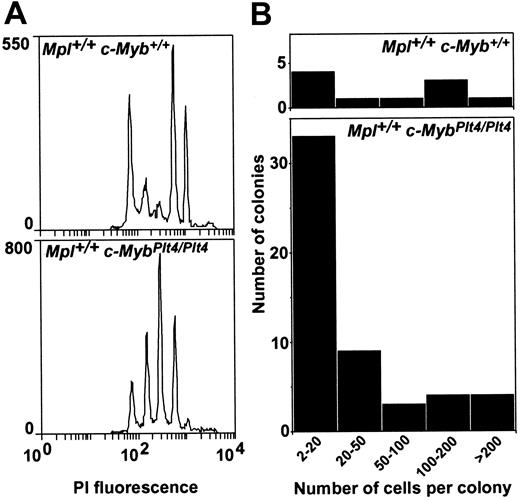
As previously described, Mpl-/- c-MybPlt3/Plt3 and Mpl-/- c-MybPlt4/Plt4 mice exhibit platelet counts 40 times higher than those in Mpl-/- c-Myb+/+ mice and 3 to 4 times higher than in Mpl+/+ c-Myb+/+ mice, and the thrombocytosis in these mice is accompanied by significant increases in the numbers of megakaryocytes in the bone marrow and spleen.12 To further explore the basis of this thrombocytosis, the DNA ploidy of megakaryocytes in mice bearing the Plt3 or Plt4 mutations was assessed. As shown in Figure 1 and Table 1, the frequency of 16N and 32N cells was lower in Mpl-/- c-MybPlt3/Plt3, Mpl-/- c-MybPlt4/Plt4, and Mpl+/+ c-MybPlt4/Plt4 marrow cells compared with their respective c-Myb+/+ controls, with a corresponding increase in the frequency of 4N and 8N cells in these populations.
Megakaryocytopoiesis in Mpl+/+ c-MybPlt4/Plt4 mice. (A) Reduced modal ploidy in megakaryocytes from Mpl+/+ c-MybPlt4/Plt4 mice. The propidium iodide (PI) fluorescence intensity, representing cellular DNA content, in CD41+ bone marrow cells is shown. (B) Reduced size of megakaryocyte colonies in cultures of Mpl+/+ c-MybPlt4/Plt4 bone marrow. The number of cells in each acetylcholinesterase-positive megakaryocyte colony was scored by microscopic examination of stained cultures.
Megakaryocytopoiesis in Mpl+/+ c-MybPlt4/Plt4 mice. (A) Reduced modal ploidy in megakaryocytes from Mpl+/+ c-MybPlt4/Plt4 mice. The propidium iodide (PI) fluorescence intensity, representing cellular DNA content, in CD41+ bone marrow cells is shown. (B) Reduced size of megakaryocyte colonies in cultures of Mpl+/+ c-MybPlt4/Plt4 bone marrow. The number of cells in each acetylcholinesterase-positive megakaryocyte colony was scored by microscopic examination of stained cultures.
DNA ploidy in megakaryocytes in c-MybPlt3/Plt3 and c-MybPlt4/Plt4 bone marrow
. | Percent of megakaryocytes in ploidy class . | . | . | . | . | ||||
|---|---|---|---|---|---|---|---|---|---|
| Mouse . | 2N . | 4N . | 8N . | 16N . | 32N . | ||||
| Mpl-/-c-Myb+/+ | 16 ± 4 | 14 ± 3 | 25 ± 5 | 39 ± 7 | 6 ± 1 | ||||
| Mpl-/-c-MybPlt3/Plt3 | 9 ± 2 | 11 ± 1 | 47 ± 5 | 30 ± 2 | 3 ± 1 | ||||
| Mpl-/-c-MybPlt4/Plt4 | 10 ± 2 | 22 ± 4 | 47 ± 2 | 19 ± 3 | 2 ± 1 | ||||
| Mpl+/+c-Myb+/+ | 18 ± 5 | 11 ± 3 | 11 ± 3 | 40 ± 5 | 20 ± 4 | ||||
| Mpl+/+c-MybPlt4/Plt4 | 15 ± 5 | 22 ± 5 | 37 ± 7 | 22 ± 7 | 4 ± 2 | ||||
Data are the mean ± standard deviation from analysis of 2 to 7 mice of each genotype.
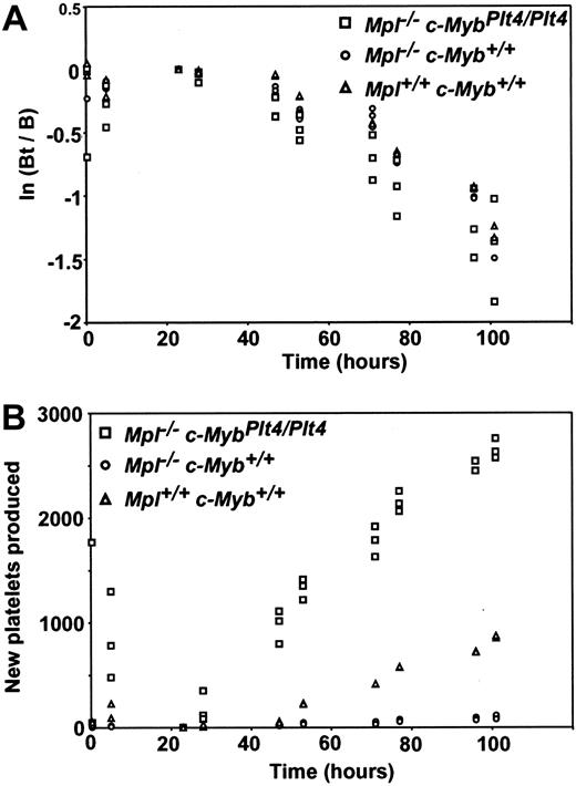
To determine whether alterations in platelet half-life contributed to thrombocytosis in mice with c-Myb mutations, in vivo labeling studies were performed in Mpl-/- c-MybPlt4/Plt4 mice (see “Materials and methods”). There was no difference in the rate of platelet clearance from the circulation in Mpl-/- c-MybPlt4/Plt4 mice compared with Mpl-/- c-Myb+/+ or Mpl+/+ c-Myb+/+ controls (Figure 2). In contrast, consistent with deregulated platelet production in these mutant mice, the rate of emergence of new unlabeled platelets was significantly greater in mice bearing the Plt4 mutation relative to control mice (Figure 2).
Clearance of platelets in Mpl-/- c-MybPlt4/Plt4 mice. Platelets were biotinylated in vivo, and then blood samples were collected at the times indicated. Platelets were identified flow cytometrically on the basis of size and CD41 positivity. (A) To measure platelet clearance, the number of labeled platelets at a given time (Bt) was divided by the initial number of labeled platelets (B, at 24 hours after biotin injection), and the logarithm of this ratio was plotted against time. (B) To monitor platelet production, the number of new unlabeled platelets (the number of unlabeled platelets at a given time less the initial number of unlabeled platelets) was plotted against time. Each point represents data from 2 to 3 mice of each genotype. □ indicates Mpl-/ c-MybPlt4/Plt4; ○, Mpl-/- c-Myb+/+; and ▵, Mpl+/+ c-Myb+/+.
Clearance of platelets in Mpl-/- c-MybPlt4/Plt4 mice. Platelets were biotinylated in vivo, and then blood samples were collected at the times indicated. Platelets were identified flow cytometrically on the basis of size and CD41 positivity. (A) To measure platelet clearance, the number of labeled platelets at a given time (Bt) was divided by the initial number of labeled platelets (B, at 24 hours after biotin injection), and the logarithm of this ratio was plotted against time. (B) To monitor platelet production, the number of new unlabeled platelets (the number of unlabeled platelets at a given time less the initial number of unlabeled platelets) was plotted against time. Each point represents data from 2 to 3 mice of each genotype. □ indicates Mpl-/ c-MybPlt4/Plt4; ○, Mpl-/- c-Myb+/+; and ▵, Mpl+/+ c-Myb+/+.
Hematopoietic progenitor cells
In cultures of Mpl-/- c-MybPlt3/Plt3 or Mpl-/- c-MybPlt4/Plt4 bone marrow cells, no colonies or clusters developed in the absence of added cytokine stimulus. In response to GM-CSF, normal numbers of granulocytic, granulocyte-macrophage, and macrophage colonies of normal size and shape developed, but eosinophil colonies were depleted or absent and, strikingly, megakaryocyte colonies were consistently observed (Table 2). Responsiveness to G-CSF by granulocyte colony formation was normal in cultures of Mpl-/- c-MybPlt3/Plt3 and Mpl-/- c-MybPlt4/Plt4 cells but, in contrast, cultures stimulated by IL-6, which developed similar numbers of small granulocytic colonies in cultures of Mpl-/- c-Myb+/+ marrow cells, failed to exhibit granulocyte colony formation in most cultures of Mpl-/- c-MybPlt3/Plt3 or Mpl-/- c-MybPlt4/Plt4 bone marrow, although, again, a few small megakaryocyte colonies did develop (Table 2). Mpl-/- c-MybPlt3/Plt3 or Mpl-/- c-MybPlt4/Plt4 bone marrow failed to develop megakaryocyte colonies in cultures stimulated by TPO, confirming the absence of c-Mpl receptors. However, the cultures developed a 10-fold increased number of megakaryocyte colonies when stimulated by IL-3 or EPO or the potent combination of SCF plus IL-3 plus EPO (Table 2).
Progenitor cell content of c-MybPlt3/Plt3 and c-MybPlt4/Plt4 bone marrow
Mouse and Stimulus . | No. of colonies per 2.5 × 10⁴ bone marrow cells . | . | . | . | . | . | |||||
|---|---|---|---|---|---|---|---|---|---|---|---|
| . | Blast . | G . | GM . | M . | Eo . | Meg . | |||||
| Mpl-/- c-MybPlt3/Plt3 | |||||||||||
| GM-CSF | 0.3 ± 0.6 | 14 ± 3 | 15 ± 6 | 25 ± 6 | 0 | 14 ± 8 | |||||
| G-CSF | 0 | 10 ± 2 | 0 | 0 | 0 | 0 | |||||
| M-CSF | 0 | 8 ± 2 | 8 ± 4 | 54 ± 15 | 0 | 0 | |||||
| IL-3 | 12 ± 6 | 15 ± 3 | 18 ± 1 | 16 ± 3 | 0 | 64 ± 16 | |||||
| EPO | 0 | 0 | 0 | 0 | 0 | 15 ± 6 | |||||
| SCF | 8 ± 5 | 16 ± 6 | 1 ± 2 | 0.3 ± 0.6 | 0 | 2 ± 2 | |||||
| TPO | 0 | 0 | 0 | 0 | 0 | 0 | |||||
| S3E | 21 ± 4 | 11 ± 5 | 31 ± 9 | 17 ± 5 | 0 | 96 ± 24 | |||||
| IL-6 | 0 | 0.5 ± 0.9 | 0 | 0 | 0 | 1 ± 1 | |||||
| Mpl-/- c-MybPlt4/Plt4 | |||||||||||
| GM-CSF | 0.5 ± 1.0 | 24 ± 12 | 21 ± 14 | 42 ± 19 | 0.6 ± 1.3 | 8 ± 3 | |||||
| G-CSF | 0 | 6 ± 1 | 0 | 0 | 0 | 0 | |||||
| M-CSF | 0 | 0.5 ± 1.0 | 5 ± 0 | 55 ± 19 | 0 | 0 | |||||
| IL-3 | 16 ± 6 | 45 ± 12 | 22 ± 11 | 16 ± 5 | 0 | 64 ± 24 | |||||
| EPO | 0 | 0 | 0 | 0 | 0 | 26 ± 17 | |||||
| SCF | 7 ± 7 | 20 ± 12 | 0.6 ± 1.2 | 0 | 0 | 0 | |||||
| TPO | 0 | 0 | 0 | 0 | 0 | 0 | |||||
| S3E | 17 ± 4 | 44 ± 15 | 19 ± 9 | 14 ± 12 | 0 | 113 ± 16 | |||||
| IL-6 | 0 | 0.6 ± 1.3 | 0 | 0 | 0 | 0 | |||||
| Mpl-/- c-Myb+/+ | |||||||||||
| GM-CSF | 0 | 15 ± 5 | 7 ± 4 | 29 ± 10 | 2 ± 1 | 0 | |||||
| G-CSF | 0 | 6 ± 3 | 0 | 0 | 0 | 0 | |||||
| M-CSF | 0 | 2 ± 1 | 2 ± 1 | 33 ± 14 | 0 | 0 | |||||
| IL-3 | 3 ± 2 | 11 ± 4 | 9 ± 2 | 9 ± 4 | 1 ± 1 | 5 ± 3 | |||||
| EPO | 0 | 0 | 0 | 0 | 0 | 5 ± 4 | |||||
| SCF | 2 ± 2 | 10 ± 1 | 0.4 ± 0.5 | 0.6 ± 0.8 | 0 | 0 | |||||
| TPO | 0 | 0 | 0 | 0 | 0 | 0 | |||||
| S3E | 4 ± 3 | 12 ± 4 | 9 ± 4 | 7 ± 3 | 0.9 ± 0.9 | 9 ± 6 | |||||
| IL-6 | 0 | 7 ± 3 | 0.7 ± 0.5 | 0 | 0 | 0 | |||||
| Mpl+/+ c-Myb+/+ | |||||||||||
| GM-CSF | 0 | 26 ± 9 | 12 ± 5 | 31 ± 9 | 4 ± 2 | 0 | |||||
| G-CSF | 0 | 11 ± 4 | 0 | 0 | 0 | 0 | |||||
| M-CSF | 0 | 1 ± 1 | 3 ± 2 | 42 ± 12 | 0 | 0 | |||||
| IL-3 | 7 ± 3 | 21 ± 4 | 11 ± 4 | 11 ± 3 | 3 ± 2 | 7 ± 1 | |||||
| EPO | 0 | 0 | 0 | 0 | 0 | 7 ± 2 | |||||
| SCF | 4 ± 2 | 15 ± 3 | 0.1 ± 0.1 | 0.4 ± 0.5 | 0 | 0 | |||||
| TPO | 0 | 0 | 0 | 0 | 0 | 3 ± 1 | |||||
| S3E | 11 ± 4 | 19 ± 4 | 12 ± 2 | 8 ± 5 | 3 ± 3 | 21 ± 6 | |||||
| IL-6 | 0 | 10 ± 3 | 1.4 ± 0.5 | 0 | 0 | 0.3 ± 0.8 | |||||
| Mpl+/+ c-MybPlt4/Plt4 | |||||||||||
| GM-CSF | 0 | 10 ± 0 | 15 ± 6 | 29 ± 5 | 0.5 ± 0.7 | 5.5 ± 0.7 | |||||
| G-CSF | 0 | 2.0 ± 0 | 0 | 0 | 0 | 0 | |||||
| M-CSF | 0 | 1.5 ± 0.7 | 3.0 ± 2.8 | 44 ± 10 | 0 | 0 | |||||
| IL-3 | 7.5 ± 2.1 | 17 ± 6 | 11 ± 9 | 9.0 ± 4.2 | 0 | 49 ± 14 | |||||
| EPO | 0 | 0 | 0 | 0 | 0 | 19 ± 8 | |||||
| SCF | 3.0 ± 1.4 | 7.5 ± 0.7 | 0.5 ± 0.7 | 0 | 0 | 0 | |||||
| TPO | 0 | 0 | 0 | 0 | 0 | 3.8 ± 2.4 | |||||
| S3E | 12 ± 6 | 23 ± 20 | 27 ± 0.7 | 14 ± 1.4 | 0 | 96 ± 25 | |||||
| IL-6 | 0 | 0 | 0 | 0 | 0 | 0 | |||||
Data are the mean ± standard deviation from analysis of 2 to 7 mice of each genotype.
G indicates granulocyte; GM, granulocyte-macrophage; M, macrophage; Eo, eosinophil, Meg, megakaryocyte; S3E, SCF + IL-3 + EPO.
In cultures of Mpl-/- c-MybPlt3/Plt3 and Mpl-/- c-MybPlt4/Plt4 bone marrow, there was a striking relative increase in the frequency of megakaryocytic colonies containing small numbers of cells. This is shown graphically in a typical analysis of colony size in Figure 1. Because megakaryocytic colonies containing low numbers of large cells are presumed to be formed by the progeny of megakaryocyte colony-forming cells, this pattern of colony formation suggests the presence of very active megakaryocytopoiesis in vivo, supporting the high megakaryocyte counts observed in the marrow and spleen.
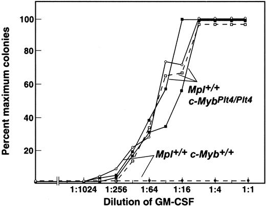
Titration studies using Mpl-/- c-MybPlt4/Plt4 bone marrow cells showed a normal quantitative responsiveness of granulocyte and/or macrophage progenitors to GM-CSF or SCF. However, megakaryocyte colony formation was as responsive to GM-CSF as granulocyte-macrophage colony formation (Figure 3). GM-CSF can stimulate megakaryocyte colony formation by normal marrow cells but only at 200-fold the maximum concentration used in the present experiments.15 Mpl-/- c-MybPlt4/Plt4 megakaryocyte colony-forming cells were also 4-fold more responsive to the combination of SCF plus IL-3 plus EPO than corresponding Mpl-/- c-Myb+/+ cells (data not shown).
Hyperresponsiveness of Mpl+/+ c-MybPlt4/Plt4 mutant megakaryocyte progenitor cells, but not granulocyte and/or macrophage progenitors, to GM-CSF. The unbroken lines depict similar GM-CSF dose response curves for granulocyte-macrophage colony formation from 2 Mpl+/+ c-MybPlt4/Plt4 and a control Mpl+/+ c-Myb+/+ bone marrow. In contrast, for megakaryocyte colony formation, Mpl+/+ c-MybPlt4/Plt4 marrow exhibited hyperresponsiveness (broken line), with maximal stimulation at doses similar to that for granulocyte-macrophage colony formation. No megakaryocyte colonies were evident from Mpl+/+ c-Myb+/+ marrow at the doses shown; 1:1 represents a GM-CSF concentration of 10 ng/mL.
Hyperresponsiveness of Mpl+/+ c-MybPlt4/Plt4 mutant megakaryocyte progenitor cells, but not granulocyte and/or macrophage progenitors, to GM-CSF. The unbroken lines depict similar GM-CSF dose response curves for granulocyte-macrophage colony formation from 2 Mpl+/+ c-MybPlt4/Plt4 and a control Mpl+/+ c-Myb+/+ bone marrow. In contrast, for megakaryocyte colony formation, Mpl+/+ c-MybPlt4/Plt4 marrow exhibited hyperresponsiveness (broken line), with maximal stimulation at doses similar to that for granulocyte-macrophage colony formation. No megakaryocyte colonies were evident from Mpl+/+ c-Myb+/+ marrow at the doses shown; 1:1 represents a GM-CSF concentration of 10 ng/mL.
Mpl-/- c-MybPlt3/Plt3 and Mpl-/- c-MybPlt4/Plt4 bone marrow cultures also exhibited an elevated frequency of blast colonies when stimulated either by IL-3 or the synergistic combination of SCF plus G-CSF, and these colonies differed in shape and size from normal blast colonies. No multicentric colonies developed, and the colonies were large, uniform, and composed of large, healthy cells. Although Mpl-/- c-MybPlt3/Plt3 and Mpl-/- c-MybPlt4/Plt4 blast colonies appeared larger than control blast colonies, cell counts on pooled colonies from cultures stimulated by G-CSF plus SCF were similar, and the apparent larger size was due to the more dispersed nature of the colonies.
As shown in Table 3, cultures of Mpl-/- c-MybPlt3/Plt3 and Mpl-/- c-MybPlt4/Plt4 spleen cells also showed a consistent pattern of abnormalities—an overall increased frequency of blast, granulocytic, and macrophage colonies, an absence of eosinophil colonies, and a 20- to 100-fold increased frequency of megakaryocytic colonies.
Progenitor cell content of c-MybPlt3/Plt3 and c-MybPlt4/Plt4 spleens
. | No. of colonies per 5 × 10⁴ spleen cells . | . | . | . | . | . | |||||
|---|---|---|---|---|---|---|---|---|---|---|---|
| Mouse . | Blast . | G . | GM . | M . | Eo . | Meg . | |||||
| Mpl-/- c-MybPlt3/Plt3 | |||||||||||
| IL-3 | 4 ± 1 | 4 ± 1 | 3 ± 4 | 3 ± 1 | 0 | 72 ± 76 | |||||
| S3E | 3 ± 4 | 4 ± 3 | 3 ± 3 | 4 ± 7 | 0 | 91 ± 105 | |||||
| Mpl-/- c-MybPlt4/Plt4 | |||||||||||
| IL-3 | 4 ± 3 | 11 ± 6 | 6 ± 2 | 7 ± 1 | 0 | 70 ± 28 | |||||
| S3E | 5 ± 4 | 12 ± 5 | 7 ± 5 | 5 ± 4 | 0 | 100 ± 23 | |||||
| Mpl-/- c-Myb+/+ | |||||||||||
| IL-3 | 0.2 ± 0.3 | 0.3 ± 0.3 | 0 | 0 | 0 | 0.6 ± 0.4 | |||||
| S3E | 0.1 ± 0.2 | 0.4 ± 0.5 | 0.5 ± 0.8 | 0.4 ± 0.8 | 0.1 ± 0.4 | 4 ± 5 | |||||
| Mpl+/+ c-Myb+/+ | |||||||||||
| IL-3 | 0.9 ± 1.0 | 0.5 ± 0.4 | 0.1 ± 0.2 | 0.1 ± 0.2 | 0 | 1.1 ± 0.9 | |||||
| S3E | 1 ± 1 | 0.6 ± 0.8 | 0.2 ± 0.4 | 0.2 ± 0.4 | 0 | 7 ± 7 | |||||
| Mpl+/+ c-MybPlt4/Plt4 | |||||||||||
| IL-3 | 4.5 ± 0.7 | 14 ± 11 | 11.0 ± 6.3 | 9.5 ± 2.1 | 0 | 84 ± 49 | |||||
| S3E | 2.0 ± 1.4 | 8.0 ± 1.4 | 12.0 ± 5.7 | 15.0 ± 7.8 | 0 | 126 ± 14 | |||||
Data are the mean ± standard deviation from analysis of 2 to 7 mice of each genotype.
G indicates granulocyte; GM, granulocyte-macrophage; M, macrophage; Eo, eosinophil, Meg, megakaryocyte; S3E, SCF + IL-3 + EPO.
Multipotential hematopoietic cells
To investigate whether the excess of megakaryocyte progenitor cells in the c-Myb mutant mice might result from anomalous activity of multipotential hematopoietic cells, the potential of blast colony-forming cells (BL-CFCs) and spleen colony-forming unit (CFU-S) cells from Plt4 mutants was assessed.
As described in “Hematopoietic progenitor cells,” above, the numbers of BL-CFCs, which form colonies containing committed progenitor cells, were elevated in Plt4 mutant mice. To examine the types of committed progenitors developing in mutant blast colonies, 40 sequential blast colonies from cultures of Mpl+/+ c-MybPlt4/Plt4 and control Mpl+/+ c-Myb+/+ marrow cells stimulated by SCF plus G-CSF were picked and replated in semisolid medium. As shown in Table 4, the Mpl+/+ c-MybPlt4/Plt4 blast colonies contained approximately 10-fold fewer CFCs than did control Mpl+/+ c-Myb+/+ blast colonies, even though the Mpl+/+ c-MybPlt4/Plt4 colonies were healthy and contained similar numbers of cells to the control blast colonies. In addition, although wild-type blast colonies yield relatively few megakaryocyte-committed progenitor cells, it was notable that no secondary megakaryocyte colonies developed in cultures of Mpl+/+ c-MybPlt4/Plt4 blast colony cells, even when using what should have been an optimal stimulus of SCF plus IL-3 plus EPO. These data suggest that the excessive numbers of megakaryocyte colony-forming cells in these mice are unlikely to be the consequence of excess production via blast colony-forming cells.
Colony-forming cell content of blast colonies
. | No. of colony-forming cells . | . | . | . | . | . | |||||
|---|---|---|---|---|---|---|---|---|---|---|---|
| Recloning stimulus . | Blast . | G . | GM . | M . | Eo . | Meg . | |||||
| M-CSF | |||||||||||
| Mpl+/+c-MybPlt4/Plt4 | 0 | 0.4 ± 1.8 | 0.8 ± 3.0 | 90 ± 168 | 0 | 0 | |||||
| Mpl+/+c-Myb+/+ | 0 | 15 ± 30 | 29 ± 42 | 826 ± 983 | 0 | 0 | |||||
| S3E | |||||||||||
| Mpl+/+c-Myblt4/lt4 | 0.4 ± 1.8 | 3.4 ± 7.9 | 2.4 ± 6.6 | 14 ± 35 | 0.2 ± 1.3 | 0 | |||||
| Mpl+/+c-Myb+/+ | 0.4 ± 1.8 | 14 ± 23 | 86 ± 135 | 180 ± 210 | 0.5 ± 1.9 | 1.3 ± 4.3 | |||||
| SCF + G-CSF | |||||||||||
| Mpl+/+c-MybPlt4/Plt4 | 0 | 0 | 0 | 0 | 0 | 0 | |||||
| Mpl+/+c-Myb+/+ | 0 | 55 ± 77 | 14 ± 43 | 11 ± 23 | 0 | 0 | |||||
Mean values ± standard deviations of calculated total colony-forming cells per colony in secondary cultures of 40 primary blast colonies of each genotype.
G indicates granulocyte; GM, granulocyte-macrophage; M, macrophage; Eo, eosinophil, Meg, megakaryocyte; S3E, SCF + IL-3 + EPO.
To further investigate the possible origin of the increased numbers of megakaryocyte progenitor cells in the mutant mice, an analysis was performed of the progenitor cell content of the spleens of irradiated wild-type mice 12 days after transplantation of Mpl-/- c-MybPlt4/Plt4 or Mpl+/+ c-MybPlt4/Plt4 bone marrow cells. As previously reported, the size of the Plt4 mutant-derived spleen colonies was abnormally small,12 making it difficult to accurately dissect out individual spleen colonies for culture analysis. Accordingly, cultures were performed on suspensions of cells derived from entire spleens collected from mice that underwent transplantation.
As shown in Table 5, cells capable of colonizing the spleen in recipients of Mpl-/- c-MybPlt4/Plt4 and Mpl+/+ c-MybPlt4/Plt4 bone marrow generated several-fold more progenitor cells of all types than did an equivalent number of transplanted marrow cells from control Mpl-/- c-Myb+/+ or Mpl+/+ c-Myb+/+ mice, respectively. However, this was particularly evident for committed megakaryocyte progenitor cells, which were 10-fold more abundant in spleens of mice that received c-MybPlt4/Plt4 marrow. These data suggest that the excess numbers of committed megakaryocyte progenitors in the mutant mice result from production of CFU-Ss that have unusually high megakaryocytic potential.
Colony-forming cell content of spleens following bone marrow transplantation
. | . | Colonies per recipient spleen . | . | . | . | . | . | |||||
|---|---|---|---|---|---|---|---|---|---|---|---|---|
| Donor . | Recipient spleen cellularity, × 10-5 . | Blast . | G . | GM . | M . | Eo . | Meg . | |||||
| Mpl+/+c-Myb+/+ | 321 ± 134 | 116 ± 329 | 122 ± 344 | 0 | 159 ± 345 | 0 | 456 ± 430 | |||||
| Mpl+/+c-MybPlt4/Plt4 | 27 ± 14 | 44 ± 56 | 467 ± 441 | 358 ± 433 | 640 ± 941 | 0 | 5 457 ± 9 385 | |||||
| Mpl-/-c-Myb+/+ | 292 ± 173 | 62 ± 197 | 1270 ± 1436 | 863 ± 1250 | 1074 ± 1253 | 0 | 1 772 ± 2 244 | |||||
| Mpl-/-c-MybPlt4/Plt4 | 73 ± 78 | 80 ± 178 | 2895 ± 3233 | 1572 ± 1006 | 2362 ± 1571 | 0 | 22 150 ± 30 122* | |||||
Mean values ± standard deviations of calculated total colony-forming cells per recipient spleen. Two to 3 donor mice of each genotype were used to perform transplantation in 4 to 5 recipients each.
G indicates granulocyte; GM, granulocyte-macrophage; M, macrophage; Eo, eosinophil, Meg, megakaryocyte.
P < .05 for comparison with data from Mpl-/-c-Myb+/+ mice
Organ histopathology
Observations have been restricted to tissues from animals aged up to 3 months, all of which exhibited good health despite their abnormally high platelet levels. As previously noted,12 the spleen and marrow of mice bearing the Plt3 or Plt4 mutations contained elevated numbers of megakaryocytes, and these were of smaller than normal size. With the exception of excess numbers of megakaryocytes, no other abnormalities were observed in the morphology of the bone marrow, and histologic sections of lymph node, salivary gland, pancreas, brain, liver, lungs, heart, muscle, bladder, skeletal muscle, or skin were normal in the mutant mice.
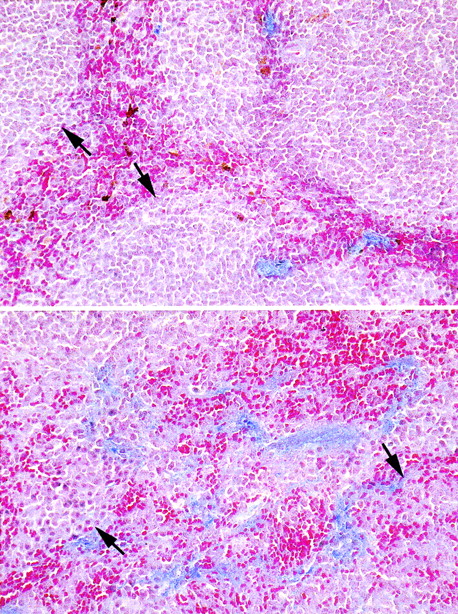
In contrast, the spleen exhibited striking abnormalities. Consistent with previous observations, the lymphoid follicles were small and usually lacked germinal centers. The red pulp contained excess numbers of mature erythrocytes, yet at the same time the red pulp often contained prominent megakaryocytes and nonhematopoietic cells resembling endothelial cells or fibroblasts. Staining of the spleen for collagen revealed a thick network of collagen fibers in the red pulp (Figure 4), and often these surrounded individual lymphoid follicles. While fibrosis has been associated with the presence of excess numbers of megakaryocytes, it was noteworthy that, even in areas of the red pulp where there were massive accumulations of megakaryocytes, there was no collagen immediately surrounding such cells. The histology of the spleen was consistent with a diagnosis of myelosclerosis.
Altered spleen architecture in Mpl-/- c-MybPlt4/Plt4 mice. Histologic section of spleens from Mpl-/- c-MybPlt3/Plt3 (lower panel) and control Mpl-/- c-Myb+/+ mice (upper panel) showing the increased network of collagen fibers in the red pulp (blue) and lymphoid follicles (arrows), which were were small and usually lacked germinal centers in mutant mice. Sections were photographed to Kodachrome ASA64 film (Eastman Kodak, Rochester, NY) using a Zeiss Axiophot microscope (Zeiss, Thornwood, NY) and camera with a Zeiss 20× objective lens, 0.60 numerical aperture. The scanned images were processed using Adobe Photoshop software (Adobe, San Jose, CA).
Altered spleen architecture in Mpl-/- c-MybPlt4/Plt4 mice. Histologic section of spleens from Mpl-/- c-MybPlt3/Plt3 (lower panel) and control Mpl-/- c-Myb+/+ mice (upper panel) showing the increased network of collagen fibers in the red pulp (blue) and lymphoid follicles (arrows), which were were small and usually lacked germinal centers in mutant mice. Sections were photographed to Kodachrome ASA64 film (Eastman Kodak, Rochester, NY) using a Zeiss Axiophot microscope (Zeiss, Thornwood, NY) and camera with a Zeiss 20× objective lens, 0.60 numerical aperture. The scanned images were processed using Adobe Photoshop software (Adobe, San Jose, CA).
Discussion
Mice bearing the Plt3 or Plt4 mutations in the c-Myb gene exhibit a striking thrombocytosis. The half-life of platelets in these mice was normal, and the elevated platelet levels resulted from excess megakaryocytopoiesis. The numbers of megakaryocyte progenitor cells and maturing megakaryocytes were grossly elevated in the hematopoietic tissues of Mpl-/- c-MybPlt3/Plt3 and Mpl-/- c-MybPlt4/Plt4 mice, and in vivo labeling studies confirmed an enhanced rate of platelet production in Plt4 mutant mice relative to mice lacking c-Myb mutations.
Previous studies have suggested that c-Myb activity may influence the balance between megakaryocyte differentiation and that of other hematopoietic lineages.3,11 We therefore examined multipotential hematopoietic cells in Plt4 mutant mice using both in vitro and in vivo assays. The mutant marrow populations contained elevated numbers of BL-CFCs, and these formed large, apparently healthy colonies with the usual stimuli. However, mutant blast colony cells failed to exhibit excessive megakaryocyte colony-forming capacity, suggesting that the excess numbers of megakaryocyte progenitor cells in Plt4 mutant mice are unlikely to be derived from BL-CFCs. In contrast, CFU-Ss in Plt4 mutant mice appeared to harbor significantly elevated capacity to generate megakaryocyte progenitor cells. Upon transplantation of marrow from mutant mice into irradiated recipients, the day 12 spleen colonies were present in increased numbers relative to c-Myb+/+ controls, but they were abnormally small. Nevertheless, cells from Plt4 mutant bone marrow generated excessive numbers of committed megakaryocyte progenitor cells in the spleens of recipients, at a level greater than the modest increase in granulocyte and macrophage progenitors that was observed relative to cells from control donor mice. Thus, in Plt4 mutant mice, altered c-Myb function appears to have skewed differentiation commitment in CFU-Ss in favor of excessive megakaryocytopoiesis.
While these data suggest that anomalous hematopoietic commitment contributes to excessive megakaryocytopoiesis in c-Myb mutant mice, a remarkable aspect of the phenotype in Mpl-/-c-MybPlt3/Plt3 and Mpl-/- c-MybPlt4/Plt4 mice is that proliferation of megakaryocyte progenitor cells and the production and differentiation of megakaryocytes are independent of signaling through c-Mpl, the receptor for TPO. The excess numbers of megakaryocytes and their progenitors as well as the degree of thrombocytosis itself were not exacerbated when the Plt3 or Plt4 mutations were established on a Mpl+/+ background. Indeed, while the numbers of megakaryocyte progenitor cells responsive to IL-3 or GM-CSF were elevated in Mpl+/+ c-MybPlt4/Plt4 mice, the number of TPO-responsive progenitor cells was no greater than in Mpl+/+ c-Myb+/+ controls. Because circulating TPO concentration is inversely proportional to the number of megakaryocytes and platelets,16 this latter observation may be due to a paucity of available TPO in the severely thrombocytotic mutant mice. Nevertheless, this observation reinforces the TPO-independent character of megakaryocytopoiesis in Plt3 and Plt4 mutant mice. TPO is the major physiological regulator of megakaryocytopoiesis,17-19 but previous studies suggested that significant platelet production is possible in the absence of TPO.20 However, the TPO-independent supraphysiological levels observed in Mpl-/- c-MybPlt3/Plt3 and Mpl-/- c-MybPlt4/Plt4 mice were unexpected and are similar to those observed in mice treated with maximal stimulating doses of TPO.21 This finding supports the possibility that c-Myb, like v-Myb in AMV, may act to inhibit megakaryocyte and platelet production and that down-regulation of c-Myb may be an important action of the TPO signaling cascade.
While the thrombocytosis in Mpl-/- c-MybPlt3/Plt3 and Mpl-/- c-MybPlt4/Plt4 mice appears not to depend on TPO, other cytokines may be important stimuli of the excessive megakaryocytopoiesis evident in these mice. A consistent anomaly in cultures of bone marrow from mice bearing the c-MybPlt3 or c-MybPlt4 mutations was the ability of GM-CSF to stimulate megakaryocyte colony formation. Previous studies have shown that GM-CSF can stimulate normal marrow cells to form megakaryocyte colonies but only at concentrations 200-fold higher than those stimulating maximum numbers of granulocyte-macrophage colonies to develop.15 While the quantitative responsiveness of granulocyte-macrophage colony formation by Plt3 and Plt4 mutant cells to stimulation by GM-CSF was normal, megakaryocyte colony formation showed similar dose responsiveness. This observation implies that megakaryocytic progenitor cells carrying the Plt3 or Plt4 mutant c-Myb genes are significantly more sensitive to GM-CSF than cells expressing the normal c-Myb gene. Interestingly, IL-6 was able to stimulate small numbers of megakaryocyte colonies to develop from mutant marrow, an activity not usually observed in cultures of wild-type cells, and hyperresponsiveness to the combination of SCF plus IL-3 plus EPO was also evident. However, expansion of megakaryocytopoiesis by excessive stimulation by cytokines such as GM-CSF or IL-6 is usually accompanied by megakaryocyte hypermaturation—increased megakaryocyte volume and DNA ploidy22 —the opposite of that we observed in Plt4 mutant mice. Thus, while altered responsiveness to cytokines may contribute to the expanded production of megakaryocytes and their progenitors, further studies will be required to define their role. Recent reports have also implicated chemokines, including stromal derived factor-1 (SDF-1) as well as fibroblast growth factor-4 (FGF-4), as mediators of interactions between progenitor cells and the marrow microenvironment that promote thrombopoiesis, and this activity is independent of TPO.23 Chemokines may therefore also contribute to the excessive platelet production in Plt3 and Plt4 mutant mice.
The phenotypes of mice carrying the Plt3 or Plt4 mutant allele of c-Myb are remarkably similar. Each of the anomalies in megakaryocytopoiesis described here, as well as those evident in other lineages,12 was almost indistinguishable in mice carrying these independent mutations despite the fact that the Plt3 and Plt4 mutations affect distinct domains of the c-Myb protein (DNA-binding and leucine zipper domains, respectively). Moreover, several aspects of the phenotype are similar to those in mice carrying a hypomorphic allele of c-Myb, in which an unmutated form of the protein is expressed at lower than normal levels.11 Mice expressing an altered form of p300 that cannot bind c-Myb also display a similar phenotype.13 These similarities suggest that a number of changes that result in reduced levels of c-Myb activity or expression can produce a common set of phenotypic changes. Nevertheless, differences in the viability of these various mutants are apparent (Emambokus et al11 and M.R.C., unpublished data, December 2004), and other subtle differences are also likely to emerge with further study. Thus, these mice provide an opportunity to dissect the contributions of various c-Myb domains to hematopoietic control and also to establish whether the myelodysplasia displayed by the mice will evolve into leukemia.
Prepublished online as Blood First Edition Paper, January 21, 2005; DOI 10.1182/blood-2004-12-4806.
Supported by grants from the Australian National Health and Medical Research Council (program no. 257500), the Cancer Council Victoria, the JD and L Harris Trust, and MuriGen Proprietary Limited.
An Inside Blood analysis of this article appears in the front of this issue.
The publication costs of this article were defrayed in part by page charge payment. Therefore, and solely to indicate this fact, this article is hereby marked “advertisement” in accordance with 18 U.S.C. section 1734.
We thank Jason Corbin and Janelle Lochland for excellent technical assistance, Jaclyn Cushen and Kristy Vella for animal husbandry, and Steven Mihajlovic for histology.


This feature is available to Subscribers Only
Sign In or Create an Account Close Modal